. 2013 Apr 19;8(4):e62391.
doi: 10.1371/journal.pone.0062391. Print 2013.

Neurotensin co-expressed in orexin-producing neurons in the lateral hypothalamus plays an important role in regulation of sleep/wakefulness states

Affiliations

Neurotensin co-expressed in orexin-producing neurons in the lateral hypothalamus plays an important role in regulation of sleep/wakefulness states

Naoki Furutani et al. PLoS One. .

Abstract

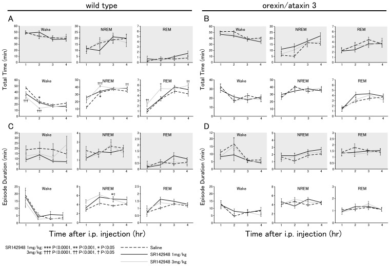
Both orexin and neurotensin are expressed in the lateral hypothalamic area (LHA) and have been implicated in the regulation of feeding, motor activity and the reward system. A double label immunofluorescence and in situ hybridization studies showed that neurotensin colocalizes with orexin in neurons of the LHA. Pharmacological studies suggested that neurotensin excites orexin-producing neurons (orexin neurons) through activation of neurotensin receptor-2 (NTSR-2) and non-selective cation channels. In situ hybridization study showed that most orexin neurons express neurotensin receptor-2 mRNA but not neurotensin receptor-1 (Ntsr-1) mRNA. Immunohistochemical studies showed that neurotensin-immunoreactive fibers make appositions to orexin neurons. A neurotensin receptor antagonist decreased Fos expression in orexin neurons and wakefulness time in wild type mice when administered intraperitoneally. However, the antagonist did not evoke any effect on these parameters in orexin neuron-ablated mice. These observations suggest the importance of neurotensin in maintaining activity of orexin neurons. The evidence presented here expands our understanding of the regulatory mechanism of orexin neurons.


Conflict of interest statement

Competing Interests: The authors have declared that no competing interests exist.

Figures

Figure 1
Figure 1. Neurotensin colocalizes with orexin in LHA neurons.
A, Immunostaining of neurotensin in a coronal section of mouse brain. Dense staining of neurotensin-like immunoreactivity is observed in the central nucleus of the amygdala (CeA), lateral hypothalamic area (LHA) and zona incerta (ZI). B, Double immunofluorescence study showing that neurotensin-containing neurons also express orexin in the LHA but not in the arcuate nucleus (ARC). Upper panels, neurotensin (green)- and orexin (red)-like immunoreactivity in the LHA (upper panels) and arcuate nucleus (lower pannels). Scale bar indicates 20 µm. C, Double-label in situ hybridization histochemistry showing distribution of orexin (brown) and neurotensin (blue) mRNAs. Left panel, Representative low power image of the LHA region (bregma −1.46 mm) of a coronal section of C57BL/6 mouse brain. Right panel, higher power view of the region indicated by a black rectangle in the left panel. Yellow arrowheads show prepro-orexin mRNA, and blue arrowheads show neurotensin mRNA. Red arrowheads show neurons expressing both neurotensin and orexin mRNAs. Rectangles show typical examples of orexin-single-positive, neurotensin-single-positive, and double-positive cells. Scale bar indicates 20 µm. D, Numbers of neurotensin- or orexin-positive neurons in LHA of wild-type (Wild-Type, n = 5) and orexin-ataxin-3 (Ataxin-3, n = 5) mice revealed by double labeling in situ hybridization. White, grey and black bars show neurotensin-, orexin- and double-positive neurons, respectively.
Figure 2
Figure 2. Neurotensin activates orexin neurons.
A, Whole cell current clamp recording of orexin neurons shows that neurotensin (100 nM) depolarized orexin neurons in the absence (top) or presence (bottom) of TTX (1 µM). B, Concentration dependence of neurotensin-induced depolarization (mV) in the presence of TTX. EC50 and Emax were 3.84±0.02 nM and 7.60±0.01 mV, respectively (n = 3–5). C, Effects of NTSR-1 or NTSR-2 antagonist on effects of neurotensin on orexin neurons. Both SR142948 (n = 7, 10 µM), a non-selective antagonist, and levocabastine (n = 7, 1 µM), an NTSR-2 preferential antagonist, almost completely blocked the effects of neurotensin-induced inward current in orexin neurons. Left panel, a typical trace. Right panel, effect of neurotensin-induced current in orexin neurons in the presence of SR142948 or lebocavastine. Extracellular solution was used as vehicle control. D, Double-labeling in situ hybridization shows most neurons expressing orexin mRNA (red) also express Ntsr-2 mRNA (blue). Black arrowheads show colocalization. Scale bar indicates 20 µm. E, Effect of extracellular Na+ on neurotensin-induced depolarization. NaCl was replaced by an equimolar concentration of choline chloride in the presence of TTX (1 µM) (Na+-free). Neurotensin (10 nM)-induced depolarization was markedly decreased in Na+-free solution (n = 4). F, Effect of extracellular Ca2+ on neurotensin-induced inward current. Neurotensin (10 nM) induced an inward current in the presence of extracellular calcium. This inward current was markedly increased in Ca2+-free extracellular solution (n = 7). Lower panel shows the fold-increase in inward current (pA) in Ca2+-free solution. G, Current-voltage relationship obtained by the current step protocol using a CsCl pipette in K+- and Ca2+-free extracellular solution. The steady state potential, at the end of the current step (marked by circle in left panel), is plotted in the current-voltage relationship in the right panel. I-V curve shows that the reversal potential of the neurotensin (10 nM)-induced current was 9.82 mV (n = 6–10). Neurons were current-clamped and the current was stepped from –240 pA to +280 pA (with increments of 40 pA with a duration of 100 ms). Open circle indicates control and filled circle indicates neurotensin (10 nM) application. H, Effects of non-selective cation channel blocker, SKF96365. Left panel, SKF96365 (10 µM) inhibited neurotensin-induced depolarization. Right panel, Bar graph indicates that SKF96365 inhibited neurotensin-induced depolarization (n = 4). Values are mean±S.E.M. *, p<0.05.
Figure 3
Figure 3. Neurotensinergic fibers appose orexin neurons.
A, Double immunofluorescence study shows that many varicose terminals with neurotensin-like immunoreactivity (green) make appositions to orexin neurons (red) in the LHA (Left panels). Right panel, high power view of rectangular region in left panel. B, Photomicrograph showing distribution of Fos (black nuclear label) and orexin (brown label) expression in LHA. Scale bar indicates 40 µm. Upper panels, left panel, control (saline injection); right panel, SR142948 injection at 20:45. Animals were sacrificed at 21:45 and subjected to immunostaining. Lower panels, left panel, control (saline injection); right panel, SR142948 injection at 8:45. Animals were sacrificed at 9:45 and subjected to immunostaining. C, SR142948 administration decreased Fos immunoreactivity of orexin neurons, but not in MCH neurons.
Figure 4
Figure 4. A neurotensin receptor antagonist SR142948 dose-dependently decreased wakefulness time and duration during the lights on period in wild-type mice, but not in orexin neuron-ablated mice.
Hourly amounts (A, B) and average episode duration (C, D) of awake, non-REM, and REM sleep states (mean±SE) plotted over 4 hr after administration of saline (dotted line) or SR142948 (solid line) in wild-type (n = 27, 14 and 7 for saline, SR 1 mg/kg, and 3 mg/kg, respectively) (A, C) and orexin/ataxin-3 mice (n = 14, 14 and 7 for saline, SR 1 mg/kg, and 3 mg/kg, respectively) (B, D). Data for the dark phase are displayed in shaded panels. SR142948 was administered at the start of light or dark periods (8:45 or 20:45).

References

    1. Dicou E, Vincent JP, Mazella J (2004) Neurotensin receptor-3/sortilin mediates neurotensin-induced cytokine/chemokine expression in a murine microglial cell line. J Neurosci Res 78: 92–99. - PubMed
    1. Martin S, Vincent JP, Mazella J (2003) Involvement of the neurotensin receptor-3 in the neurotensin-induced migration of human microglia. J Neurosci 23: 1198–1205. - PMC - PubMed
    1. Chabry J, Labbe-Jullie C, Gully D, Kitabgi P, Vincent JP, et al. (1994) Stable expression of the cloned rat brain neurotensin receptor into fibroblasts: binding properties, photoaffinity labeling, transduction mechanisms, and internalization. J Neurochem 63: 19–27. - PubMed
    1. Najimi M, Gailly P, Maloteaux JM, Hermans E (2002) Distinct regions of C-terminus of the high affinity neurotensin receptor mediate the functional coupling with pertussis toxin sensitive and insensitive G-proteins. FEBS Lett 512: 329–333. - PubMed
    1. Chalon P, Vita N, Kaghad M, Guillemot M, Bonnin J, et al. (1996) Molecular cloning of a levocabastine-sensitive neurotensin binding site. FEBS Lett 386: 91–94. - PubMed

Publication types

MeSH terms

LinkOut - more resources