. 2013 Oct;19(19-20):2201-14.
doi: 10.1089/ten.TEA.2012.0650. Epub 2013 Jun 5.

Marine collagen scaffolds for nasal cartilage repair: prevention of nasal septal perforations in a new orthotopic rat model using tissue engineering techniques

Affiliations

Marine collagen scaffolds for nasal cartilage repair: prevention of nasal septal perforations in a new orthotopic rat model using tissue engineering techniques

Christian Bermueller et al. Tissue Eng Part A. 2013 Oct.

Abstract

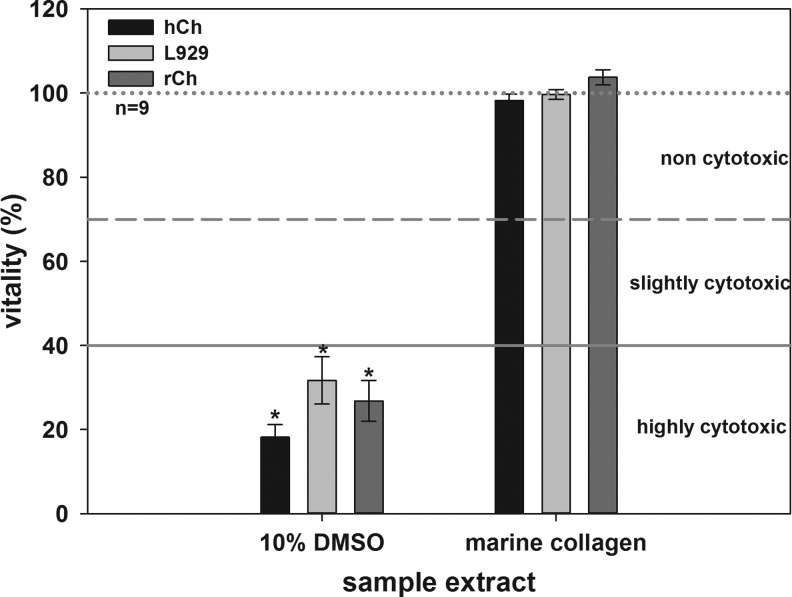
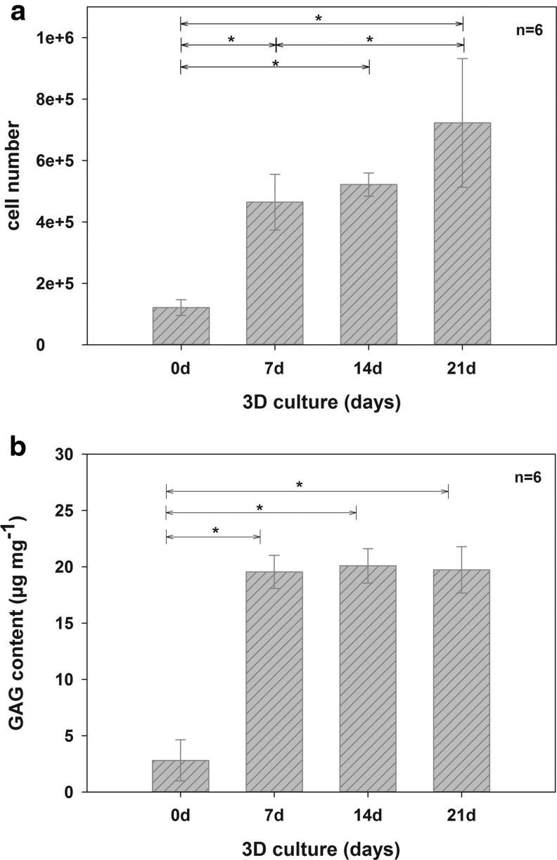
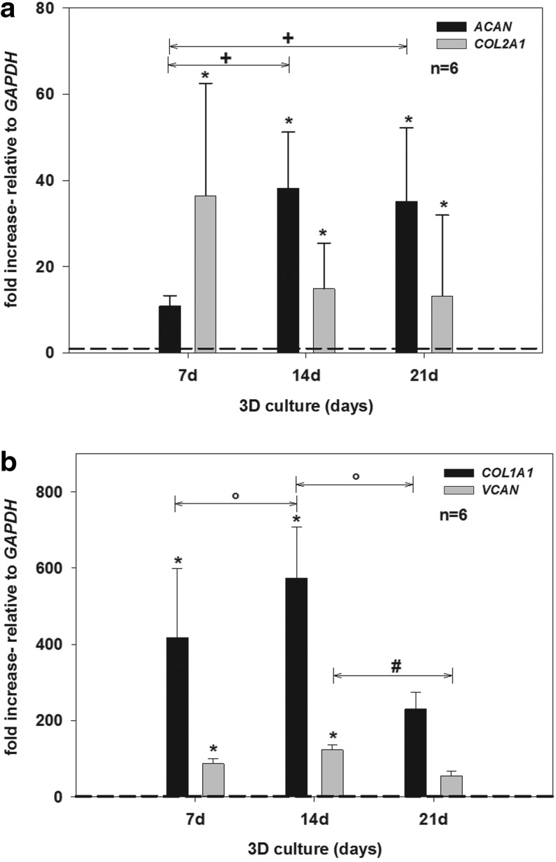
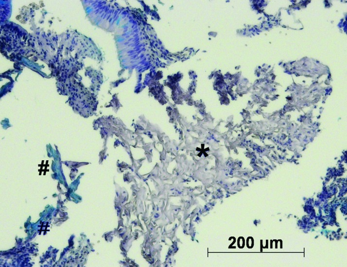
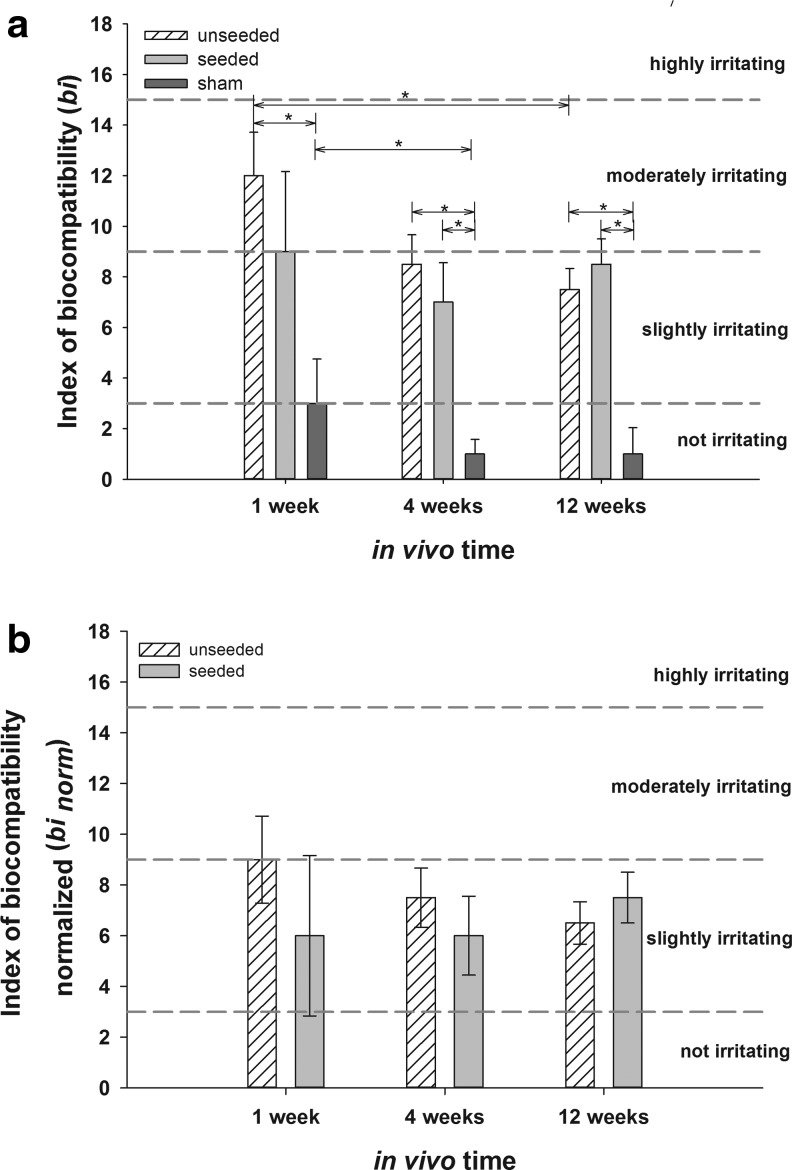
Autologous grafts are frequently needed for nasal septum reconstruction. Because they are only available in limited amounts, there is a need for new cartilage replacement strategies. Tissue engineering based on the use of autologous chondrocytes and resorbable matrices might be a suitable option. So far, an optimal material for nasal septum reconstruction has not been identified. The aim of our study was to provide the first evaluation of marine collagen for use in nasal cartilage repair. First, we studied the suitability of marine collagen as a cartilage replacement matrix in the context of in vitro three dimensional cultures by analyzing cell migration, cytotoxicity, and extracellular matrix formation using human and rat nasal septal chondrocytes. Second, we worked toward developing a suitable orthotopic animal model for nasal septum repair, while simultaneously evaluating the biocompatibility of marine collagen. Seeded and unseeded scaffolds were transplanted into nasal septum defects in an orthotopic rat model for 1, 4, and 12 weeks. Explanted scaffolds were histologically and immunohistochemically evaluated. Scaffolds did not induce any cytotoxic reactions in vitro. Chondrocytes were able to adhere to marine collagen and produce cartilaginous matrix proteins, such as collagen type II. Treating septal cartilage defects in vivo with seeded and unseeded scaffolds led to a significant reduction in the number of nasal septum perforations compared to no replacement. In summary, we demonstrated that marine collagen matrices provide excellent properties for cartilage tissue engineering. Marine collagen scaffolds are able to prevent septal perforations in an autologous, orthotopic rat model. This newly described experimental surgical procedure is a suitable way to evaluate new scaffold materials for their applicability in the context of nasal cartilage repair.


Figures

FIG. 1.
FIG. 1.
For orthotopic nasal septum replacement, the nasofrontal (*) and nasomaxillary (>) sutures were exposed and opened, whereas the internasal suture (°) was maintained (a). After detaching the mucosa, the complete nasal septal cartilage was removed (b). Color images available online at www.liebertpub.com/tea
FIG. 2.
FIG. 2.
SEM analysis of marine collagen scaffolds reveals high porosity and homogenous pore distribution on the scaffold surface (a) and within the complete scaffold matrix (b). Both views, the superficial top view (a) and the vertical cross section, demonstrate the distinct interconnectivity of the scaffold matrix.
FIG. 3.
FIG. 3.
In vitro cytotoxicity test of marine collagen. L929, rCh, and hCh were used as indicator cells. All cells cultured in negative control (dotted line, 100%), or in undiluted sample extracts demonstrate a high viability and reflect the noncytotoxic effect of the extracted marine collagen. No significant differences between negative controls and extracts were detectable. Compared to these results, the cytotoxic effect of 10% DMSO, used as positive control, is significant (each *p<0.05).
FIG. 4.
FIG. 4.
Changes in cell number (a) and content of sGAG (b) on marine collagen scaffolds after 0, 7, 14, and 21 days of three dimensional (3D) culture with hCh. Marine collagen scaffolds were initially seeded with 1×10⁶ hCh. During 3D culture, the cell number increased significantly. GAG neo synthesis per mg dry weight of unseeded compared to seeded and cultivated scaffolds. GAG synthesis significantly increased during the cultivation period of 21 days, due to enhanced GAG accumulation by human chondrocytes (*p<0.05). sGAG, sulfated glycosaminoglycan
FIG. 5.
FIG. 5.
Relative gene expression of chondrogenic, cartilage-specific marker genes (ACAN and COL2A1) (a), and marker for dedifferentiation (COL1A1 and VCAN) (b), in 3D culture for 0, 7, 14, and 21 days using hCh. Relative gene expression was calculated by means of the 2-ΔΔCT formula. GAPDH was used as reference gene and values for COL2A1, ACAN, COL1A1, and VCAN expression on day 0 (monolayer culture) were set to 1 (dashed threshold line). COL2A1 and ACAN (a) were expressed at significantly higher levels compared with day 0 (each *p<0.05). With proceeding culture time a significant increase in gene expression of ACAN was detected (+p<0.05) while expression of COL2A1 remained on a stable level. Compared to the bench mark of day 0, VCAN and COL1A1 (b) expression increased significantly (each *p<0.05) during the first 14 days of cultivation. Subsequently, gene expression of both dedifferentiation markers significantly decreased during 3D culture (°COL1A1 p<0.05; #VCAN p<0.05).
FIG. 6.
FIG. 6.
Histological AB (a–c) and immunohistochemical staining for detection of ECM neo synthesis (Agg, collagen type I, and II; d–l) in marine collagen scaffolds seeded with hCh starting from day 7 (a, d, g, j) until day 14 (b, e, h, k) and 21 (c, f, i, l). The AB (a–c) staining reflects enhanced GAG deposition during 3D culture. 7 days after seeding (a) GAG accumulation was detectable and increased until day 14 (b) and 21 (c). A visible increase of GAG and aggrecan accumulation from day 7 (d) to day 14 (e) and 21 (f) was demonstrated. Neo synthesis of collagen type II (g–i) started after the first week and visibly increased during further culture after 14 (h) and 21 days (i). Collagen type I was present within the whole scaffold starting within one week after initial seeding (j) and increasing to day 14 (k). Distribution of collagen type I proceeded into the center of the scaffolds after 21 days. However, the intensity of the staining remained at a comparable level (l). *periphery of scaffold; °center of scaffold;→scaffold fibers; formula image 
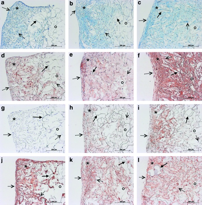
cell nuclei. Color images available online at
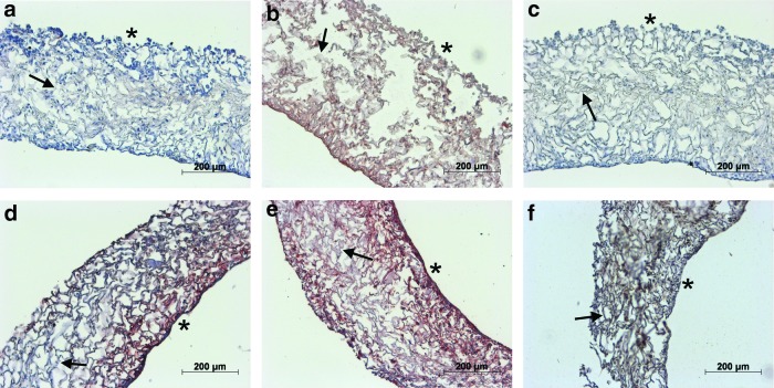
 www.liebertpub.com/tea
FIG. 7.
FIG. 7.
Marine collagen scaffolds seeded with rCh. While after one day (a) aggrecan was not detected, aggrecan deposition (red) became visible after 7 days (d). Collagen type I synthesis visibly increased (red) from day 1 (b) to day 7 (e). Within the tightly seeded marine collagen scaffold slight accumulation of collagen type II (brown) (c,f) was detectable after 7 days (f).→scaffold fibers; *seeded surface. Color images available online at www.liebertpub.com/tea
FIG. 8.
FIG. 8.
Presence of GAG in unseeded marine collagen and in scaffolds seeded with rCh. GAG content significantly increased during the cultivation period of 7 days (*p<0.05).
FIG. 9.
FIG. 9.
AB staining of remaining scaffold material (*) of unseeded marine collagen scaffolds after one week in vivo. Unseeded collagen scaffolds, demonstrate a slight accumulation of GAGs (#). Color images available online at www.liebertpub.com/tea
FIG. 10.
FIG. 10.
bi and binorm of seeded and unseeded scaffolds after 1, 4, and 12 weeks as median value±median deviation compared to sham group (bi, a) and after subtraction of the bi of the sham group (binorm, b) (*p<0.05). All detected bi values are within the slightly irritating range (Classification of bi: not irritating 0.0–2.9, slightly irritating 3.0–8.9, moderately irritating 9.0–15.0, and highly irritating >15.1).

References

    1. Rotter N. Haisch A. Bucheler M. Cartilage and bone tissue engineering for reconstructive head and neck surgery. Eur Arch Otorhinolaryngol. 2005;262:539. - PubMed
    1. Mandl E.W. Jahr H. Koevoet J.L. van Leeuwen J.P. Weinans H. Verhaar J.A. Van Osch G.J. Fibroblast growth factor-2 in serum-free medium is a potent mitogen and reduces dedifferentiation of human ear chondrocytes in monolayer culture. Matrix Biol. 2004;23:231. - PubMed
    1. Rotter N. Bucheler M. Haisch A. Wollenberg B. Lang S. Cartilage tissue engineering using resorbable scaffolds. J Tissue Eng Regen Med. 2007;1:411. - PubMed
    1. Nagata S. Modification of the stages in total reconstruction of the auricle: Part I. Grafting the three-dimensional costal cartilage framework for lobule-type microtia. Plast Reconstr Surg. 1994;93:221. - PubMed
    1. Ohara K. Nakamura K. Ohta E. Chest wall deformities and thoracic scoliosis after costal cartilage graft harvesting. Plast Reconstr Surg. 1997;99:1030. - PubMed

Publication types

LinkOut - more resources