

Recombinant adeno-associated virus integration sites in murine liver after ornithine transcarbamylase gene correction
- PMID: 23621841
- PMCID: PMC3655627
- DOI: 10.1089/hum.2012.112
Recombinant adeno-associated virus integration sites in murine liver after ornithine transcarbamylase gene correction
Abstract
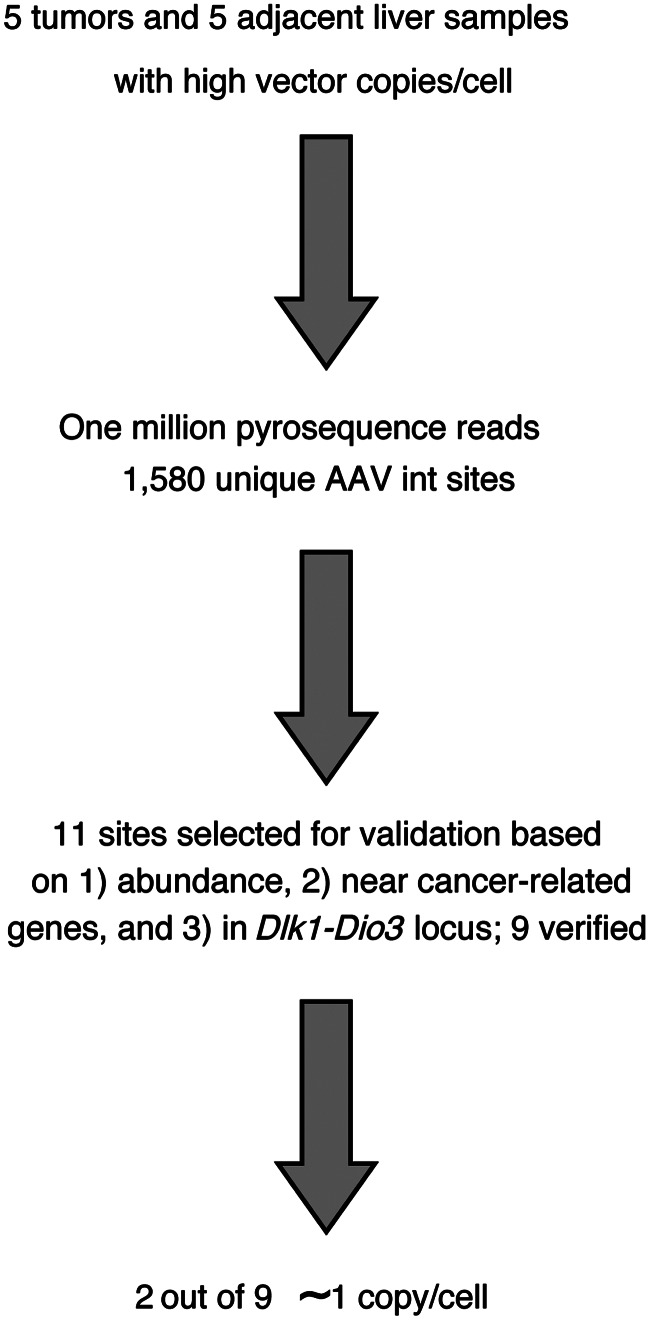
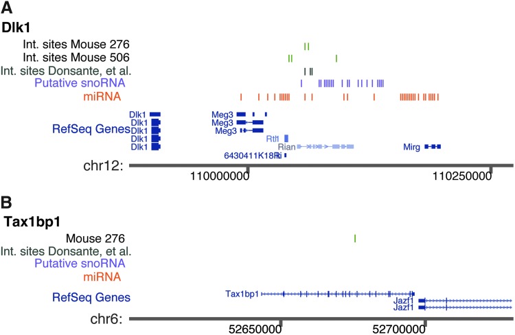
Recombinant adeno-associated viruses (rAAVs) have been tested in humans and other large mammals without adverse events. However, one study of mucopolysaccharidosis VII correction in mice showed repeated integration of rAAV in cells from hepatocellular carcinoma (HCC) in the Dlk1-Dio3 locus, suggesting possible insertional mutagenesis. In contrast, another study found no association of rAAV integration with HCC, raising questions about the generality of associations between liver transformation and integration at Dlk1-Dio3. Here we report that in rAAV-treated ornithine transcarbamylase (Otc)-deficient mice, four examples of integration sites in Dlk1-Dio3 could be detected in specimens from liver nodule/tumors, confirming previous studies of rAAV integration in the Dlk1-Dio3 locus in the setting of another murine model of metabolic disease. In one case, the integrated vector was verified to be present at about one copy per cell, consistent with clonal expansion. Another verified integration site in liver nodule/tumor tissue near the Tax1bp1 gene was also detected at about one copy per cell. The Dlk1-Dio3 region has also been implicated in human HCC and so warrants careful monitoring in ongoing human clinical trials with rAAV vectors.
Figures
Similar articles
-
Evaluating the state of the science for adeno-associated virus integration: An integrated perspective.Mol Ther. 2022 Aug 3;30(8):2646-2663. doi: 10.1016/j.ymthe.2022.06.004. Epub 2022 Jun 10. Mol Ther. 2022. PMID: 35690906 Free PMC article. Review.
-
DLK1-DIO3 genomic imprinted microRNA cluster at 14q32.2 defines a stemlike subtype of hepatocellular carcinoma associated with poor survival.J Biol Chem. 2011 Sep 2;286(35):30706-30713. doi: 10.1074/jbc.M111.229831. Epub 2011 Jul 7. J Biol Chem. 2011. PMID: 21737452 Free PMC article.
-
AAV-encoded OTC activity persisting to adulthood following delivery to newborn spf(ash) mice is insufficient to prevent shRNA-induced hyperammonaemia.Gene Ther. 2013 Dec;20(12):1184-7. doi: 10.1038/gt.2013.51. Epub 2013 Oct 10. Gene Ther. 2013. PMID: 24108150
-
AAV vector integration sites in mouse hepatocellular carcinoma.Science. 2007 Jul 27;317(5837):477. doi: 10.1126/science.1142658. Science. 2007. PMID: 17656716
-
[Integration of AAV vectors and insertional mutagenesis].Med Sci (Paris). 2016 Feb;32(2):167-74. doi: 10.1051/medsci/20163202010. Epub 2016 Mar 2. Med Sci (Paris). 2016. PMID: 26936174 Review. French.
Cited by
-
Pathogenesis of Hepatic Tumors following Gene Therapy in Murine and Canine Models of Glycogen Storage Disease.Mol Ther Methods Clin Dev. 2019 Nov 11;15:383-391. doi: 10.1016/j.omtm.2019.10.016. eCollection 2019 Dec 13. Mol Ther Methods Clin Dev. 2019. PMID: 31890731 Free PMC article.
-
Integrated vector genomes may contribute to long-term expression in primate liver after AAV administration.Nat Biotechnol. 2024 Aug;42(8):1232-1242. doi: 10.1038/s41587-023-01974-7. Epub 2023 Nov 6. Nat Biotechnol. 2024. PMID: 37932420 Free PMC article.
-
A long-term study of AAV gene therapy in dogs with hemophilia A identifies clonal expansions of transduced liver cells.Nat Biotechnol. 2021 Jan;39(1):47-55. doi: 10.1038/s41587-020-0741-7. Epub 2020 Nov 16. Nat Biotechnol. 2021. PMID: 33199875 Free PMC article.
-
Evaluating the state of the science for adeno-associated virus integration: An integrated perspective.Mol Ther. 2022 Aug 3;30(8):2646-2663. doi: 10.1016/j.ymthe.2022.06.004. Epub 2022 Jun 10. Mol Ther. 2022. PMID: 35690906 Free PMC article. Review.
-
AAV Infection: Protection from Cancer.Hum Gene Ther. 2017 Apr;28(4):323-327. doi: 10.1089/hum.2016.147. Epub 2016 Nov 10. Hum Gene Ther. 2017. PMID: 27832705 Free PMC article.
References
-
- Bell P. Moscioni A.D. Mccarter R.J. Wu D., et al. Analysis of tumors arising in male B6C3F1 mice with and without AAV vector delivery to liver. Mol. Ther. 2006;14:34–44. - PubMed
-
- Donsante A. Miller D.G. Li Y. Vogler C., et al. AAV vector integration sites in mouse hepatocellular carcinoma. Science. 2007;317:477. - PubMed
Publication types
MeSH terms
Substances
Grants and funding
LinkOut - more resources
Full Text Sources
Other Literature Sources
