. 2013 Apr 27:14:98.
doi: 10.1186/1471-2369-14-98.

Dipeptidyl peptidase IV inhibitor attenuates kidney injury in rat remnant kidney

Affiliations

Dipeptidyl peptidase IV inhibitor attenuates kidney injury in rat remnant kidney

Kwon Wook Joo et al. BMC Nephrol. .

Abstract

Background: The inhibition of dipeptidyl peptidase (DPP) IV shows protective effects on tissue injury of the heart, lung, and kidney. Forkhead box O (FoxO) transcriptional factors regulate cellular differentiation, growth, survival, the cell cycle, metabolism, and oxidative stress. The aims of this study were to investigate whether the DPP IV inhibitor sitagliptin could attenuate kidney injury and to evaluate the status of FoxO3a signaling in the rat remnant kidney model.

Methods: Rats were received two-step surgery of 5/6 renal mass reduction and fed on an oral dose of 200 mg/kg/day sitagliptin for 8 weeks. Before and after the administration of sitagliptin, physiologic parameters were measured. After 8 weeks of treatment, the kidneys were harvested.

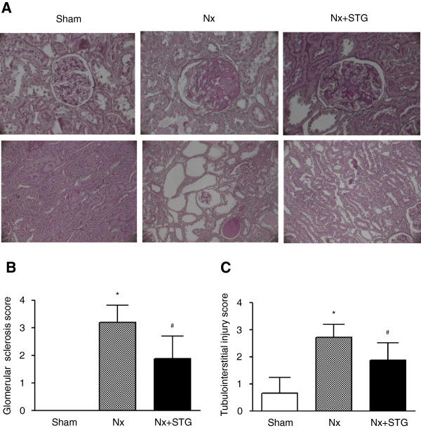
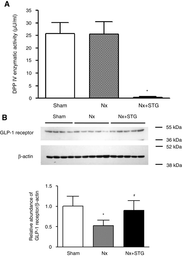
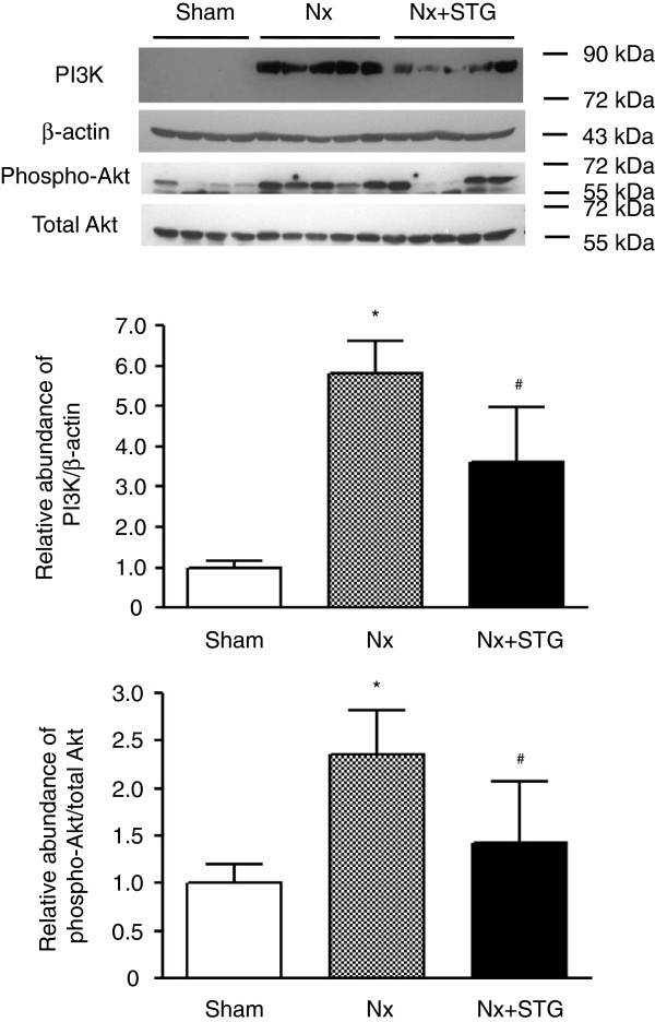
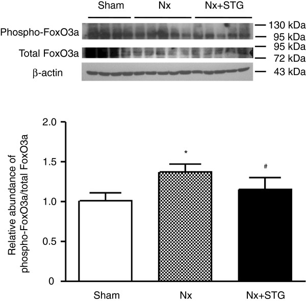
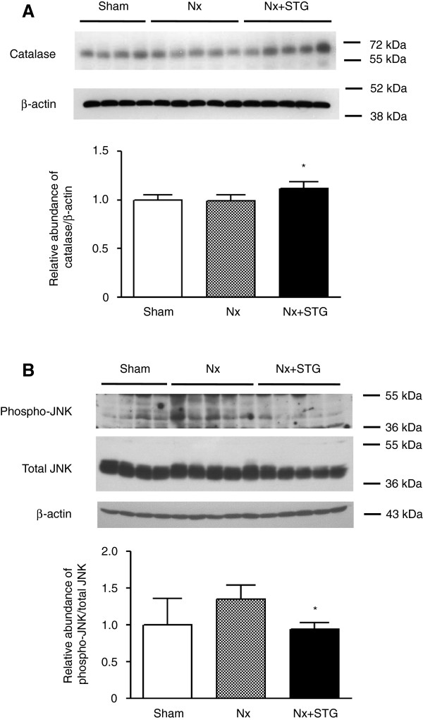
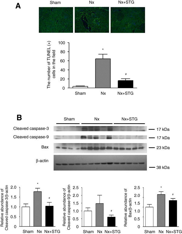
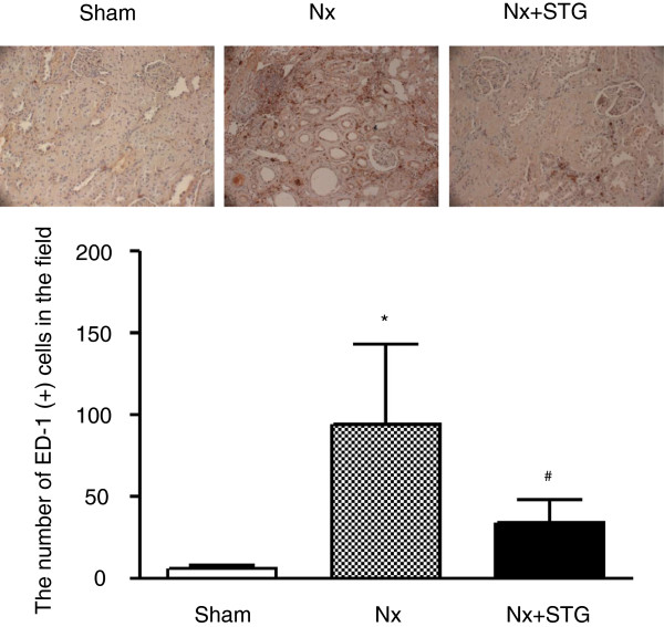
Results: The sitagliptin treatment attenuated renal dysfunction. A histological evaluation revealed that glomerulosclerosis and tubulointerstitial injury were significantly decreased by sitagliptin. Sitagliptin decreased DPP IV activity and increased the renal expression of glucagon-like peptide-1 receptor (GLP-1R). The subtotal nephrectomy led to the activation of phosphatidylinositol 3-kinase (PI3K)-Akt and FoxO3a phosphorylation, whereas sitagliptin treatment reversed these changes, resulting in PI3K-Akt pathway inactivation and FoxO3a dephosphorylation. The renal expression of catalase was increased and the phosphorylation of c-Jun N-terminal kinase (JNK) was decreased by sitagliptin. Sitagliptin treatment reduced apoptosis by decreasing cleaved caspase-3 and -9 and Bax levels and decreased macrophage infiltration.

Conclusions: In rat remnant kidneys, DPP IV inhibitor attenuated renal dysfunction and structural damage. A reduction of apoptosis, inflammation and an increase of antioxidant could be suggested as a renoprotective mechanism together with the activation of FoxO3a signaling. Therefore, DPP IV inhibitors might provide a promising approach for treating CKD, but their application in clinical practice remains to be investigated.


Figures

Figure 1
Figure 1
Effects of sitagliptin on renal histology. Representative images of H&E-stained renal tissues (A). Histological evaluation of glomerular sclerosis (B) and tubulointerstitial injury (C). The extent of glomerular sclerosis and tubulointerstitial injury was evaluated and scored. The results are expressed as the mean ± s.d. *Significantly different with respect to the sham-operated rats; #significantly different with respect to the nephrectomized rats; *#P < 0.05. Sham, sham operation; Nx, nephrectomy; Nx+STG, nephrectomy and sitagliptin treatment. Magnification × 400 (upper panel) and x 200 (lower panel).
Figure 2
Figure 2
Effects of sitagliptin on DPP IV activity and GLP-1 receptor expression. DPP IV enzymatic activity in serum of rats from each group (A). Representative western blot and group data showing GLP-1 receptor protein abundance in the kidneys of rats from each group (B). The intensity of the bands corresponding to the GLP-1 receptor was corrected by β-actin levels to obtain relative measures of GLP-1 receptor expression among all samples. The results are expressed as the mean ± s.d. *Significantly different with respect to the sham-operated rats; #significantly different with respect to the nephrectomized rats; *#P < 0.05. Sham, sham operation; Nx, nephrectomy; Nx+STG, nephrectomy and sitagliptin treatment.
Figure 3
Figure 3
Effects of sitagliptin on PI3K and Akt activity. Representative western blot and group data showing PI3K protein abundance and Akt phosphorylation in the kidneys of rats from each group. The intensity of the bands corresponding to PI3K was corrected by β-actin levels, and phospho-Akt intensity was corrected by total Akt levels. The results are expressed as the mean ± s.d. *Significantly different with respect to the sham-operated rats; #significantly different with respect to the nephrectomized rats; *#P < 0.05. Sham, sham operation; Nx, nephrectomy; Nx+STG, nephrectomy and sitagliptin treatment.
Figure 4
Figure 4
Effects of sitagliptin on FoxO3a phosphorylation. Representative western blot and group data showing FoxO3a phosphorylation in the kidneys of rats from each group. The intensity of the bands corresponding to phospho-FoxO3a was corrected by total FoxO3a levels to obtain relative measures of FoxO3a phosphorylation among all samples. The results are expressed as the mean ± s.d. *Significantly different with respect to the sham-operated rats; #significantly different with respect to the nephrectomized rats; *#P < 0.05. Sham, sham operation; Nx, nephrectomy; Nx+STG, nephrectomy and sitagliptin treatment.
Figure 5
Figure 5
Effects of sitagliptin on oxidative stress. Representative western blot and group data showing catalase expression (A) and JNK phosphorylation (B) in the kidneys of rats from each group. The intensity of the bands corresponding to catalase was corrected by β-actin levels, and phospho-JNK intensity was corrected by total JNK levels. The results are expressed as the mean ± s.d. *Significantly different with respect to the nephrectomized rats; *P < 0.05. Sham, sham operation; Nx, nephrectomy; Nx+STG, nephrectomy and sitagliptin treatment; JNK, c-Jun N-terminal kinase.
Figure 6
Figure 6
Effects of sitagliptin on apoptosis. Apoptotic cells detected in the kidneys stained with TUNEL (A). Representative western blot and group data showing cleaved caspase-3, cleaved caspase-9, and Bax protein abundance in the kidneys of rats from each group (B). The intensity of the bands corresponding to cleaved caspase-3, cleaved caspase-9, and Bax was corrected by β-actin levels. The results are expressed as the mean ± s.d. *Significantly different with respect to the sham-operated rats; #significantly different with respect to the nephrectomized rats; *#P < 0.05. Sham, sham operation; Nx, nephrectomy; Nx+STG, nephrectomy and sitagliptin treatment. Magnification × 200.
Figure 7
Figure 7
Effects of sitagliptin on the infiltration of macrophages. Immunohistochemical stainings for ED-1 in representative kidneys of experimental animals. The number of ED-1-positive cells per field was counted in each group. The results are expressed as the mean ± s.d. *Significantly different with respect to the sham-operated rats; #significantly different with respect to the nephrectomized rats; *#P < 0.05. Sham, sham operation; Nx, nephrectomy; Nx+STG, nephrectomy and sitagliptin treatment. Magnification × 200.

References

    1. Baggio LL, Drucker DJ. Biology of incretins: GLP-1 and GIP. Gastroenterology. 2007;132:2131–2157. doi: 10.1053/j.gastro.2007.03.054. - DOI - PubMed
    1. Mentlein R. Dipeptidyl-peptidase IV (CD26)–role in the inactivation of regulatory peptides. Regul Pept. 1999;85:9–24. doi: 10.1016/S0167-0115(99)00089-0. - DOI - PubMed
    1. Drucker DJ, Nauck MA. The incretin system: glucagon-like peptide-1 receptor agonists and dipeptidyl peptidase-4 inhibitors in type 2 diabetes. Lancet. 2006;368:1696–1705. doi: 10.1016/S0140-6736(06)69705-5. - DOI - PubMed
    1. Park CW, Kim HW, Ko SH, Lim JH, Ryu GR, Chung HW, Han SW, Shin SJ, Bang BK, Breyer MD, Chang YS. Long-term treatment of glucagon-like peptide-1 analog exendin-4 ameliorates diabetic nephropathy through improving metabolic anomalies in db/db mice. J Am Soc Nephrol. 2007;18:1227–1238. doi: 10.1681/ASN.2006070778. - DOI - PubMed
    1. Kodera R, Shikata K, Kataoka HU, Takatsuka T, Miyamoto S, Sasaki M, Kajitani N, Nishishita S, Sarai K, Hirota D, Sato C, Ogawa D, Makino H. Glucagon-like peptide-1 receptor agonist ameliorates renal injury through its anti-inflammatory action without lowering blood glucose level in a rat model of type 1 diabetes. Diabetologia. 2011;54:965–978. doi: 10.1007/s00125-010-2028-x. - DOI - PubMed

Publication types