. 2013 Apr 28:13:211.
doi: 10.1186/1471-2407-13-211.

Evidence for tankyrases as antineoplastic targets in lung cancer

Affiliations

Evidence for tankyrases as antineoplastic targets in lung cancer

Alexander M Busch et al. BMC Cancer. .

Abstract

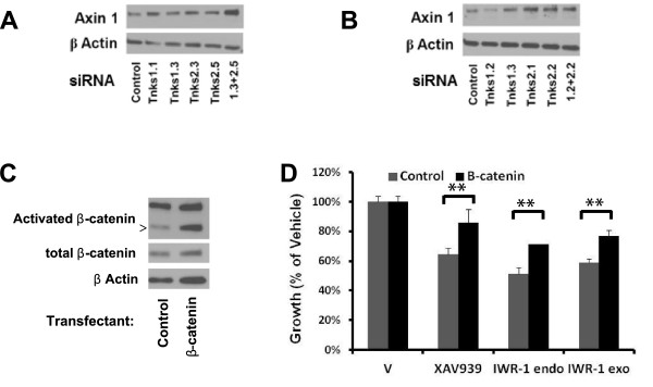
Background: New pharmacologic targets are urgently needed to treat or prevent lung cancer, the most common cause of cancer death for men and women. This study identified one such target. This is the canonical Wnt signaling pathway, which is deregulated in cancers, including those lacking adenomatous polyposis coli or β-catenin mutations. Two poly-ADP-ribose polymerase (PARP) enzymes regulate canonical Wnt activity: tankyrase (TNKS) 1 and TNKS2. These enzymes poly-ADP-ribosylate (PARsylate) and destabilize axin, a key component of the β-catenin phosphorylation complex.

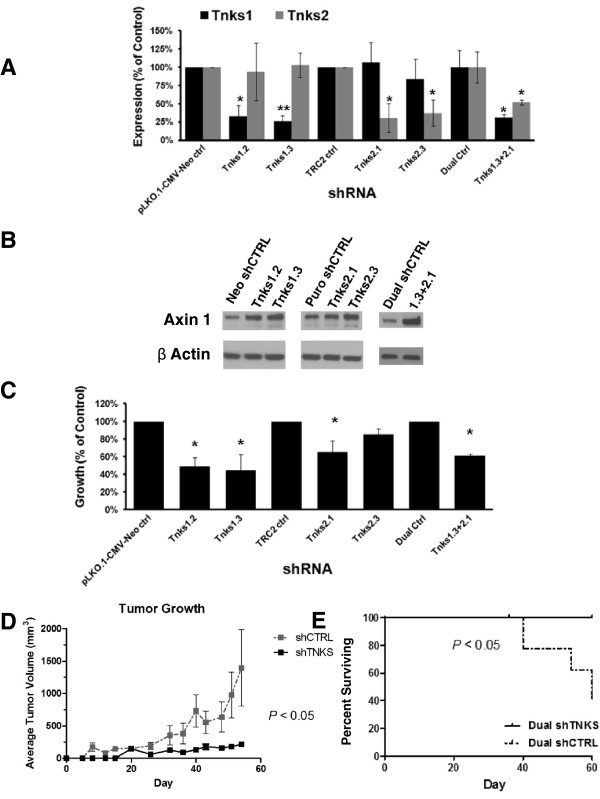
Methods: This study used comprehensive gene profiles to uncover deregulation of the Wnt pathway in murine transgenic and human lung cancers, relative to normal lung. Antineoplastic consequences of genetic and pharmacologic targeting of TNKS in murine and human lung cancer cell lines were explored, and validated in vivo in mice by implantation of murine transgenic lung cancer cells engineered with reduced TNKS expression relative to controls.

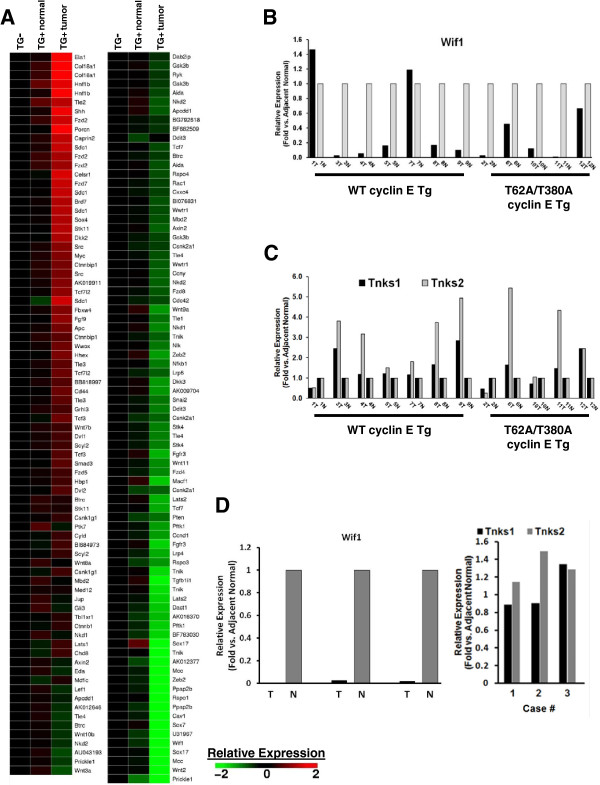
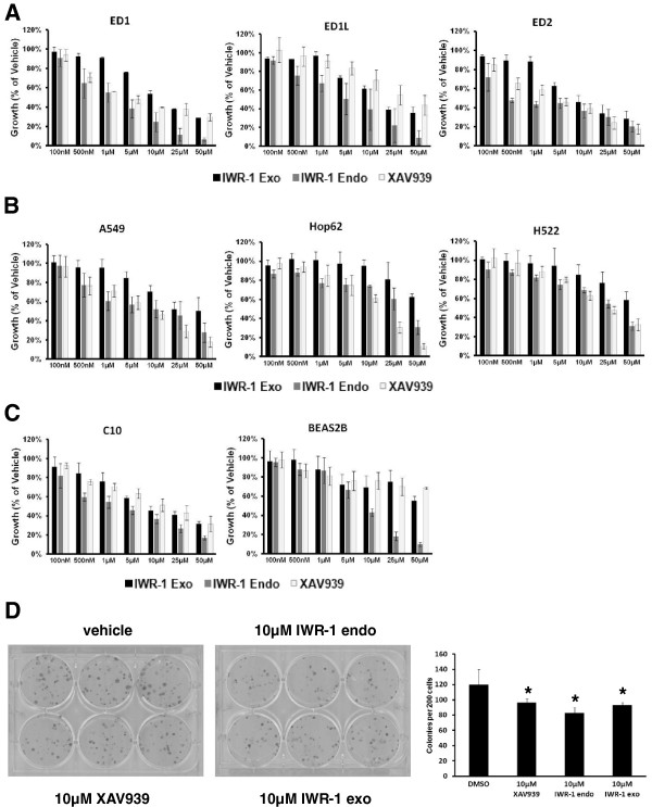
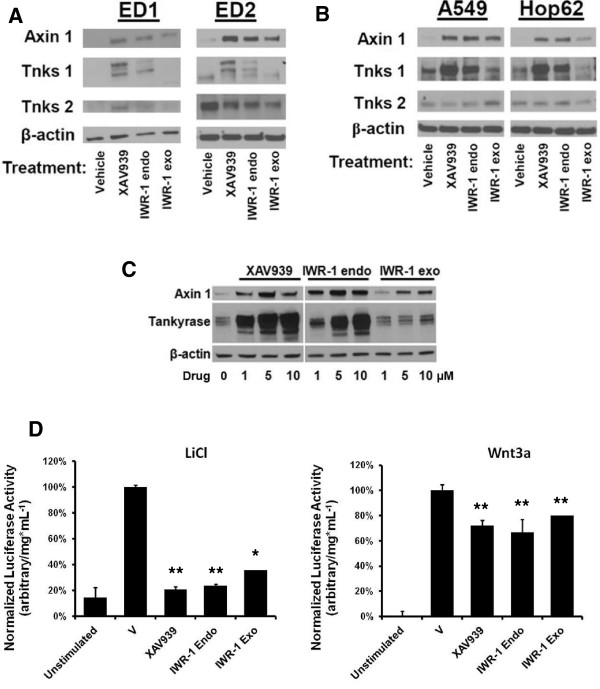
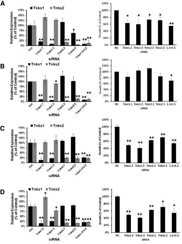
Results: Microarray analyses comparing Wnt pathway members in malignant versus normal tissues of a murine transgenic cyclin E lung cancer model revealed deregulation of Wnt pathway components, including TNKS1 and TNKS2. Real-time PCR assays independently confirmed these results in paired normal-malignant murine and human lung tissues. Individual treatments of a panel of human and murine lung cancer cell lines with the TNKS inhibitors XAV939 and IWR-1 dose-dependently repressed cell growth and increased cellular axin 1 and tankyrase levels. These inhibitors also repressed expression of a Wnt-responsive luciferase construct, implicating the Wnt pathway in conferring these antineoplastic effects. Individual or combined knockdown of TNKS1 and TNKS2 with siRNAs or shRNAs reduced lung cancer cell growth, stabilized axin, and repressed tumor formation in murine xenograft and syngeneic lung cancer models.

Conclusions: Findings reported here uncovered deregulation of specific components of the Wnt pathway in both human and murine lung cancer models. Repressing TNKS activity through either genetic or pharmacological approaches antagonized canonical Wnt signaling, reduced murine and human lung cancer cell line growth, and decreased tumor formation in mouse models. Taken together, these findings implicate the use of TNKS inhibitors to target the Wnt pathway to combat lung cancer.


Figures

Figure 1
Figure 1
Wnt pathway deregulation in murine and human NSCLC. Comprehensive gene expression microarrays and qPCR assays reveal deregulation of specific components of the Wnt signaling pathway in murine and human NSCLC. (A)161 probes on the array representing 117 unique genes defined by Gene Ontology under the classification Wnt receptor signaling pathway are significantly (ANOVA, P ≤ 0.05) over- or under-expressed in murine cyclin-E transgenic lung cancers as compared to adjacent normal or non-transgenic mouse lung. (B) The qPCR-based assays of Wif1 expression levels in a panel of paired malignant (samples labeled T) and normal (samples labeled N) lung tissues from 12 transgenic cyclin E mice, both wild type (WT) and proteasome-degradation resistant (T62A/T380A). (C) The qPCR-based measurements of TNKS1 and TNKS2 expression levels in the same panel of paired malignant and normal murine lung tissues. (D) The qPCR-based measurement of Wif1 (left panel) and TNKS1 and TNKS2 (right panel) expression levels in three human lung adenocarcinoma tumor samples versus adjacent normal lung tissue.
Figure 2
Figure 2
Antineoplastic effects of TNKS inhibitors in vitro. Treatments with TNKS inhibitors XAV939, IWR-1 endo, and IWR-1 exo exerted antineoplastic effects against NSCLC cell lines in vitro, as compared to vehicle controls. (A) Cell proliferation dose-response curves for the three TNKS inhibitors against ED1 (left panel), ED1L (middle panel), and ED2 (right panel) murine lung cancer cell lines as compared to vehicle control are shown, as measured by luminescent cell viability assay after 3 days. (B) Cell proliferation dose-response relationships for the three TNKS inhibitors against A549 (left panel), Hop62 (middle panel), and H522 human NSCLC cell lines as compared to vehicle control are shown, using the same luminescent cell viability assay and 3 day time frame. (C) Dose-response curves show antiproliferative effects of the three TNKS inhibitors against the immortalized bronchial epithelial cell lines C-10 (murine) and BEAS-2B (human) versus vehicle control. (D) Ten-day colony formation is shown for the ED1 cell line following individual treatment with 10 μM of each TNKS inhibitor or vehicle control (left two images), and quantified (right panel). Error bars represent standard deviations of three experiments in triplicate. (* p ≤ 0.05).
Figure 3
Figure 3
TNKS inhibition antagonizes canonical Wnt signaling in lung cancer. (A) Immunoblots for Wnt pathway components axin 1, TNKS1, and TNKS2 are shown in ED1 (left panel) and ED2 (right panel) murine lung cancer cell lines following 3 days treatment with TNKS inhibitors or vehicle. (B) Immunoblots are shown for Wnt pathway components, as in panel A, in A549 (left panel) and Hop62 (right panel) human NSCLC cell lines following 3 days treatment with TNKS inhibitors or vehicle. (C) Dose-response of Wnt pathway component stabilization is shown in ED1L cells following 3 days treatment with TNKS inhibitors or vehicle at doses shown. (D) Activity of a lentiviral Wnt-responsive luciferase construct stably expressed in the ED1 cell line was measured following 16 hours cotreatment with TNKS inhibitors and a Wnt activator: 20 mM LiCl (left panel) or 25 ng/mL recombinant murine Wnt3a (right panel). Luciferase activity was normalized to total protein concentrations in each sample and compared to vehicle control. Error bars represent SD of three experiments in triplicate. (* p ≤ 0.05).
Figure 4
Figure 4
Transient TNKS knockdown in lung cancer cell lines. Antineoplastic consequences of specific genetic knockdown of the TNKS via siRNAs are shown. Two independent siRNAs targeting respectively each of TNKS1 or TNKS2, or a non-targeting control were transfected into (A) ED1, (B) ED2, (C) A549, and (D) Hop62 cells, alone or in combination. Gene expression levels of TNKS1 and TNKS2, measured by qPCR assays, are shown following knockdown for 24 hours (left panels) as compared to control. Proliferative consequences were measured by luminescent cell viability assay after 3 days culture (right panels). Error bars represent SD of three experiments in triplicate. (* p ≤ 0.05, ** p ≤ 0.01).
Figure 5
Figure 5
In vitro effects of TNKS knockdown and rescue of Wnt pathway inhibition. (A) Protein expression levels of axin 1 following transient TNKS knockdown are shown by immunoblot analysis in ED1. (B) Protein expression levels of axin 1 following transient TNKS knockdown are shown by immunoblot analysis in Hop62. (C) Expression of activated β-catenin and total β-catenin in ED1 cells following stable infection with constitutively active β-catenin or empty vector. (D) Growth inhibition of ED1 cells infected as in C by TNKS inhibitors for 3 days at 10 μM is shown by luminescent cell viability assay. Error bars represent SD of three experiments in triplicate. (* p ≤ 0.05, ** p ≤ 0.01).
Figure 6
Figure 6
In vivo consequences of TNKS knockdown. (A) ED1 cells were infected with lentiviral shRNA constructs targeting TNKS1 or TNKS2 at two independent sites each, or the combination, and were selected with G418 (TNKS1 constructs and pLKO.1-CMV-Neo control), puromycin (TNKS2 constructs and TRC2 control), or the combination (dual control and dual shRNA). mRNA expression levels of the indicated species are detected by qPCR. (B) Axin 1 protein levels following stable TNKS knockdown are shown by immunoblot analysis. (C) Consequences on proliferation of stable TNKS knockdown in ED1 cells are shown in vitro as measured by luminescent cell viability assay 3 days after plating. (D) ED1 TNKS1 and TNKS2 shRNA dual transductants (or control) were injected into the flanks of athymic nude mice and tumor diameters measured twice weekly. Tumor growth rates are shown, N = 10 in each arm +/- SEM. (E) Time to the specified endpoint (designated as percent surviving) for the xenograft study is shown. Error bars represent SD of the mean. (* p ≤ 0.05).

Similar articles

Cited by

References

    1. Siegel R, Naishadham D, Jemal A. Cancer statistics, 2012. CA Cancer J Clin. 2012;62:10–29. - PubMed
    1. Jemal A, Bray F, Center MM, Ferlay J, Ward E, Forman D. Global cancer statistics. CA Cancer J Clin. 2011;61:69–90. - PubMed
    1. Moolgavkar SH, Holford TR, Levy DT, Kong CY, Foy M, Clarke L, Jeon J, Hazelton WD, Meza R, Schultz F, McCarthy W, Boer R, Gorlova O, Gazelle GS, Kimmel M, McMahon PM, De Koning HJ, Feuer EJ. Impact of reduced tobacco smoking on lung cancer mortality in the united states during 1975–2000. J Natl Cancer Inst. 2012;104:541–548. - PMC - PubMed
    1. Aberle DR, Adams AM, Berg CD, Black WC, Clapp JD, Fagerstrom RM, Gareen IF, Gatsonis C, Marcus PM, Sicks JD. Reduced lung-cancer mortality with low-dose computed tomographic screening. N Engl J Med. 2011;365:395–409. - PMC - PubMed
    1. Yang P. Clinical features of 5,628 primary lung cancer patients: experience at Mayo Clinic from 1997 to 2003. Chest. 2005;128:452–462. - PubMed

Publication types

MeSH terms