. 2013 Apr 29;15(2):R36.
doi: 10.1186/bcr3419.

Parity induces differentiation and reduces Wnt/Notch signaling ratio and proliferation potential of basal stem/progenitor cells isolated from mouse mammary epithelium

Parity induces differentiation and reduces Wnt/Notch signaling ratio and proliferation potential of basal stem/progenitor cells isolated from mouse mammary epithelium

Fabienne Meier-Abt et al. Breast Cancer Res. .

Abstract

Introduction: Early pregnancy has a strong protective effect against breast cancer in humans and rodents, but the underlying mechanism is unknown. Because breast cancers are thought to arise from specific cell subpopulations of mammary epithelia, we studied the effect of parity on the transcriptome and the differentiation/proliferation potential of specific luminal and basal mammary cells in mice.

Methods: Mammary epithelial cell subpopulations (luminal Sca1-, luminal Sca1+, basal stem/progenitor, and basal myoepithelial cells) were isolated by flow cytometry from parous and age-matched virgin mice and examined by using a combination of unbiased genomics, bioinformatics, in vitro colony formation, and in vivo limiting dilution transplantation assays. Specific findings were further investigated with immunohistochemistry in entire glands of parous and age-matched virgin mice.

Results: Transcriptome analysis revealed an upregulation of differentiation genes and a marked decrease in the Wnt/Notch signaling ratio in basal stem/progenitor cells of parous mice. Separate bioinformatics analyses showed reduced activity for the canonical Wnt transcription factor LEF1/TCF7 and increased activity for the Wnt repressor TCF3. This finding was specific for basal stem/progenitor cells and was associated with downregulation of potentially carcinogenic pathways and a reduction in the proliferation potential of this cell subpopulation in vitro and in vivo. As a possible mechanism for decreased Wnt signaling in basal stem/progenitor cells, we found a more than threefold reduction in the expression of the secreted Wnt ligand Wnt4 in total mammary cells from parous mice, which corresponded to a similar decrease in the proportion of Wnt4-secreting and estrogen/progesterone receptor-positive cells. Because recombinant Wnt4 rescued the proliferation defect of basal stem/progenitor cells in vitro, reduced Wnt4 secretion appears to be causally related to parity-induced alterations of basal stem/progenitor cell properties in mice.

Conclusions: By revealing that parity induces differentiation and downregulates the Wnt/Notch signaling ratio and the in vitro and in vivo proliferation potential of basal stem/progenitor cells in mice, our study sheds light on the long-term consequences of an early pregnancy. Furthermore, it opens the door to future studies assessing whether inhibitors of the Wnt pathway may be used to mimic the parity-induced protective effect against breast cancer.


Figures
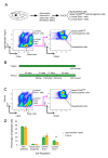
Figure 1
Figure 1
The CD24/Sca1 and CD49fHigh/CD24 flow-cytometry profiles of parous and age-matched virgin mice are similar. (A) Schematic illustration of the cell-isolation strategy and representative flow-cytometry pseudocolor plots of mammary cells from age-matched virgin control mice. After depletion of CD45+ white blood cells, luminal and basal mammary epithelial cells were separated on the basis of CD24 and Sca1 expression. Further separation of basal cells into myoepithelial and basal stem/progenitor cell subpopulations was based on the expression of CD24 and CD49f. The isolated mammary epithelial cell subpopulations included luminal Sca1+ (CD24+HighSca1+) cells, luminal Sca1- (CD24+HighSca1-) cells, basal CD49fHigh (CD24+LowSca1-CD49fHigh) or basal stem/progenitor cells, and basal myoepithelial (CD24+LowSca1-CD49fLow) cells. (B) Outline of the mouse mating, parturition, weaning, and involution protocol. (C) Representative flow-cytometry pseudocolor plots of mammary cells from parous mice. The gates applied were the same as those for age-matched virgin controls. (D) Bar graph showing the distribution of mammary epithelial cell subpopulations comparing cells from parous with age-matched virgin control mice. Data represent the mean ± SEM of seven cell-isolation experiments with a minimum of 10 mice per experiment. The proportion of luminal Sca1+ cells was reduced by approximately 50% in parous mice (P = 0.02 with a two-tailed unpaired Student t test).
Figure 2
Figure 2
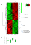
Parity-induced gene signature in total mammary cell suspensions. (A) Heat-map and cluster analysis of differential gene expression in total isolated mammary cells from age-matched virgin control and parous mice. Gene expression is presented as normalized Z-scores, defined as Z = (x-μ)/sd to allow visualization. A cut-off of P < 0.05 was applied. If multiple probe sets existed for the same gene, the probe set with the largest change in expression was selected. Four independent experiments were performed with 10 mice (five virgins; five parous) per experiment. (B) qPCR validation of pregnancy-induced gene-expression changes in total mammary cell suspensions. Fold changes are shown in relation to expression in cell suspensions from age-matched virgin control mice. Ct values were normalized to the reference gene Hprt. As control for luminal epithelial cell number, qPCR for Krt8 was performed. Data represent the mean ± SEM of four independent experiments with 10 mice (five virgins; five parous) per experiment.
Figure 3
Figure 3
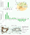
Parity leads to differentiation and decreases the Wnt/Notch signaling ratio in basal stem/progenitor cells. (A) Bar graph depicting the number of gene-expression changes in FACS-sorted mammary stromal and epithelial cell subpopulations from parous mice compared with age-matched virgin control mice by using a cut-off of fold change > 1.5 and an adjusted P value < 0.05. By far the most gene-expression changes were observed in basal stem/progenitor cells. Three independent experiments were performed with 10 mice (five virgins; five parous) per experiment. (B) Schematic illustration of prominent gene-expression changes in FACS-sorted basal stem/progenitor cells from parous as compared with age-matched virgin control mice. Fold changes are shown in parentheses with upregulated genes denoted as positive (+), and downregulated genes, as negative (-). Differentiation genes were upregulated (blue), Wnt target genes were downregulated (green), Wnt inhibitor Sfrp1 was upregulated (green) and overall Notch signaling (orange) was increased in basal stem/progenitor cells from parous mice. (C) qPCR validation of the changes in gene expression in basal stem/progenitor cells of parous mice. All classic Wnt target genes were downregulated, including Lgr5, Axin2, and versican (Vcan), whereas the more ubiquitously regulated target Myc was unchanged. In all cases, fold changes are shown relative to cells from age-matched virgin control mice. Ct values were normalized to the reference genes Hprt and Ubc [63]. Data represent the mean ± SEM of three independent experiments with 10 mice (five virgins; five parous) per experiment. (D) Representative images and quantification of immunostaining for the Wnt target gene versican in mammary gland sections from age-matched virgin and parous mice in estrus. Scale bar, 50 μm. Quantitative data represent the mean ± SEM from 60 randomly selected images from three virgin and three parous mice.
Figure 4
Figure 4
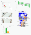
Effects of parity on Wnt transcription-factor activities and nuclear β-catenin in basal mammary epithelial cells. (A) Wnt transcription factor LEF1/TCF7 motif activity in basal stem/progenitor cells from parous as compared with virgin control mice, as predicted by MARA [32]. The binding motif of the LEF1/TCF7 transcription factor is shown in color. (B) Representative images of immunostaining for β-catenin in mammary gland sections from age-matched virgin and parous mice in estrus. Arrow, basal mammary epithelial cells with nuclear β-catenin. Arrowhead, basal mammary epithelial cells lacking nuclear β-catenin. Scale bar, 10 μm. (C) Bar graph representing the quantification of nuclear β-catenin in mammary gland sections of virgin and parous mice. Data represent the mean ± SD (virgin mice: n = 3; parous mice: n = 3). P = 0.004 with the two-tailed unpaired Student t test. (D) Transcription-factor activities in basal stem/progenitor cells, predicted on the basis of target gene expression by Ingenuity IPA [40]. Activity is reported as Z scores (positive Z score, upregulation; negative Z score, downregulation) by using a cut-off of linear fold change > 1.5 and P value < 0.05. (E) BioFunctions most strongly downregulated (blue) or upregulated (orange) in basal stem/progenitor cells, as calculated by Ingenuity IPA [40]. The color of the fields represents the Z score; the size of the fields represents the number of genes. A stringent cut-off of linear fold change > 2 and P value < 0.01 was used to minimize the number of false positives.
Figure 5
Figure 5
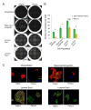
Parity reduces the progenitor potential of mammary epithelial cell subpopulations. (A) Representative images of individual wells with colonies formed by the specified cell subpopulations from age-matched virgin and parous mice. Scale bar, 4 mm. (B) Bar graph comparing the colony-forming capacities of myoepithelial cells, basal stem/progenitor cells, luminal Sca1-, and luminal Sca1+ cells of age-matched virgin and parous mice. Data are from three independent experiments and represent the mean ± SEM of colonies per well; 18 wells were assessed per cell type. *P < 0.015, NS: not significant (P = 0.08), by using two-tailed unpaired Student t test. (C) Representative images for the immunophenotyping of 5-day-old colonies grown from myoepithelial cells, basal stem/progenitor cells, luminal Sca1-, and luminal Sca1+ cells. The colonies were stained for luminal Krt18 (green) and basal Krt14 (red) expression. Hoechst 33342 (blue) was used to distinguish nuclei and to label feeder cells (negative control). Scale bar, 200 μm.
Figure 6
Figure 6
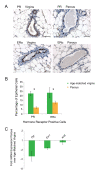
Early pregnancy decreases the proportion of progesterone receptor (PR) and estrogen receptor α (ERα)-positive cells. (A) Representative images of immunostaining for PR and ERα in mammary gland sections from age-matched virgin and parous mice in estrus. Scale bar, 50 μm. (B) Bar graph comparing the relative frequency of estrogen- and progesterone receptor-positive cells between mammary glands of virgin and parous mice. Data represent the mean ± SD (virgin mice, n = 6; parous mice, n = 5). For PR, P = 3.70E-07; for ERα, P = 0.003, by using two-tailed unpaired Student t test. (C) qPCR for progesterone receptor (Pgr), estrogen receptor alpha (Esr1), and the luminal marker keratin 8 (Krt8) genes in total mammary cell suspensions. Data are expressed as the mean ± SEM from four groups of a minimum of five parous and five age-matched virgin control mice.
Figure 7
Figure 7
Effect of Wnt4 on the proliferation capacity of basal stem/progenitor cells after early pregnancy in mice. (A) Recombinant Wnt4 rescues the parity-induced in vitro proliferation defect in basal mammary epithelial cells. Selected mammary epithelial cells from parous mice were cultured in the absence or presence of recombinant Wnt4. Three independent experiments were performed. Data represent the mean ± SEM of colonies per well, with six to nine wells assessed per cell type. *P ≤ 0.02 (two-tailed unpaired Student t test). (B) Mechanistic model illustrating the parity-induced decrease in hormone-sensing and Wnt4-secreting luminal cells on the Wnt/Notch signaling pathways and the proliferation/differentiation potential in basal stem/progenitor cells.

Similar articles

Cited by

References

    1. MacMahon B, Cole P, Lin TM, Lowe CR, Mirra AP, Ravnihar B, Salber EJ, Valaoras VG, Yuasa S. Age at first birth and breast cancer risk. Bull World Health Org. 1970;43:209–221. - PMC - PubMed
    1. Albrektsen G, Heuch I, Hansen S, Kvale G. Breast cancer risk by age at birth, time since birth and time intervals between births: exploring interaction effects. Br J Cancer. 2005;92:167–175. doi: 10.1038/sj.bjc.6602302. - DOI - PMC - PubMed
    1. Medina D. Mammary developmental fate and breast cancer risk. Endocr Relat Cancer. 2005;12:483–495. doi: 10.1677/erc.1.00804. - DOI - PubMed
    1. Rajkumar L, Kittrell FS, Guzman RC, Brown PH, Nandi S, Medina D. Hormone-induced protection of mammary tumorigenesis in genetically engineered mouse models. Breast Cancer Res. 2007;9:R12. doi: 10.1186/bcr1645. - DOI - PMC - PubMed
    1. Thordarson G, Jin E, Guzman RC, Swanson SM, Nandi S, Talamantes F. Refractoriness to mammary tumorigenesis in parous rats: is it caused by persistent changes in the hormonal environment or permanent biochemical alterations in the mammary epithelia? Carcinogenesis. 1995;16:2847–2853. doi: 10.1093/carcin/16.11.2847. - DOI - PubMed

Publication types

MeSH terms