Review
. 2013 Jul;22(3):545-61.
doi: 10.1016/j.soc.2013.02.009. Epub 2013 Mar 13.

Novel approaches to treatment of hepatocellular carcinoma and hepatic metastases using thermal ablation and thermosensitive liposomes

Affiliations
Review

Novel approaches to treatment of hepatocellular carcinoma and hepatic metastases using thermal ablation and thermosensitive liposomes

Mark W Dewhirst et al. Surg Oncol Clin N Am. 2013 Jul.

Abstract

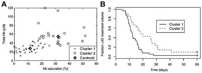
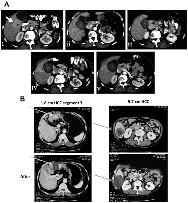
Because of the limitations of surgical resection, thermal ablation is commonly used for the treatment of hepatocellular carcinoma and liver metastases. Current methods of ablation can result in marginal recurrences of larger lesions and in tumors located near large vessels. This review presents a novel approach for extending treatment out to the margins where temperatures do not provide complete treatment with ablation alone, by combining thermal ablation with drug-loaded thermosensitive liposomes. A history of the development of thermosensitive liposomes is presented. Clinical trials have shown that the combination of radiofrequency ablation and doxorubicin-loaded thermosensitive liposomes is a promising treatment.


Conflict of interest statement

Conflict of Interest Statement: Dr Dewhirst holds stock in the Celsion Corporation

Figures

Fig. 1
Fig. 1
Percent of Dox released for Doxil and DPPC/MSPC (10%) at t = 4 minutes. Comparison of doxorubicin release after 4 minutes of heating across a range of temperatures for LTSL-Dox (DPPC/MSPC (10%)) versus Doxil. LTSL-Dox releases drug in the temperature range between 39.5°C and 42°C, which is in the range that can be achieved for routine application of hyperthermia. Maximal release occurs at the phase transition temperature of 41.3°C. DPPC - Dipalmitoylphosphatidylcholine; MSPC - Monostearoylphosphatidylcholine.
Fig. 2
Fig. 2
Performance of cationic thermosensitive liposome. Using the dorsal skin-fold window chamber, the localization and extravasation of the liposomes can be monitored over time. (Upper panel) Selected images of cationic thermosensitive liposome accumulation in endothelial cells lining vessel walls of B16 melanoma tumors, 2 hours after administration. Some evidence for extravasation is also observed. (Bottom panel) Appearance of window chamber after 1 hour of heating at 43°C. Green represents the presence of carboxyfluorescein, which was previously loaded into the liposomes. The appearance of green signal shows that the contents have been released. Note the lack of green signal in the images taken before heating, which reflects quenching of the fluorescence when calcein is encapsulated inside the liposome (upper panel). (Reprinted from Dicheva BM, Hagen TL, Li L, et al. Cationic thermosensitive liposomes: a novel dual targeted heat-triggered drug delivery approach for endothelial and tumor cells. Nano Lett 2012; [Epub ahead of print]. Copyright (2012) American Chemical Society; with permission.)
Fig. 3
Fig. 3
Comparison of drug penetration distances from nearest blood vessel for free drug ± 42°C heating for 1 hour, LTSL-Dox + heat and Doxil + heat (A, B). Green = CD31 for endothelial cells, red = doxorubicin, and yellow = EF5 hypoxia marker. The difference in total amount of drug delivered for free drug versus LTSL drug is obvious, but more importantly is the drug coverage around tumor blood vessels and the encroachment into hypoxic areas. The drug penetration distance was doubled for P = .0106, between LTSL + HT and Doxil + HT compared with the other treatment groups (C, D). The differences were highly significant. (From Manzoor AA, Lindner LH, Landon CD, et al. Overcoming limitations in nanoparticle drug delivery: triggered, intravascular release to improve drug penetration into tumors. Cancer Res 2012;72(21):5573; with permission.)
Fig. 4
Fig. 4
Relationship between the concentration of doxorubicin achieved in tumor tissue and the tumor growth time for free drug and 3 liposomal formulations. The Gaber formulation is a PEGylated version of the original Yatvin formulation. The open and closed symbols represent replicate experiments. Data were obtained by removing a cohort of animals at the end of treatment from each group and having the tumor analyzed for total doxorubicin concentration, using high-performance liquid chromatography. The remaining animals in each group were followed for tumor regrowth. (Adapted from Kong G, Anyarambhatla G, Petros WP, et al. Efficacy of liposomes and hyperthermia in a human tumor xenograft model: importance of triggered drug release. Cancer Res 2000;60:6954; with permission.)
Fig. 5
Fig. 5
Relationship between perfusion and extent of tumor hypoxia and duration of local tumor control after treatment with LTSL-Dox and hyperthermia. (A) Total hemoglobin (Hb) and Hb saturation (Hbsat) reflect perfusion and extent of hypoxia in individual tumors as measured using a noninvasive optical spectroscopy method. These same tumors were followed for growth time. Cluster analysis revealed 2 separate populations of tumors, which were characterized by relatively low Hb and Hbsat versus the second group, which had higher values of these 2 parameters. (B) The time to reach 3 times treatment volume was linked to these values, indicating that the more poorly perfused and hypoxic tumors responded less favorably to the treatment. (Reproduced from Palmer GM, Boruta RJ, Viglianti BL, et al. Non-invasive monitoring of intra-tumor drug concentration and therapeutic response using optical spectroscopy. J Control Release 2010;142(3):463; with permission.)
Fig. 6
Fig. 6
Contrast-enhanced computed tomography scans of hepatic lesions, before and after RF ablation, combined with LTLD. (A) Appearance of metastatic adrenal cell carcinoma, before and several months after thermal ablation. Note that the ablation zone enlarges and stabilizes after treatment. (i) Before treatment (arrow), (ii) 3 days after treatment, (iii) 4 weeks after treatment, (iv) 11 weeks after treatment, (v) 20 weeks after treatment. (B) Appearance of 2 primary HCCs, before and after thermal ablation. (Courtesy of Brad Wood and Celsion Corporation, Lawrenceville, NJ; Images (i), (ii), and (v) in (A) Reprinted from Wood BJ, Poon RT, Locklin JK, et al. Phase I study of heat-deployed liposomal doxorubicin during radiofrequency ablation for hepatic malignancies. J Vasc Interv Radiol 2012;23(2):248–55.e7, with permission from author and publisher; and Images (iii) and (iv) in (A) were kindly provided by the Celsion Corporation.)

Similar articles

Cited by

References

    1. Poon R, Borys N. Lyso-thermosensitive liposomal doxorubicin: a novel approach to enhance efficacy of thermal ablation of liver cancer. Expert Opin Pharmacother. 2009;10(2):333–43. - PubMed
    1. Lencioni R, Crocetti L, Cioni D, et al. Percutaneous radiofrequency ablation of hepatic colorectal metastases–technique, indications, results, and new promises. Invest Radiol. 2004;39(11):689–97. - PubMed
    1. Solbiati L, Livraghi T, Goldberg SN, et al. Percutaneous radio-frequency ablation of hepatic metastases from colorectal cancer: long-term results in 117 patients. Radiology. 2001;221(1):159–66. - PubMed
    1. Stauffer PR, Diederich CJ, Seegenschmiedt MH. Interstitial heating technologies. In: Seegenschmiedt MH, Fessenden P, Vernon CC, editors. Thermoradiotherapy and thermochemotherapy: volume 1, biology, physiology and physics. Berlin, New York: Springer-Verlag; 1995. pp. 279–320.
    1. Stauffer PR, Goldberg SN. Introduction: thermal ablation therapy. Int J Hyperthermia. 2004;20(7):671–7. - PubMed

Publication types