Review
. 2013 Jun;144(6):1170-9.
doi: 10.1053/j.gastro.2013.01.074.

Control of cell identity in pancreas development and regeneration

Affiliations
Review

Control of cell identity in pancreas development and regeneration

Ben Z Stanger et al. Gastroenterology. 2013 Jun.

Abstract

The endocrine and exocrine cells in the adult pancreas are not static, but can change their differentiation state in response to injury or stress. This concept of cells in flux means that there may be ways to generate certain types of cells (such as insulin-producing β-cells) and prevent formation of others (such as transformed neoplastic cells). We review different aspects of cell identity in the pancreas, discussing how cells achieve their identity during embryonic development and maturation, and how this identity remains plastic, even in the adult pancreas.


Figures

Figure 1
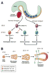
Figure 1. Developmental of the Mammalian Pancreas
(A) Molecular regulation of pancreas development in the mouse. Following gastrulation, 2 patches of endoderm (yellow)—1 in the ventral foregut and 1 in the dorsal midgut—receive signals from adjacent structures resulting in pancreatic specification (purple). Cells in both regions express the Pdx1 transcription factor whereas cells located in the ventral foregut patch also express the Sox17 transcription factor. Over time, Pdx1+/Sox17+ cells resolve into a Sox17 single-positive population (which gives rise to the extrahepatic biliary tree) and a Pdx1 single-positive population (which gives rise to the ventral pancreas). Subsequently, the ventral and dorsal pancreatic buds merge during rotation of the gut. (B) Cells become polarized within the growing pancreatic buds, forming microlumens which fuse to form a tubular plexus. During the secondary transition, a period marked by a large increase in endocrine and exocrine differentiation, the tubular plexus resolves into better defined ductal structures. The tips of these primitive ducts initially remain multipotent, having the capacity to give rise to all pancreatic cell types including acinar cells, while the trunks are committed to ductal and endocrine fates. Endocrine progenitor cells, detectable by the expression of Ngn3, delaminate from the trunks, eventually aggregating as Islets of Langerhans.
Figure 2
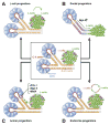
Figure 2. Cellular Plasticity in the Adult Pancreas
Depiction of the structural elements of the exocrine pancreas, including acinar, centro-acinar (C–A), and duct cells, and the endocrine islet, including α- and β-cells in the center of the schematic. (A) β-cells serve as progenitors for β-cells and expand via proliferation. (B) Upon injury, duct or duct-associated cells begin to express Ngn3, which could result in the formation of endocrine cells, including β-cells. (C) Viral expression of Pdx-1, Ngn-3 and MafA in adult acinar cells promotes reprogramming towards β-cells. (D) α-cells can transdifferentiate into β-cells following their depletion, whereas β-cells can become α-like cells or undifferentiated cells during stress or following deletion of Foxo1. (Adapted from Puri & Hebrok, 2010.)
Figure 3
Figure 3. Loss of Cell Identity in the Adult Pancreas
(A) β-cells undergo a process of “de-differentiation” or “un-differentiation,” either via genetic mutation (e.g. Foxo1 elimination) or physiological changes, (e.g. aging, T2D). These empty β-cells have reduced expression of markers of mature β-cells and increased expression of markers found in progenitors, including Ngn3, and Oct4. Further progression results in non-β-cell endocrine cells that express other endocrine hormones. (Adapted from Puri & Hebrok, 2012). (B) Exocrine cells, including acinar, centro-acinar, and duct cells, are considered to be progenitors to PanINs and PDA. (Adapted from Morris et al, 2010).

References

    1. Pavlov I. The Work of the Digestive Glands. Charles Griffin; 1902.
    1. Teff KL. How neural mediation of anticipatory and compensatory insulin release helps us tolerate food. Physiol Behav. 2011;103:44–50. - PMC - PubMed
    1. Heller RS. The comparative anatomy of islets. Adv Exp Med Biol. 2010;654:21–37. - PubMed
    1. Madsen OD. Pancreas phylogeny and ontogeny in relation to a ‘pancreatic stem cell’. C R Biol. 2007;330:534–7. - PMC - PubMed
    1. Hesselson D, Anderson RM, Stainier DY. Suppression of Ptf1a activity induces acinar-to-endocrine conversion. Curr Biol. 2011;21:712–7. - PMC - PubMed

Publication types

MeSH terms