

Acute disseminated encephalomyelitis
- PMID: 23622336
- PMCID: PMC7152359
- DOI: 10.1016/B978-0-444-52910-7.00048-9
Acute disseminated encephalomyelitis
Abstract
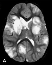
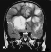
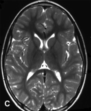
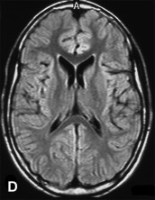
The advent of magnetic resonance imaging (MRI) has contributed to increase the interest and awareness in childhood white matter disorders. Pediatric inflammatory demyelinating diseases of the central nervous system (CNS) are clinically heterogeneous with respect to their mode of presentation, clinical severity, rate of progression, and prognosis. Acute disseminated encephalomyelitis (ADEM) is an immune-mediated inflammatory disorder of the CNS, typically transitory and self-limiting. The highest incidence of ADEM is observed during childhood. It is characterized by an acute encephalopathy with polyfocal neurological deficits. In the absence of specific biological markers the diagnosis of ADEM is still based on clinical features and MRI evidence of widespread demyelination, after ruling out other possible explanations for an acute encephalopathy. Over the past decade, many retrospective patient studies have focused on clinical and neuroimaging features, in an attempt to define specific diagnostic criteria. The occurrence of relapses in children with ADEM poses diagnostic difficulties in its differentiation from multiple sclerosis (MS) and neuromyelitis optica (NMO). With the widespread use of high-dose steroids, the long-term prognosis of ADEM with regard to functional and cognitive recovery is favorable. This chapter summarizes the available literature on ADEM in children, including the proposed consensus definitions for its monophasic and relapsing variants.
Copyright © 2013 Elsevier B.V. All rights reserved.
Figures
Similar articles
-
Comparison of deep gray matter lesions on magnetic resonance imaging among adults with acute disseminated encephalomyelitis, multiple sclerosis, and neuromyelitis optica.Mult Scler. 2014 Apr;20(4):418-23. doi: 10.1177/1352458513499420. Epub 2013 Jul 25. Mult Scler. 2014. PMID: 23886831
-
Acute disseminated encephalomyelitis.Neurology. 2007 Apr 17;68(16 Suppl 2):S23-36. doi: 10.1212/01.wnl.0000259404.51352.7f. Neurology. 2007. PMID: 17438235 Review.
-
Multiple sclerosis and acute disseminated encephalomyelitis diagnosed in children after long-term follow-up: comparison of presenting features.Dev Med Child Neurol. 2009 Jun;51(6):480-6. doi: 10.1111/j.1469-8749.2008.03136.x. Epub 2008 Oct 24. Dev Med Child Neurol. 2009. PMID: 19018840 Free PMC article.
-
Pediatric central nervous system inflammatory demyelination: acute disseminated encephalomyelitis, clinically isolated syndromes, neuromyelitis optica, and multiple sclerosis.Curr Opin Neurol. 2009 Jun;22(3):233-40. doi: 10.1097/wco.0b013e32832b4c47. Curr Opin Neurol. 2009. PMID: 19434783 Review.
-
Disseminated encephalomyelitis in children.Clin Neurol Neurosurg. 2008 Nov;110(9):928-38. doi: 10.1016/j.clineuro.2007.12.018. Epub 2008 Feb 12. Clin Neurol Neurosurg. 2008. PMID: 18272282 Free PMC article. Review.
Cited by
-
Spinal Cord Involvement in MS and Other Demyelinating Diseases.Biomedicines. 2020 May 22;8(5):130. doi: 10.3390/biomedicines8050130. Biomedicines. 2020. PMID: 32455910 Free PMC article. Review.
-
Cognitive and psychopathological outcomes in acute disseminated encephalomyelitis.BMJ Neurol Open. 2024 May 8;6(1):e000640. doi: 10.1136/bmjno-2024-000640. eCollection 2024. BMJ Neurol Open. 2024. PMID: 38736586 Free PMC article. Review.
-
Sex differences in risk factors for Alzheimer dementia encephalopathy patients.Front Dement. 2025 May 26;4:1593788. doi: 10.3389/frdem.2025.1593788. eCollection 2025. Front Dement. 2025. PMID: 40491970 Free PMC article.
-
Pediatric Neuroimaging of Multiple Sclerosis and Neuroinflammatory Diseases.Tomography. 2024 Dec 20;10(12):2100-2127. doi: 10.3390/tomography10120149. Tomography. 2024. PMID: 39728911 Free PMC article. Review.
-
MRI characteristics of Japanese macaque encephalomyelitis: Comparison to human diseases.J Neuroimaging. 2021 May;31(3):480-492. doi: 10.1111/jon.12868. Epub 2021 Apr 30. J Neuroimaging. 2021. PMID: 33930224 Free PMC article.
References
-
- Anlar B., Basaran C., Kose G. Acute disseminated encephalomyelitis in children: outcome and prognosis. Neuropediatrics. 2003;34:194–199. - PubMed
-
- Balasubramanya K.S., Kovoor J.M., Jayakumar P.N. Diffusion-weighted imaging and proton MR spectroscopy in the characterization of acute disseminated encephalomyelitis. Neuroradiology. 2007;49:177–183. - PubMed
-
- Banwell B., Tenembaum S., Lennon V.A. Neuromyelitis optica-IgG in childhood inflammatory demyelinating CNS disorders. Neurology. 2008;70:344–352. - PubMed
-
- Banwell B., Kennedy J., Sadovnick D. Incidence of acquired demyelination of the CNS in Canadian children. Neurology. 2009;72:232–239. - PubMed
Publication types
MeSH terms
LinkOut - more resources
Full Text Sources
Other Literature Sources
Medical
Miscellaneous
