Morphological polymorphism of Trypanosoma copemani and description of the genetically diverse T. vegrandis sp. nov. from the critically endangered Australian potoroid, the brush-tailed bettong (Bettongia penicillata (Gray, 1837))
- PMID: 23622560
- PMCID: PMC3641998
- DOI: 10.1186/1756-3305-6-121
Morphological polymorphism of Trypanosoma copemani and description of the genetically diverse T. vegrandis sp. nov. from the critically endangered Australian potoroid, the brush-tailed bettong (Bettongia penicillata (Gray, 1837))
Abstract
Background: The trypanosome diversity of the Brush-tailed Bettong (Bettongia penicillata), known locally as the woylie, has been further investigated. At a species level, woylies are critically endangered and have declined by 90% since 1999. The predation of individuals made more vulnerable by disease is thought to be the primary cause of this decline, but remains to be proven.
Methods: Woylies were sampled from three locations in southern Western Australia. Blood samples were collected and analysed using fluorescence in situ hybridization, conventional staining techniques and microscopy. Molecular techniques were also used to confirm morphological observations.
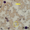
Results: The trypanosomes in the blood of woylies were grouped into three morphologically distinct trypomastigote forms, encompassing two separate species. The larger of the two species, Trypanosoma copemani exhibited polymorphic trypomastigote forms, with morphological phenotypes being distinguishable, primarily by the distance between the kinetoplast and nucleus. The second trypanosome species was only 20% of the length of T. copemani and is believed to be one of the smallest recorded trypanosome species from mammals. No morphological polymorphism was identified for this genetically diverse second species. We described the trypomastigote morphology of this new, smaller species from the peripheral blood of the woylie and proposed the name T. vegrandis sp. nov. Temporal results indicate that during T. copemani Phenotype 1 infections, the blood forms remain numerous and are continuously detectable by molecular methodology. In contrast, the trypomastigote forms of T. copemani Phenotype 2 appear to decrease in prevalence in the blood to below molecular detectable levels.
Conclusions: Here we report for the first time on the morphological diversity of trypanosomes infecting the woylie and provide the first visual evidence of a mixed infection of both T. vegrandis sp. nov and T. copemani. We also provide supporting evidence that over time, the intracellular T. copemani Phenotype 2 may become localised in the tissues of woylies as the infection progresses from the active acute to chronic phase. As evidence grows, further research will be necessary to investigate whether the morphologically diverse trypanosomes of woylies have impacted on the health of their hosts during recent population declines.
Figures


Similar articles
-
Temporal and spatial dynamics of trypanosomes infecting the brush-tailed bettong (Bettongia penicillata): a cautionary note of disease-induced population decline.Parasit Vectors. 2014 Apr 7;7:169. doi: 10.1186/1756-3305-7-169. Parasit Vectors. 2014. PMID: 24708757 Free PMC article.
-
Trypanosomes in a declining species of threatened Australian marsupial, the brush-tailed bettong Bettongia penicillata (Marsupialia: Potoroidae).Parasitology. 2008 Sep;135(11):1329-35. doi: 10.1017/S0031182008004824. Epub 2008 Aug 28. Parasitology. 2008. PMID: 18752704
-
Morphological and molecular characterization of Trypanosoma copemani n. sp. (Trypanosomatidae) isolated from Gilbert's potoroo ( Potorous gilbertii) and quokka ( Setonix brachyurus).Parasitology. 2009 Jun;136(7):783-92. doi: 10.1017/S0031182009005927. Epub 2009 May 6. Parasitology. 2009. PMID: 19416553
-
Trypanosomes of Australian mammals: A review.Int J Parasitol Parasites Wildl. 2014 Mar 15;3(2):57-66. doi: 10.1016/j.ijppaw.2014.02.002. eCollection 2014 Aug. Int J Parasitol Parasites Wildl. 2014. PMID: 25161902 Free PMC article. Review.
-
Epidemiology of Trypanosomiasis in Wildlife-Implications for Humans at the Wildlife Interface in Africa.Front Vet Sci. 2021 Jun 14;8:621699. doi: 10.3389/fvets.2021.621699. eCollection 2021. Front Vet Sci. 2021. PMID: 34222391 Free PMC article. Review.
Cited by
-
Field and experimental evidence of a new caiman trypanosome species closely phylogenetically related to fish trypanosomes and transmitted by leeches.Int J Parasitol Parasites Wildl. 2015 Oct 21;4(3):368-78. doi: 10.1016/j.ijppaw.2015.10.005. eCollection 2015 Dec. Int J Parasitol Parasites Wildl. 2015. PMID: 26767165 Free PMC article.
-
The phylogeography of trypanosomes from South American alligatorids and African crocodilids is consistent with the geological history of South American river basins and the transoceanic dispersal of Crocodylus at the Miocene.Parasit Vectors. 2013 Oct 29;6(1):313. doi: 10.1186/1756-3305-6-313. Parasit Vectors. 2013. PMID: 24499634 Free PMC article.
-
The Troublesome Ticks Research Protocol: Developing a Comprehensive, Multidiscipline Research Plan for Investigating Human Tick-Associated Disease in Australia.Pathogens. 2022 Nov 3;11(11):1290. doi: 10.3390/pathogens11111290. Pathogens. 2022. PMID: 36365042 Free PMC article.
-
Trypanosoma livingstonei: a new species from African bats supports the bat seeding hypothesis for the Trypanosoma cruzi clade.Parasit Vectors. 2013 Aug 3;6(1):221. doi: 10.1186/1756-3305-6-221. Parasit Vectors. 2013. PMID: 23915781 Free PMC article.
-
High prevalence of Trypanosoma vegrandis in bats from Western Australia.Vet Parasitol. 2015 Dec 15;214(3-4):342-7. doi: 10.1016/j.vetpar.2015.10.016. Epub 2015 Oct 20. Vet Parasitol. 2015. PMID: 26541211 Free PMC article.
References
Publication types
MeSH terms
LinkOut - more resources
Full Text Sources
Other Literature Sources
Research Materials

