

Hippocampal shape abnormalities of patients with childhood-onset schizophrenia and their unaffected siblings
- PMID: 23622854
- PMCID: PMC3812431
- DOI: 10.1016/j.jaac.2013.02.003
Hippocampal shape abnormalities of patients with childhood-onset schizophrenia and their unaffected siblings
Abstract
Objective: The hippocampus has been implicated in the pathogenesis of schizophrenia, and hippocampal volume deficits have been a consistently reported abnormality, but the subregional specificity of the deficits remains unknown. The authors explored the nature and developmental trajectory of subregional shape abnormalities of the hippocampus in patients with childhood-onset schizophrenia (COS), their healthy siblings, and healthy volunteers.
Method: Two hundred twenty-five anatomic brain magnetic resonance images were obtained from 103 patients with COS, 169 from their 79 healthy siblings, and 255 from 101 age- and sex-matched healthy volunteers (age range = 9-29 years). The hippocampus was segmented using FreeSurfer automated image analysis software, and hippocampal shape was evaluated by comparing subjects at more than 6,000 vertices on the left and right hippocampal surfaces. Longitudinal data were examined using mixed model regression analysis.
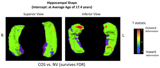
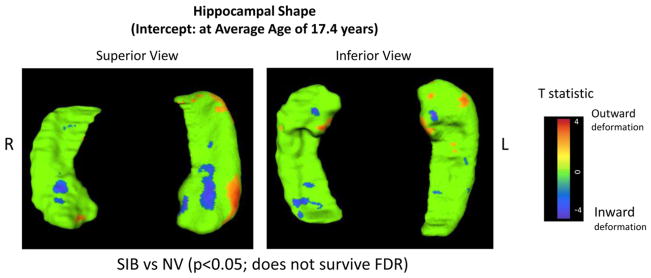
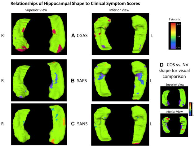
Results: Patients with COS showed significant bilateral inward deformation in the anterior hippocampus. Healthy siblings also showed a trend for anterior inward deformation. However, the trajectory of shape change did not differ significantly between the groups. Inward deformations in the anterior hippocampus were positively related to positive symptom severity, whereas outward surface displacement was positively related to overall functioning.
Conclusion: This is the first and largest longitudinal three-way analysis of subregional hippocampal shape abnormalities in patients with COS and their healthy siblings compared with healthy controls. The anterior hippocampal abnormalities in COS suggest the pathophysiologic importance of this subregion in schizophrenia. The trend level and overlapping shape abnormalities in the healthy siblings suggest a more subtle, subregionally specific neuroanatomic endophenotype.
Published by Elsevier Inc.
Conflict of interest statement
Disclosure: Drs. Wang, Greenstein, Clasen, Lalonde, Miller, Rapoport, and Gogtay, Ms. Johnson, and Ms. Alpert report no biomedical financial interests or potential confiicts of interest.
Figures
Similar articles
-
Absence of anatomic corpus callosal abnormalities in childhood-onset schizophrenia patients and healthy siblings.Psychiatry Res. 2013 Jan 30;211(1):11-6. doi: 10.1016/j.pscychresns.2012.09.013. Epub 2012 Nov 13. Psychiatry Res. 2013. PMID: 23154096 Free PMC article.
-
At the boundary of the self: the insular cortex in patients with childhood-onset schizophrenia, their healthy siblings, and normal volunteers.Int J Dev Neurosci. 2014 Feb;32:58-63. doi: 10.1016/j.ijdevneu.2013.05.010. Epub 2013 Jun 11. Int J Dev Neurosci. 2014. PMID: 23770018
-
Shared hippocampal abnormalities in sporadic temporal lobe epilepsy patients and their siblings.Epilepsia. 2020 Apr;61(4):735-746. doi: 10.1111/epi.16477. Epub 2020 Mar 20. Epilepsia. 2020. PMID: 32196657
-
Decreased left middle temporal gyrus volume in antipsychotic drug-naive, first-episode schizophrenia patients and their healthy unaffected siblings.Schizophr Res. 2013 Mar;144(1-3):37-42. doi: 10.1016/j.schres.2012.12.018. Epub 2013 Jan 27. Schizophr Res. 2013. PMID: 23360727 Review.
-
Neuroimaging findings from childhood onset schizophrenia patients and their non-psychotic siblings.Schizophr Res. 2016 Jun;173(3):124-131. doi: 10.1016/j.schres.2015.03.003. Epub 2015 Mar 26. Schizophr Res. 2016. PMID: 25819937 Free PMC article. Review.
Cited by
-
Hippocampal Shape Maturation in Childhood and Adolescence.Cereb Cortex. 2019 Aug 14;29(9):3651-3665. doi: 10.1093/cercor/bhy244. Cereb Cortex. 2019. PMID: 30272143 Free PMC article.
-
Hippocampal Subregions Across the Psychosis Spectrum.Schizophr Bull. 2018 Aug 20;44(5):1091-1099. doi: 10.1093/schbul/sbx160. Schizophr Bull. 2018. PMID: 29272467 Free PMC article.
-
Thalamic Shape Abnormalities Differentially Relate to Cognitive Performance in Early-Onset and Adult-Onset Schizophrenia.Front Psychiatry. 2022 Apr 11;13:803234. doi: 10.3389/fpsyt.2022.803234. eCollection 2022. Front Psychiatry. 2022. PMID: 35479490 Free PMC article.
-
Volumetric and Shape Analysis of the Subcortical Regions in Schizophrenia Patients: A Pilot Study.J Clin Imaging Sci. 2019 Jan 31;9:1. doi: 10.4103/jcis.JCIS_61_18. eCollection 2019. J Clin Imaging Sci. 2019. PMID: 30788185 Free PMC article.
-
Hippocampal morphology mediates biased memories of chronic pain.Neuroimage. 2018 Feb 1;166:86-98. doi: 10.1016/j.neuroimage.2017.10.030. Epub 2017 Nov 6. Neuroimage. 2018. PMID: 29080714 Free PMC article.
References
-
- Bogerts B, Falkai P, Haupts M, et al. Post-mortem volume measurements of limbic system and basal ganglia structures in chronic schizophrenics. Initial results from a new brain collection. Schizophr Res. 1990;3:295–301. - PubMed
-
- Davatzikos C, Shen D, Gur RC, et al. Whole-brain morphometric study of schizophrenia revealing a spatially complex set of focal abnormalities. Arch Gen Psychiatry. 2005;62:1218–1227. - PubMed
-
- Narr KL, Thompson PM, Szeszko P, et al. Regional specificity of hippocampal volume reductions in first-episode schizophrenia. Neuroimage. 2004;21:1563–1575. - PubMed
-
- Adriano F, Caltagirone C, Spalletta G. Hippocampal volume reduction in first-episode and chronic schizophrenia: a review and meta-analysis. Neuroscientist. 2012;18:180–200. - PubMed
Publication types
MeSH terms
Grants and funding
LinkOut - more resources
Full Text Sources
Other Literature Sources
