

Characteristics of incident geographic atrophy in the complications of age-related macular degeneration prevention trial
- PMID: 23622873
- PMCID: PMC3728180
- DOI: 10.1016/j.ophtha.2013.01.049
Characteristics of incident geographic atrophy in the complications of age-related macular degeneration prevention trial
Abstract
Objective: To characterize the size, location, conformation, and features of incident geographic atrophy (GA) as detected by annual stereoscopic color photographs and fluorescein angiograms (FAs).
Design: Retrospective cohort study within a larger clinical trial.
Participants: Patients with bilateral large drusen in whom GA developed during the course of the Complications of Age-related Macular Degeneration Prevention Trial (CAPT).
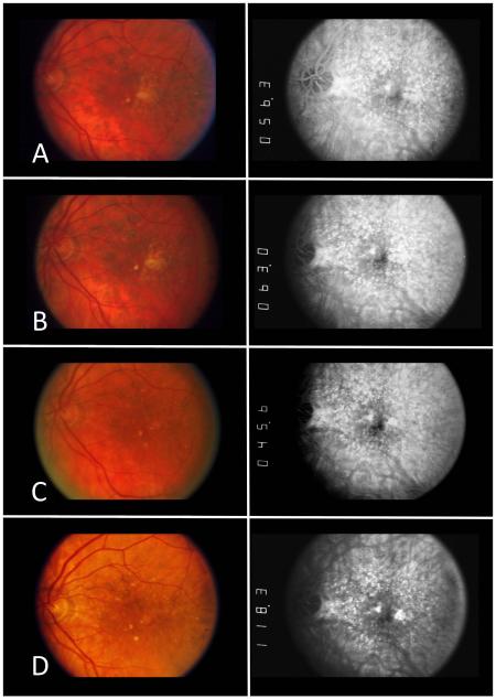
Methods: Annual stereoscopic color photographs and FAs were reviewed from 114 CAPT patients in whom GA developed in the untreated eye during 5 to 6 years of follow-up. Geographic atrophy was defined according to the Revised GA Criteria for identifying early GA.(23) Color-optimized fundus photographs were viewed concurrently with the FAs during grading.
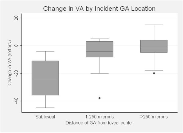
Main outcome measures: Size and distance from the fovea of individual GA lesions, number of areas of atrophy, and change in visual acuity (VA) when GA first developed in an eye.
Results: At presentation, the median total GA area was 0.26 mm(2) (0.1 disc area). Geographic atrophy presented as a single lesion in 89 (78%) eyes. The median distance from the fovea was 395 μm. Twenty percent of incident GA lesions were subfoveal and an additional 18% were within 250 μm of the foveal center. Development of GA was associated with a mean decrease of 7 letters from the baseline VA level compared with 1 letter among matched early age-related macular degeneration eyes without GA. Geographic atrophy that formed in areas previously occupied by drusenoid pigment epithelial detachments on average were larger (0.53 vs. 0.20 mm(2); P = 0.0001), were more central (50 vs. 500 μm from the center of the fovea; P<0.0001), and were associated with significantly worse visual outcome (20/50 vs. 20/25; P = 0.0003) than GA with other drusen types as precursors.
Conclusions: Incident GA most often appears on color fundus photographs and FAs as a small, singular, parafoveal lesion, although a large minority of lesions are subfoveal or multifocal at initial detection. The characteristics of incident GA vary with precursor drusen types. These data can facilitate design of future clinical trials of therapies for GA.
Financial disclosure(s): The author(s) have no proprietary or commercial interest in any materials discussed in this article.
Copyright © 2013 American Academy of Ophthalmology. Published by Elsevier Inc. All rights reserved.
Figures
References
-
- Eye Diseases Prevalence Research Group Prevalence of age-related macular degeneration in the United States. Arch Ophthalmol. 2004;122:564–72. - PubMed
-
- Klein R, Chou CF, Klein BE, et al. Prevalence of age-related macular degeneration in the US population. Arch Ophthalmol. 2011;129:75–80. - PubMed
-
- Klein R, Klein BE, Knudtson MD, et al. Fifteen-year cumulative incidence of age-related macular degeneration: the Beaver Dam Eye Study. Ophthalmology. 2007;114:253–62. - PubMed
-
- Klein ML, Ferris FL, III, Armstrong J, et al. AREDS Research Group Retinal precursors and the development of geographic atrophy in age-related macular degeneration. Ophthalmology. 2008;115:1026–31. - PubMed
-
- Klein R, Klein BE, Tomany SC, et al. Ten-year incidence and progression of age-related maculopathy: the Beaver Dam Eye Study. Ophthalmology. 2002;109:1767–79. - PubMed
Publication types
MeSH terms
Grants and funding
LinkOut - more resources
Full Text Sources
Other Literature Sources
Medical
Research Materials
Miscellaneous
