Review
. 2013 Jun;23(3):345-56.
doi: 10.1016/j.sbi.2013.04.003. Epub 2013 Apr 25.

Expression of recombinant glycoproteins in mammalian cells: towards an integrative approach to structural biology

Affiliations
Review

Expression of recombinant glycoproteins in mammalian cells: towards an integrative approach to structural biology

A Radu Aricescu et al. Curr Opin Struct Biol. 2013 Jun.

Abstract

Mammalian cells are rapidly becoming the system of choice for the production of recombinant glycoproteins for structural biology applications. Their use has enabled the structural investigation of a whole new set of targets including large, multi-domain and highly glycosylated eukaryotic cell surface receptors and their supra-molecular assemblies. We summarize the technical advances that have been made in mammalian expression technology and highlight some of the structural insights that have been obtained using these methods. Looking forward, it is clear that mammalian cell expression will provide exciting and unique opportunities for an integrative approach to the structural study of proteins, especially of human origin and medically relevant, by bridging the gap between the purified state and the cellular context.


Figures

Figure 1
Figure 1
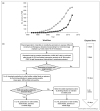
Mammalian expression technology applied to structural biology. (a) Plot of the cumulative total number of chains deposited in the PDB whose expression system was identified as either HEK-293 (Human Embryonic Kidney) or CHO (Chinese hamster ovary) cells by year of deposition. Expression data were parsed from the set of PDB files available from ftp://ftp.wwpdb.org/pub/pdb/data/structures/divided/pdb as of November 2012. Chains were counted rather than PDB entries as expression information is recorded by chain in the PDB. Note: entries marked ‘obsolete’ could not be included, which might have excluded a small number of early 1990s structures. (b) Workflow for the production of recombinant proteins in HEK-293 cells for structural biology applications.
Figure 2
Figure 2
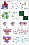
The structure and management of canonical N-linked glycans. (a) Paucimannose glycans typically produced by insect cell lines (e.g. Sf9), difficult to remove enzymatically [4]. (b) Mammalian cell glycans, in contrast, have a larger size and chemical heterogeneity. (c)(e) Glycosylation inhibitors or mutant cell lines are used to trap specific structures in the biosynthetic pathway. These include Man9GlcNAc2, upon kifunensine treatment (c), Man5GlcNAc2, in GnTI cells (d) and hybrid mannose, following treatment with swainsonine (e). (f) All these intermendiates (c)–(e) can be trimmed down to a single GlcNAc by Endo H/F1 treatment. (g) Crystal structure of the human RPTPμ trans-adhesive interaction [28]. (h) Each monomer carries 12 N-glycosylation sites (around one per 100 residues, a typical amount for cell surface receptors). Man9GlcNAc2 glycans were modeled at nine sites per monomer, to illustrate their considerable volume and coverage of the protein surface. (i) Successful crystallization could only be achieved upon EndoH glycan trimming [28]. Colour/shape coding of glycan units: GlcNAc, blue square; Man, green circle; Gal, yellow circle; NeuNAc, pink diamond; Fuc, red triangle.
Figure 3
Figure 3
Emerging concepts in cell surface signaling: examples of combined structure/function approaches that have revealed supramolecular receptor organization. Such assemblies are driven by either protein–protein interactions (e.g. the semaphoring/plexin system) or by non-protein ligands (e.g. type IIa RPTPs interactions with HSPGs and CSPGs). (a) Crystal structure of a Sema4D-PlexinB1 ectodomain complex [31••]. (b) Binding of dimeric Sema4D ligands (pink/red) to the PlexinB1 extracellular region (dark blue) triggers receptor dimerization. Note that non-ligand-dependent PlexinB1 ectodomain oligomerization has also been reported, but its extent or architecture are still unclear [31••]. (c) Crystal structure of the PlexinB1 intracellular region in complex with Rac1 [61]. (d) This interaction leads to the formation of a 3:3 receptor–ligand assembly. (e) Schematic representation of the putative impact of simultaneous Sema4D and Rac1 binding to PlexinB1. The combined 2-fold and 3-fold interactions may lead to a hexagonal ‘honeycomb’ arrangement (shown in part here) that facilitates bi-directional signaling. (f)(g) COS7 cell collapse assays were used to validate crystallographic interfaces using structure-guided mutagenesis. Scalebar: 40 μm. (h) Electrostatic potential representation (±5kT/e) of the glycosaminoglycan (GAG)-binding region of human RPTPσ [45••]. (i) A basic residue cluster, conserved in all family members, interacts with sulphated sugars (shown here is human LAR in complex with sucrose-octasulphate). (j)(k) The polymeric nature of heparan sulphate (HS) triggers receptor clustering. (l)(m) Importantly, the distribution of sulphate groups along the HS chains is not even: 12–14 sulphate-rich units (red), flanked by intermediate sulphation regions (yellow) are separated by long low-sulphation portions. This imposes an uneven distribution of receptors on the cell surface (~four receptors per cluster), which is essential to promote neuronal motility [45••]. (n) In contrast, the distribution of sulphate groups on chondroitin sulphate molecules prevents formation of RPTPσ clusters and inhibits cell motility [45••].
Figure 4
Figure 4
The impact of N-linked glycans in modulating protein complexes. (a) Crystal structure of the glycosylated complex between the IgG1-Fc region and its FcγRIIIa receptor, essential for antibody-mediated cellular cytotoxicity [72••]. (b) Antibodies lacking core fucosylation show a large increase in affinity for FcγRIIIa, due to additional glycan–glycan and glycan–protein interactions (putative hydrogen bonds are shown as dashed lines). This leads to an improved receptor-mediated effector function and forms the basis for a next generation of therapeutics, glycoengineered antibodies. (c) In the complex structure containing fucosylated IgG1-Fc, such contacts are very limited, explaining the decreased affinity for the FcγRIIIa receptor. Fuc: fucose attached to the Asn297-linked GlcNAc in IgG1-Fc. Colour coding of carbon atoms in the glycan units (sphere and stick representation) in panels (b) and (c) is as described in Figure 2.

References

    1. Reichert JM. Marketed therapeutic antibodies compendium. MAbs. 2012;4:413–415. - PMC - PubMed
    1. Nettleship JE, Assenberg R, Diprose JM, Rahman-Huq N, Owens RJ. Recent advances in the production of proteins in insect and mammalian cells for structural biology. J Struct Biol. 2010;172:55–65. - PubMed
    1. Apweiler R, Hermjakob H, Sharon N. On the frequency of protein glycosylation, as deduced from analysis of the SWISS-PROT database. Biochim Biophys Acta. 1999;1473:4–8. - PubMed
    1. Chang VT, Crispin M, Aricescu AR, Harvey DJ, Nettleship JE, Fennelly JA, Yu C, Boles KS, Evans EJ, Stuart DI, et al. Glycoprotein structural genomics: solving the glycosylation problem. Structure. 2007;15:267–273. - PMC - PubMed
    1. Stanley P. Chinese hamster ovary cell mutants with multiple glycosylation defects for production of glycoproteins with minimal carbohydrate heterogeneity. Mol Cell Biol. 1989;9:377–383. - PMC - PubMed

Publication types

LinkOut - more resources