. 2013 May 2;92(5):781-91.
doi: 10.1016/j.ajhg.2013.03.020. Epub 2013 Apr 25.

Yunis-Varón syndrome is caused by mutations in FIG4, encoding a phosphoinositide phosphatase

Affiliations

Yunis-Varón syndrome is caused by mutations in FIG4, encoding a phosphoinositide phosphatase

Philippe M Campeau et al. Am J Hum Genet. .

Abstract

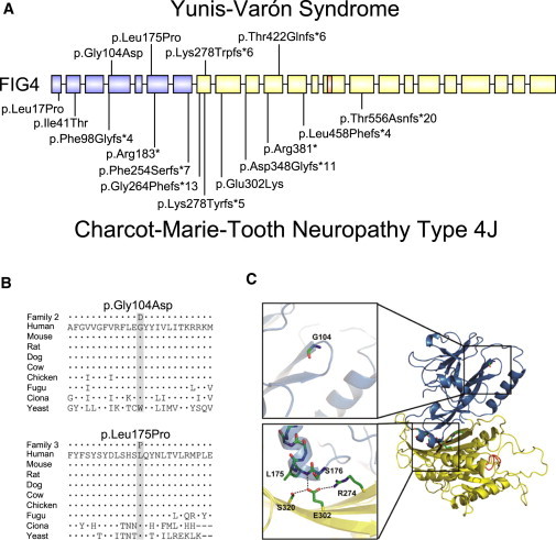
Yunis-Varón syndrome (YVS) is an autosomal-recessive disorder with cleidocranial dysplasia, digital anomalies, and severe neurological involvement. Enlarged vacuoles are found in neurons, muscle, and cartilage. By whole-exome sequencing, we identified frameshift and missense mutations of FIG4 in affected individuals from three unrelated families. FIG4 encodes a phosphoinositide phosphatase required for regulation of PI(3,5)P(2) levels, and thus endosomal trafficking and autophagy. In a functional assay, both missense substitutions failed to correct the vacuolar phenotype of Fig4-null mouse fibroblasts. Homozygous Fig4-null mice exhibit features of YVS, including neurodegeneration and enlarged vacuoles in neurons. We demonstrate that Fig4-null mice also have small skeletons with reduced trabecular bone volume and cortical thickness and that cultured osteoblasts accumulate large vacuoles. Our findings demonstrate that homozygosity or compound heterozygosity for null mutations of FIG4 is responsible for YVS, the most severe known human phenotype caused by defective phosphoinositide metabolism. In contrast, in Charcot-Marie-Tooth disease type 4J (also caused by FIG4 mutations), one of the FIG4 alleles is hypomorphic and disease is limited to the peripheral nervous system. This genotype-phenotype correlation demonstrates that absence of FIG4 activity leads to central nervous system dysfunction and extensive skeletal anomalies. Our results describe a role for PI(3,5)P(2) signaling in skeletal development and maintenance.


Figures

Figure 1
Figure 1
Abnormal Subcellular Morphology in YVS Tissues (A) Electron micrograph of cultured skin fibroblasts from individual 5 showing large, empty cytoplasmic vacuoles and electron-dense inclusion bodies. Scale bar represents 1 μm. (B–D) Muscle sections stained with Gomori trichrome (B), Periodic Acid Schiff (PAS) (C), and acid phosphatase (D) reveal variable fiber size and large vacuoles containing basophilic material stained by acid phosphatase and PAS staining. Scale bars represent 10 μm. (E and F) Electron micrographs of skeletal muscle showing vacuoles in the subsarcolemmal (E) and intramyofibrillar (F) spaces filled with amorphous, granular, or membranous material and partially degraded organelles. The vacuoles are mostly membrane limited; those located at the fiber surface abutting the extracellular space are also limited by the basal lamina (arrowheads). The mitochondria were normal in number, size, and morphology (inset). Scale bars represent 1 μm.
Figure 2
Figure 2
Segregation of FIG4 Mutations in Three Unrelated YVS-Affected Families FIG4 genotypes of family members demonstrating autosomal-recessive inheritance with sequencing chromatograms below. Numbering from RefSeq NM_014845.5.
Figure 3
Figure 3
Location of FIG4 Mutations (A) Location of mutations in FIG4 exons; introns not drawn to scale. Above, YVS; below, CMT4J. Protein-interaction domain, blue; catalytic domain, yellow; P loop containing the catalytic CX5R(T/S) motif, orange. (B) Evolutionary conservation of amino acids around the missense mutations. Alignment was performed with ClustalW2. Dots represent identity; dashes represent gaps. (C) Positions of substitutions in the 3-dimensional protein structure (courtesy of Yuxin Mao27). Same color scheme as in (A). Abbreviations used to designate amino acids are as follows: G104, p.Gly104; L175, p.Leu175; S176, p.Ser176; S320, p.Ser320; E302, p.Glu302; and R274, p.Arg274. Gly104 is located at the beginning of β sheet 7 within the noncatalytic, protein-binding domain of FIG4. p.Leu175 is located in α helix 2 of the same domain and appears to interact directly with residue p.Glu302, the site of a functionally null mutation in an individual with CMT4J. GenBank transcript used for FIG4 residue numbering was NM_014845.5. The NCBI protein sequences used for alignment were as follows: human, accession number NP_055660.1; mouse, NP_598760.1; rat, NP_001040561.1; dog, NP_001108097.1; cow, NP_001069482.1; chicken, NP_001108095.1; Fugu, CAG03571.1; Ciona intestinalis, XP_002125633.1; and yeast Saccharomyces cerevisiae, NP_014074.1.
Figure 4
Figure 4
Functional Effects of FIG4 Mutations Transfection assay for FIG4 function. Low-passage (<7) embryonic fibroblasts from E13 homozygous Fig4−/− mice (MEFs) were cotransfected with 6 μg GFP cDNA and 6 μg of the indicated Fig4 cDNA via Fugene-6 (Promega). The presence of vacuoles in fluorescent cells was assessed with an inverted Leica DMIRB microscope equipped with epifluorescence and an Olympus DP30 BW digital camera. Fluorescent cells were classified as vacuolated if they contained ≥6 vacuoles. Representative transfected cells are shown in (A) (GFP on the left). The percent of transfected (fluorescent) cells lacking vacuoles resulting from correction by the transfected Fig4 cDNA is shown in (B), with the number of nonvacuolated cells and the total number of cells assayed.
Figure 5
Figure 5
3D Reconstruction after MicroCT Scanning of Mouse Skeletons Developmental defects of the skeletons are not present in Fig4-null mice (age P21). Multiple views are provided for the cranium, clavicles, and pelvis.
Figure 6
Figure 6
MicroCT Analysis of Fig4-Null Mice (A) 3D reconstruction of L4 vertebrae of P21 littermates. (B) Trabecular bone of L4 vertebrae. (C–K) MicroCT analysis of L4 vertebral bone. Bone volume fraction, BV/TV (C); bone surface, BS (D); trabecular number, Tb.N (E); trabecular thickness, Tb.Th (F); trabecular separation, Tb.Sp (G); connectivity density, Conn.D (H); density of trabecular bone, Density of BV (I). (J) 3D reconstruction of femurs of P21 littermates, with a cut plane through the third trochanter. (K) Femoral cortical thickness, from below the third trochanter to the middiaphysis. n = 4 per group for the vertebrae and femurs; *p < 0.05. (L) Vacuolization is clearly seen in cultured primary osteoblasts from Fig4-null mice. All errors bars represent standard deviation.

Similar articles

Cited by

References

    1. Yunis E., Varón H. Cleidocranial dysostosis, severe micrognathism, bilateral absence of thumbs and first metatarsal bone, and distal aphalangia: a new genetic syndrome. Am. J. Dis. Child. 1980;134:649–653. - PubMed
    1. Hughes H.E., Partington M.W. Brief clinical report: the syndrome of Yunis and Varón—report of a further case. Am. J. Med. Genet. 1983;14:539–544. - PubMed
    1. Pfeiffer R.A., Diekmann L., Stock H.J. Aplasia of the thumbs and great toes as the outstanding feature of Yunis and Varon syndrome. A new entity. A new observation. Ann. Genet. 1988;31:241–243. - PubMed
    1. Hennekam R.C., Vermeulen-Meiners C. Further delineation of the Yunis-Varon syndrome. J. Med. Genet. 1989;26:55–58. - PMC - PubMed
    1. Garrett C., Berry A.C., Simpson R.H., Hall C.M. Yunis-Varon syndrome with severe osteodysplasty. J. Med. Genet. 1990;27:114–121. - PMC - PubMed

Publication types

MeSH terms

Substances

Supplementary concepts