Case Reports
. 2013 May 2;92(5):807-19.
doi: 10.1016/j.ajhg.2013.04.007. Epub 2013 Apr 25.

Malfunction of nuclease ERCC1-XPF results in diverse clinical manifestations and causes Cockayne syndrome, xeroderma pigmentosum, and Fanconi anemia

Affiliations
Case Reports

Malfunction of nuclease ERCC1-XPF results in diverse clinical manifestations and causes Cockayne syndrome, xeroderma pigmentosum, and Fanconi anemia

Kazuya Kashiyama et al. Am J Hum Genet. .

Abstract

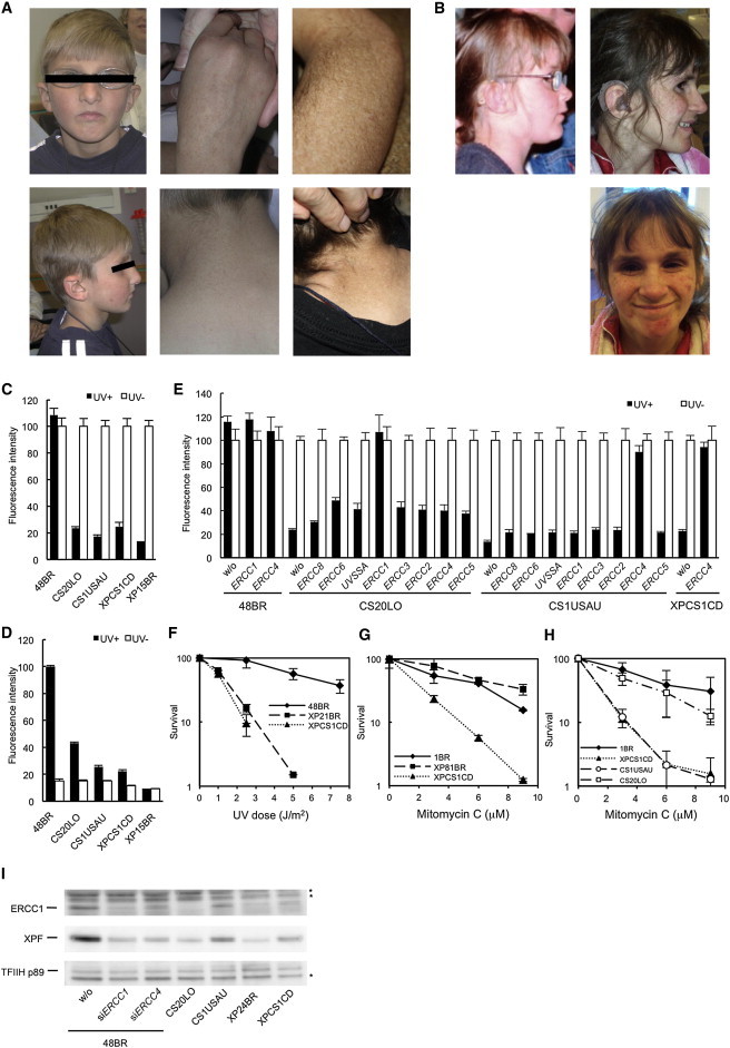
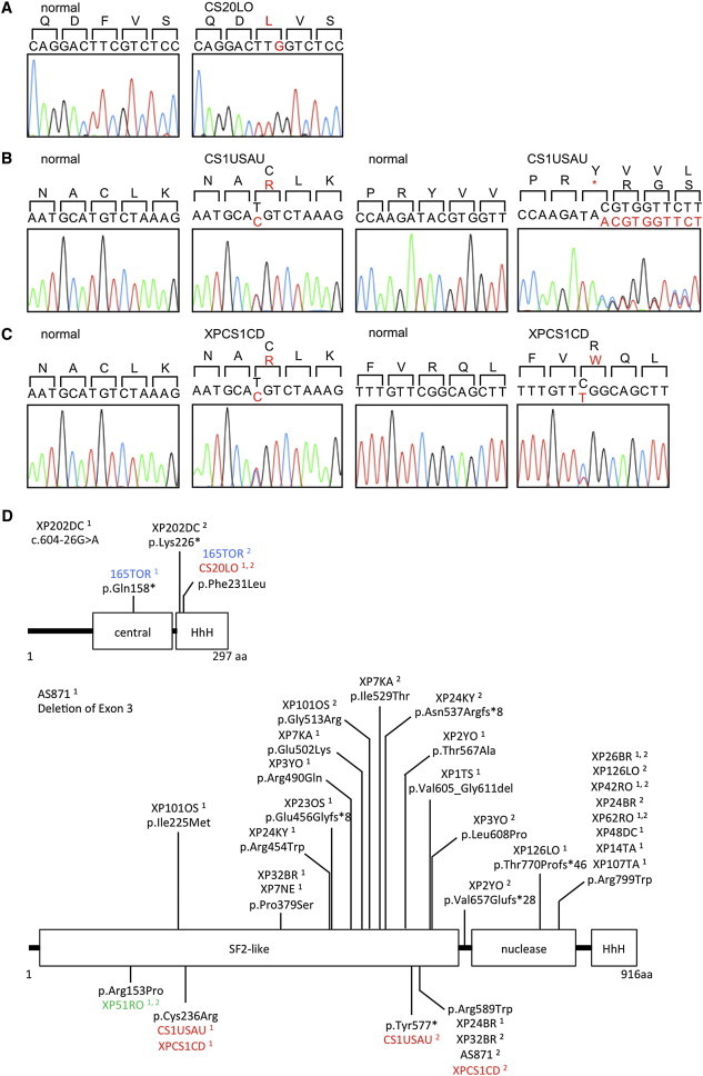
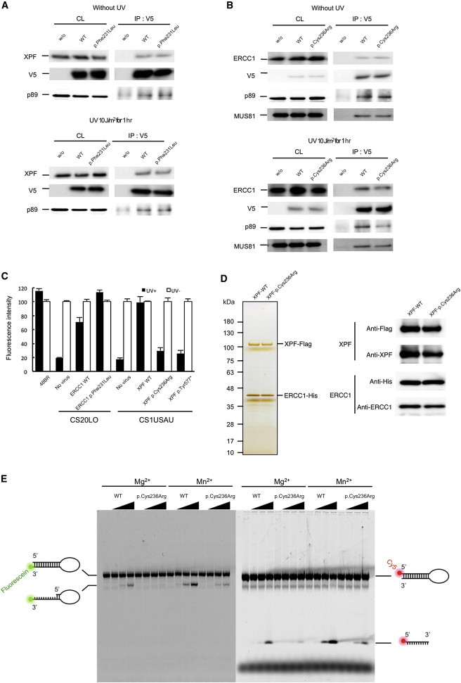
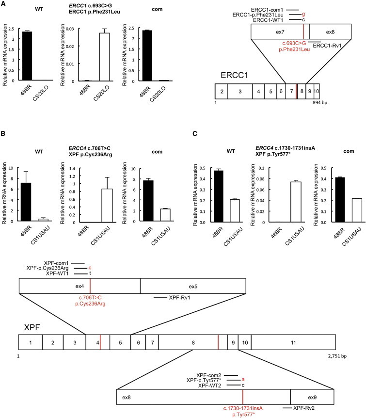
Cockayne syndrome (CS) is a genetic disorder characterized by developmental abnormalities and photodermatosis resulting from the lack of transcription-coupled nucleotide excision repair, which is responsible for the removal of photodamage from actively transcribed genes. To date, all identified causative mutations for CS have been in the two known CS-associated genes, ERCC8 (CSA) and ERCC6 (CSB). For the rare combined xeroderma pigmentosum (XP) and CS phenotype, all identified mutations are in three of the XP-associated genes, ERCC3 (XPB), ERCC2 (XPD), and ERCC5 (XPG). In a previous report, we identified several CS cases who did not have mutations in any of these genes. In this paper, we describe three CS individuals deficient in ERCC1 or ERCC4 (XPF). Remarkably, one of these individuals with XP complementation group F (XP-F) had clinical features of three different DNA-repair disorders--CS, XP, and Fanconi anemia (FA). Our results, together with those from Bogliolo et al., who describe XPF alterations resulting in FA alone, indicate a multifunctional role for XPF.


Figures

Figure 1
Figure 1
Three CS Individuals Belong to the ERCC1 and XP-F Complementation Groups (A) CS1USAU, aged 8 years (left and middle panels) and 16 years (right panels). Note the pigmentation and hypopigmented maculas. (B) XPCS1CD with features of XP, CS, and FA at 7.5 (left) and 11 years old (right). (C) Reduction of RRS rates after UV irradiation in three CS cell lines (filled bars, 11 J/m2 UVC; empty bars, no UV). RRS was measured by ethynyluridine (EU) incorporation and nuclear fluorescence detection (EU assay). 48BR is a normal control, CS20LO, CS1USAU, and XPCS1CD are CS cells, and XP15BR is affected by XP complementation group A (XP-A). (D) Reduced level of unscheduled DNA synthesis (UDS) in the CS cells (filled bars, 20 J/m2 UVC; empty bars, no UV). (E) Complete rescue of the RRS deficiency by the infection of recombinant lentivirus expressing ERCC1 cDNA in CS20LO cells and ERCC4 cDNA in CS1USAU and XPCS1CD cells (filled bars, 10 J/m2 UVC; empty bars, no UV). (F) The colony-forming ability of XPCS1CD cells after UVC irradiation was compared with that of a normal control (48BR) and that of a cell strain with XP complementation group C (XP21BR). (G) The colony-forming ability of XPCS1CD cells was compared with that of a normal control (1BR) and that of an XP-A cell strain (XP81BR) after treatment with different doses of mitomycin C (MMC) for 10 min. (H) MMC sensitivity of different cell strains was determined by MMC treatment for 10 min and measurement of their viability by their ability to incorporate 3H-thymidine (5 μCi/ml; 3 hr incubation) 3 days after treatment. (I) Immunoblotting of ERCC1 and XPF in cells from CS individuals, ERCC4-deficient XP24BR cells, and normal 48BR cells with the TFIIH-p89 (XPB) subunit as a loading control. 48BR cells were mock transfected or transfected with siRNAs targeting either ERCC1 or ERCC4. Asterisks indicate nonspecific bands. RRS was normalized to activity in nonirradiated cells. UDS activity was normalized to that of normal 48BR cells. Error bars represent the SD of medians of nuclear fluorescence measurements in quintuplicate samples in (C)–(E) and the SD of means of triplicate experiments in (F)–(H).
Figure 2
Figure 2
Locations of the ERCC1 and ERCC4 Mutations Identified in the CS Cells and in Known NER Disorders (A) The homozygous c.693C>G single-nucleotide variant (SNV) in ERCC1 exon 7 in the CS individual, CS20LO; the altered amino acid, Phe231, is shown in red. (B) The heterozygous c.706T>C SNV in ERCC4 exon 4 and the 1 base insertion, c.1730_1731insA, in ERCC4 exon 8 are identified in the CS individual, CS1USAU. The altered amino acids, Cys236 and Tyr577, are shown in red. (C) The heterozygous c.706T>C SNV in ERCC4 exon 4 and the heterozygous c.1765C>T SNV in XPCS1CD. The altered amino acids, Cys236 and Arg589, are shown in red. (D) The structure of ERCC1 and XPF and the amino acid alterations reported here and published previously. The cases shown in colors are as follows: XP, black; COFS, blue; XFE, green; and CS or XPCS, red. Superscripted 1 and 2 designate the number of discrete alleles.
Figure 3
Figure 3
ERCC1 and XPF Interaction in the CS Cells (A) V5-tagged wild-type and p.Phe231Leu altered ERCC1 interactions with the endogenous wild-type XPF were assayed by immunoprecipitation either without UV irradiation or 1 hr after 10 J/m2 of UV irradiation. (B) V5-tagged wild-type and p.Cys236Arg altered XPF were expressed in 293T cells, and interactions with the endogenous wild-type ERCC1 were assayed by immunoprecipitation from extracts of cells either without UV irradiation or 1 hr after 10 J/m2 of UV irradiation. Interactions were detected by immunoblotting with antibodies against the V5-tagged ERCC1 and XPF (1H6, MBL), MUS81 (MTA30 2G10/3, Santa Cruz), ERCC1 (8F1, Santa Cruz), and XPF (Ab-1, Thermo scientific). Abbreviations are as follows: CL, crude lysate (10% load); and IP, immunoprecipitate. (C) Rescue of the RRS deficiency was assayed by the infection of recombinant lentivirus expressing either wild-type or mutant ERCC1 and ERCC4 cDNAs (filled bars, 11 J/m2 UV; empty bars, no UV). RRS was normalized to activity in nonirradiated cells. Error bars represent the SD of medians of nuclear fluorescence measurements in quintuplicate samples. (D) Purification of the recombinant ERCC1-XPF complexes. FLAG-tagged (C terminus) wild-type and p.Cys236Arg altered XPF were coexpressed with the 6 × His-tagged ERCC1 (C terminus) in 293T cells. The cells were harvested and lysed in lysis buffer (50 mM Tris-HCl, 150 mM KCl, 1 mM EDTA, 1% NP-40, 10% glycerol, 0.25 mM PMSF, protease-inhibitor cocktail [PIC, Roche], pH 7.5). Tandem affinity purification was performed with mouse anti-FLAG IgG-conjugated beads (M2 agarose, SIGMA) followed by TALON metal affinity resin (Clontech). Cell lysates were incubated with anti-FLAG agarose beads and were subsequently washing with salt buffer (20 mM Tris-HCl, 1 M KCl, 0.1 mM EDTA, 10% glycerol, PIC, pH 7.5) and starting buffer (20 mM Tris-HCl, 800 mM KCl, 10% glycerol, 10 mM imidazole, PIC, pH 7.5). Binding proteins were eluted with starting buffer containing 1 mg/ml FLAG peptide. The eluted fractions were incubated with TALON metal affinity resin (Clontech) and were then sequentially washed with buffer A (40 mM HEPES, 1,000 mM NaCl, 10% glycerol, 10 mM Imidazole, PIC, pH 7.5) and buffer B (40 mM HEPES, 100 mM NaCl, 10% glycerol, 10 mM Imidazole, PIC, pH 7.5). The purified complex was sequentially eluted with elution buffer A (40 mM HEPES, 100 mM NaCl, 10% glycerol, 100 mM Imidazole, PIC, pH 7.5) and elution buffer B (40 mM HEPES, 100 mM NaCl, 10% glycerol, 300 mM Imidazole, pH 7.5). Purified complex was dialyzed against GF buffer (25 mM HEPES, 150 mM NaCl, 10% glycerol, 5 mM beta-mercaptoethanol, PIC, pH 8.0) and concentrated. The purified complexes were resolved by SDS-PAGE and detected by silver staining (100 ng of total protein). Purity of the complexes was assessed by immunoblotting with antibodies against the FLAG (MBL) and 6 × His (MBL) tags, as well as ERCC1 and XPF. No endogenous XPF was detected. (E) Endonuclease activity of the wild-type and altered ERCC1-XPF complexes was determined with fluorescently labeled 12 bp stem-dT22-loop DNA templates. Either 3′-fluorescein- or 5′-Cy5-labeled oligonucleotides (5′-GCCAGCGCTCGG-dT22-CCGAGCGCTGGC-3′) were purchased from SIGMA and self-annealed in TE buffer. Nuclease incision reactions were performed on 200 fmol of the DNA templates and 0, 25, 50, 100 ng of the purified ERCC1-XPF complexes in a total volume of 15 μl nuclease reaction buffer (25 mM HEPES, 40 mM NaCl, 10% glycerol, 0.5 mM beta-mercaptoethanol, and 0.1 mg/ml BSA and either 2 mM MgCl2 or 0.4 mM MnSO4, pH 8.0). The reaction mixtures were incubated at 30°C for 1 hr. Samples were separated on urea-denatured 15% PAGE, and the cleaved products were analyzed by a Typhoon imager (GE).
Figure 4
Figure 4
Expression of the Pathogenic Alleles in the CS Cells Total RNA was extracted with ISOGEN reagent (NIPPON GENE) according to the manufacturer’s instruction. One microgram of total RNA was reverse transcribed with a high-capacity RNA-to-cDNA kit (Applied Biosystems, Life Technologies). Quantitative PCR amplification and real-time detection were carried out in a Thermal Cycler Dice Real-Time System (TaKaRa Bio) with SYBR Premix Ex TaqII (TaKaRa Bio) and a QuantiTect SYBR Green PCR kit (QIAGEN). For each sample, relative mRNA levels were normalized against HPRT1 mRNA expression. Error bars represent the SD of means of triplicate experiments. (A) Selective quantitative amplification of the wild-type and the mutated c.693C>G (p.Phe231Leu) ERCC1 alleles in CS20LO cells and normal 48BR cells. Allele-specific primers selectively amplified the wild-type (c.693C) allele (ERCC1-WT1 and ERCC1-Rv1, left panel), the CS pathogenic mutant (c.693C>G) allele (ERCC1-p.Phe231Leu and ERCC1-Rv1, middle panel), and both alleles at once (ERCC1-com1 and ERCC1-Rv1, right panel). (B) Selective quantitative amplification of the wild-type and the mutated c.706T>C (p.Cys236Arg) ERCC4 alleles in CS1USAU cells and normal 48BR cells. Allele-specific primers selectively amplified the wild-type (c.706T) allele (XPF-WT1 and XPF-Rv1, left panel), the CS pathogenic mutant (c.706T>C) allele (XPF-p.Cys236Arg and XPF-Rv1, middle panel), and both alleles at once (XPF-com1 and XPF-Rv1, right panel). (C) Selective quantitative amplification of the wild-type and the c.1730_1731insA (p.Tyr577*) ERCC4 alleles. Allele-specific primers selectively amplified the wild-type (c.1731C) allele (XPF-WT2 and XPF-Rv2, left panel), the CS pathogenic mutant (c.1730_1731insA) allele (XPF-p.Tyr577* and XPF-Rv2, middle panel), and both alleles at once (XPF-com2 and XPF-Rv2). Locations of the primer sets used for the qRT-PCR experiments are depicted on the right-hand side. Transcripts from HPRT1 were used as a quantification control. Primers used for the qPCR are listed in Table 1.

References

    1. Kleijer W.J., Laugel V., Berneburg M., Nardo T., Fawcett H., Gratchev A., Jaspers N.G., Sarasin A., Stefanini M., Lehmann A.R. Incidence of DNA repair deficiency disorders in western Europe: Xeroderma pigmentosum, Cockayne syndrome and trichothiodystrophy. DNA Repair (Amst.) 2008;7:744–750. - PubMed
    1. Laugel V., Dalloz C., Tobias E.S., Tolmie J.L., Martin-Coignard D., Drouin-Garraud V., Valayannopoulos V., Sarasin A., Dollfus H. Cerebro-oculo-facio-skeletal syndrome: three additional cases with CSB mutations, new diagnostic criteria and an approach to investigation. J. Med. Genet. 2008;45:564–571. - PubMed
    1. Friedberg E.C., Walker G.C., Siede W., Wood R.D., Schultz R.A., Ellenberger T. ASM Press; Washington, DC: 2006. DNA Repair and Mutagenesis, Second Edition.
    1. Hanawalt P.C., Spivak G. Transcription-coupled DNA repair: two decades of progress and surprises. Nat. Rev. Mol. Cell Biol. 2008;9:958–970. - PubMed
    1. Laugel V., Dalloz C., Durand M., Sauvanaud F., Kristensen U., Vincent M.C., Pasquier L., Odent S., Cormier-Daire V., Gener B. Mutation update for the CSB/ERCC6 and CSA/ERCC8 genes involved in Cockayne syndrome. Hum. Mutat. 2010;31:113–126. - PubMed

Publication types

MeSH terms