. 2013 May 9;50(3):420-9.
doi: 10.1016/j.molcel.2013.03.021. Epub 2013 Apr 25.

The magic spot: a ppGpp binding site on E. coli RNA polymerase responsible for regulation of transcription initiation

Affiliations

The magic spot: a ppGpp binding site on E. coli RNA polymerase responsible for regulation of transcription initiation

Wilma Ross et al. Mol Cell. .

Abstract

The global regulatory nucleotide ppGpp ("magic spot") regulates transcription from a large subset of Escherichia coli promoters, illustrating how small molecules can control gene expression promoter-specifically by interacting with RNA polymerase (RNAP) without binding to DNA. However, ppGpp's target site on RNAP, and therefore its mechanism of action, has remained unclear. We report here a binding site for ppGpp on E. coli RNAP, identified by crosslinking, protease mapping, and analysis of mutant RNAPs that fail to respond to ppGpp. A strain with a mutant ppGpp binding site displays properties characteristic of cells defective for ppGpp synthesis. The binding site is at an interface of two RNAP subunits, ω and β', and its position suggests an allosteric mechanism of action involving restriction of motion between two mobile RNAP modules. Identification of the binding site allows prediction of bacterial species in which ppGpp exerts its effects by targeting RNAP.


Figures

Figure 1
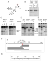
Figure 1. Mapping 32P-6-thio-ppGpp crosslinks to the E. coli RNAP β′ subunit
(A) 6-thio-ppGpp; 6-thio group is circled. (B) SDS gel with RNAP holoenzyme or core enzyme after crosslinking to 32P-6-thio-ppGpp without a competitor (0), with 1 mM GTP (GTP), or with 1 mM non-radioactive ppGpp (ppGpp). Position of the comigrating β and β′ subunits is indicated. (C) Wild-type (WT) RNAP or RNAP containing a β′-GFP fusion, crosslinked to ppGpp as in (B). Both panels are from the same gel, but intervening lanes have been deleted for clarity. (D) and (E) Undigested or thrombin-digested crosslinked RNAP. Wild-type β′ (1407 residues) contains a thrombin site at position ~900. Other RNAPs contain an engineered thrombin site at the indicated position (e.g. β′ 675th). Arrows: positions of crosslinked complete-digestion products (fragment endpoints are indicated). Comparison of phosphorimages with stained images of the same gels is shown in Fig. S1. (F) A 36 amino acid crosslinked interval (β′ 612–648; red bar) is common to all crosslinked digestion products. Black bars: thrombin products forming crosslinks. Grey bars: hydroxylamine products forming crosslinks. (G) Amino acid sequence of crosslinked interval in β′ (612–648). Positions where substitutions were constructed are underlined. See also Figure S1.
Figure 2
Figure 2. RNAP substitutions that reduce the inhibition of transcription by ppGpp
(A) Representative gel showing transcripts produced in vitro by the rRNA promoter rrnB P1 or the vector-derived RNA 1 promoter with wild-type RNAP or β′ K615A RNAP and 0–200 μM ppGpp. (B) Concentration for half-maximal inhibition of rrnB P1 transcription by ppGpp (IC50 values) with wild-type or mutant RNAPs. Values were determined from replicate experiments as illustrated in panels (A) and (C–F). (C–F) Plots of representative transcription experiments, as in (A), quantifying rrnB P1 transcript levels as a function of ppGpp concentration. Transcription with wild-type (WT) and two mutant RNAPs are shown in each panel.
Figure 3
Figure 3. RNAP substitutions eliminate the destabilizing effects of ppGpp on promoter complexes, reduce crosslinking to 32P-6-thio-ppGpp, and alter cell growth
(A) Plot of a representative promoter complex half-life experiment with WT RNAP or K615A, R417A RNAP ± 50 μM ppGpp. Plots indicate the fraction of complexes remaining as a function of time after competitor addition. Half-life values from replicate experiments with associated ranges are shown in (B). (B) Promoter complex half-lives (min) ± ppGpp with indicated RNAPs, quantified as in (A). Error bars represent the range from at least two independent experiments. (C) Representative crosslinking experiment of 32P-6-thio-ppGpp with different RNAPs as in Fig. 1B. (D) Relative ppGpp crosslinking to indicated WT or mutant RNAPs, determined as in (C). Relative crosslinking from replicate experiments with associated ranges are shown (see Experimental Procedures). Error bars represent the range from at least two independent experiments. (E) Growth curves of E. coli wild type or rpoC R417A, K615A strains grown in LB, washed, and diluted into fresh LB (black, wild-type; green, mutant) or downshifted into MOPS-glucose minimal medium (MM; blue, wild-type; red, mutant). Error ranges for six replicate determinations are shown. Efficiencies of plating (MM/LB) were 1.2 ± 0.5 (wild-type) and 1.2 ± 0.2 (mutant). The time for the mutant strain to achieve maximum exponential growth rate or to reach stationary phase was 4–5 hr longer than for the wild-type strain.
Figure 4
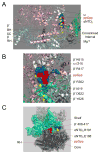
Figure 4. Location of the ppGpp Binding Site on E. coli RNAP
(A) Model of E. coli RNAP, adapted from 3LU0 (Opalka et al., 2010), with residues implicated in ppGpp function shown in blue spacefill. The crosslinked interval (β′612–648, yellow) is located between the secondary channel (SC) and α NTDII (green). The location of ppGpp (red spacefill) is predicted from the genetic and biochemical data in Figs. 2 and 3. BH, bridge helix. RH, rim helices, Mg2+, active site. (B) Close-up view rotated ~90° from the view in (A). Residues implicated in binding to ppGpp are indicated. (C) The proposed ppGpp binding site is at the junction of the rigid-body core and shelf modules of RNAP (core module, dark grey; shelf module, cyan; clamp, green) on the T. thermophilus RNAP-Gfh1 co-crystal structure (3AOI, adapted from Tagami et al., 2010; Gfh1 is not shown). ppGpp (red) is shown at the location corresponding to the proposed binding site on E. coli RNAP (ppGpp does not bind to T. thermophilus RNAP; Fig. 3D; Vrentas et al., 2008). T. thermophilus core module residues corresponding to E coli α NTDII α E188 (T. thermophilus E182) and R191 (T. thermophilus R185), shown in yellow spacefill, are in a region proposed to form a stabilizing contact with an α helix in β′ in the shelf module (blue; E. coli β′ 408–417; T. thermophilus β′ 685–696; Tagami et al., 2010). We suggest that substitutions for E. coli residues R191 and E188 would increase complex stability by disfavoring a clamp-open (ratcheted) state.
Figure 5
Figure 5. Evolutionary conservation of the proposed ppGpp binding site
(A) Three regions comprising the proposed ppGpp binding site in E. coli RNAP. Sixty amino acids from each region are illustrated on the model of E. coli RNAP (3LU0): Region 1 (β′ 600–659; yellow), Region 2 (β′ 340–399; green), and Region 3 (ω 1–60; blue). ppGpp is in red. Residues predicted to contact ppGpp are in spacefill (see Fig. 4). (B) Alignments of the E. coli RNAP sequences from the three regions illustrated in (A) with the corresponding regions from Bacillus subtilis subsp subtilis str 168 and Thermus thermophilus HB8. Residues predicted to bind ppGpp in each region of E. coli RNAP are in red. The 36 amino acid interval in Region 1 that crosslinked to 6-thio-ppGpp is in green. Alignments of the three RNAP regions from other bacterial species are provided in Fig. S2. Boxes in Regions 1 and 3 surround short region containing residues with the largest effects on ppGpp binding. Dots indicate sequence identity. See also Figure S2.

Comment in

References

    1. Artsimovitch I, Patlan V, Sekine S, Vassylyeva MN, Hosaka T, Ochi K, Yokoyama S, Vassylyev DG. Structural basis for transcription regulation by alarmone ppGpp. Cell. 2004;117:299–310. - PubMed
    1. Artsimovitch I, Svetlov V, Murakami KS, Landick R. Co-overexpression of Escherichia coli RNA polymerase subunits allows isolation and analysis of mutant enzymes lacking lineage-specific sequence insertions. J Biol Chem. 2003;278:12344–12355. - PubMed
    1. Atkinson GC, Tenson T, Hauryliuk V. The RelA/SpoT homolog (RSH) superfamily: distribution and functional evolution of ppGpp synthetases and hydrolases across the tree of life. PLoS One. 2011;6:e23479. - PMC - PubMed
    1. Baker CR, Booth LN, Sorrells TR, Johnson AD. Protein modularity, cooperative binding, and hybrid regulatory states underlie transcription network diversification. Cell. 2012;151:80–95. - PMC - PubMed
    1. Barker MM, Gaal T, Gourse RL. Mechanism of regulation of transcription initiation by ppGpp. II Models for positive control based on properties of RNAP mutants and competition for RNAP. J Mol Biol. 2001a;305:689–702. - PubMed

Publication types

MeSH terms

LinkOut - more resources