. 2013 May 9;50(3):430-6.
doi: 10.1016/j.molcel.2013.03.020. Epub 2013 Apr 25.

The mechanism of E. coli RNA polymerase regulation by ppGpp is suggested by the structure of their complex

Affiliations

The mechanism of E. coli RNA polymerase regulation by ppGpp is suggested by the structure of their complex

Yuhong Zuo et al. Mol Cell. .

Abstract

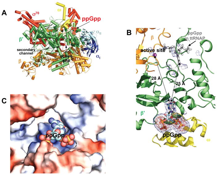
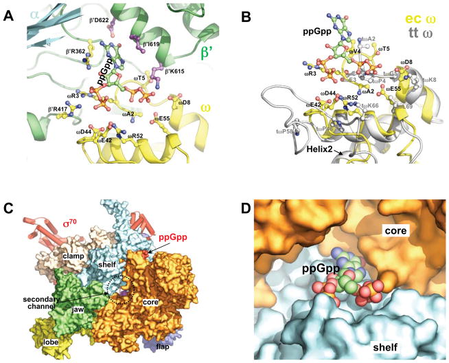
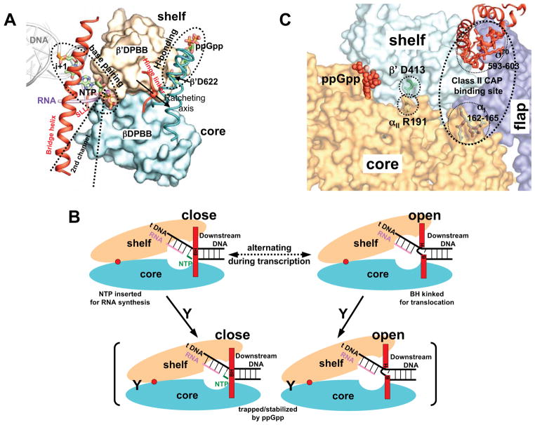
Guanosine tetraphosphate (ppGpp) is an alarmone that enables bacteria to adapt to their environment. It has been known for years that ppGpp acts directly on RNA polymerase (RNAP) to alter the rate of transcription, but its exact target site is still under debate. Here we report a crystal structure of Escherichia coli RNAP holoenzyme in complex with ppGpp at 4.5 Å resolution. The structure reveals that ppGpp binds at an interface between the shelf and core modules on the outer surface of RNAP, away from the catalytic center and the nucleic acid binding path. Bound ppGpp connects these two pivotal modules that may restrain the opening of the RNAP cleft. A detailed mechanism of action of ppGpp is proposed in which ppGpp prevents the closure of the active center that is induced by the binding of NTP, which could slow down nucleotide addition cycles and destabilize the initial transcription complexes.


Figures

Figure 1
Figure 1. ppGpp bound to E. coli RNA polymerase holoenzyme
(A) An overview of ppGpp bound to the ecRNAP holoenzyme. The ecRNAP holoenzyme is shown in a tube and arrow cartoon representation with each subunit colored differently: α I – blue; α II – light blue; β - orange; β′ – green; ω - yellow; σ70 – red. A metal ion at the active site is shown as a magenta sphere, and ppGpp is shown in red as space-filling model. (B) A close-up view of the ppGpp binding site and the active site. σA-weighted Fobs-Fcalc electron density maps contoured at 2.5σ (blue) and 5.0σ (orange) show ppGpp bound at the interface between the ω and β′ subunits. No recognizable electron density corresponding to ppGpp binding is seen near the catalytic center at the analogous site (grey model) identified previously in the ttRNAP complex with ppGpp; the σA-weighted Fobs-Fcalc electron density map is contoured at 2.5σ. The ppGpp molecules are shown in stick models, and a metal ion at the active site is shown as a magenta ball. EcRNAP subunits are shown in cartoon representation and colored as in (A). (C) Electrostatic surface potential diagram of the ppGpp-binding pocket in ecRNAP with the positive potential in blue and the negative in red. ppGpp is shown as space-filling model with distinct colors for each atom type. The electrostatic surface potential diagram of the ecRNAP holoenzyme was generated using Pymol (Delano, 2002).
Figure 2
Figure 2. ppGpp binding site
(A) Details of the residues of ecRNAP that are contacting ppGpp. EcRNAP subunits are shown as cartoon representation with each subunit colored differently as in Figure 1A. ppGpp and the side chains of the residues that contact ppGpp are shown as ball-and-stick models. (B) Structural comparison of the ω subunits of the E. coli (yellow) and Thermus thermophilus (grey) RNAPs at the ppGpp binding site. The side-chains of ecRNAP that contact ppGpp and their spatial counterparts in ttRNAP are shown as ball-and-stick models. The ω subunit structures are aligned on ω residues 17–32 in both sequences, the highly conserved second α-helix (Helix2) that is involved in interactions with the β′ subunit. The structural alignments were performed with the lsqkab program in the CCP4 suite (Winn et al., 2011). (C) ppGpp bound on a modular interface of the ecRNAP holoenzyme. EcRNAP is shown in the same orientation as in Figure 1A, with the core enzyme shown in surface representation and colored differently for individual modules. (D) A close-up view of ppGpp bound at the interface between the core and shelf modules. ppGpp is shown as a space-filling model, and the core (orange) and shelf (cyan) modules are shown as surface diagrams.
Figure 3
Figure 3. Mechanism of ppGpp action
(A) ppGpp binding in the contexts of RNAP modular movement. The core and shelf modules pack against each other on two beta barrel domains (βDPBB and β′DPBB, shown as surface diagrams), and make a ratcheting movement relative to each other on a ratcheting axis (Tagami et al., 2010). Base pairing between the incoming NTP and the template DNA at the i+1 site and the putative ppGpp-β′D622 hydrogen bonding form a leverage system hinged on the core-shelf ratcheting axis. The three linkers (bridge helix, hinge linker and a switch-like linker, SLL), which restrain the core-shelf ratcheting, are shown in red as helix and loops. The secondary channel that provides access for NTP binding and the binding site of some regulatory proteins are marked for reference. (B) Schematic representation of the proposed mechanism of ppGpp action. Dynamic cycles of ratcheting of the core and shelf modules between the open and closed states of the active site chamber that alternates between the nucleotide addition reaction and RNAP translocation. ppGpp binding on the outer surface of the cleft traps or stabilizes RNAP in certain state and slows down the ratcheting dynamics as well as RNA synthesis. ppGpp is represented by a letter Y, and the ratcheting axis is represented by a red dot. The bridge helix is BH and is separated into B and H for the kinked conformation. (C) A proposed general mechanism of transcription regulation that involves the interface between the core and shelf modules. (i) ppGpp (red, space-filling model) binds at the core-shelf interface to exert stringent response (this study); (ii) substitution of αII R191 by an alanine, which breaks a salt bridge between the side chains of a core module residue (αII R191) and a shelf module residue (β′ D413), thereby stabilizing transcription open complexes (Ross et al., accompanying manuscript); (iii) Class II CAP actions involve the interface between the core and shelf modules. In addition to interacting with the CTD of the α subunit for RNAP recruiting, CAP interacts with RNAP at two other places. It interacts with residues 162–165 of the NTD of the αI subunit and residues 593–603 of the CTD of σ70, when it binds to a class II CAP-dependent promoters (Niu et al.,1996; Lonetto et al., 1998). These two sequence segments span a region that lies at the interface between the core, shelf and flap modules. The core, shelf and flap modules of the region of interest are shown in distinct colors as surface diagrams. The σ70 factor is shown in red as ribbon diagrams. Side chains of αII R191, β′ D413, and the two CAP-interacting sequence segments are shown as sticks.

Comment in

References

    1. Artsimovitch I, Patlan V, Sekine S, Vassylyeva MN, Hosaka T, Ochi K, Yokoyama S, Vassylyev DG. Structural basis for transcription regulation by alarmone ppGpp. Cell. 2004;117:299–310. - PubMed
    1. Barker MM, Gaal T, Josaitis CA, Gourse RL. Mechanism of regulation of transcription initiation by ppGpp. I Effects of ppGpp on transcription initiation in vivo and in vitro. J Mol Biol. 2001a;305:673–688. - PubMed
    1. Barker MM, Gaal T, Gourse RL. Mechanism of regulation of transcription initiation by ppGpp. II Models for positive control based on properties of RNAP mutants and competition for RNAP. J Mol Biol. 2001b;305:689–702. - PubMed
    1. Buglino J, Shen V, Hakimian P, Lima CD. Structural and biochemical analysis of the Obg GTP binding protein. Structure. 2002;10:1581–1592. - PubMed
    1. Cashel M, Gentry DR, Hernandez VH, Vinella D. In: The stringent response Escherichia coli and Salmonella. Neidhardt FC, editor. ASM Press; Washington, DC: 1996. pp. 1458–1496.

Publication types

MeSH terms

Substances