. 2013 Jun 4;21(6):920-8.
doi: 10.1016/j.str.2013.04.002. Epub 2013 Apr 25.

Architecture of human translation initiation factor 3

Affiliations

Architecture of human translation initiation factor 3

Jordi Querol-Audi et al. Structure. .

Abstract

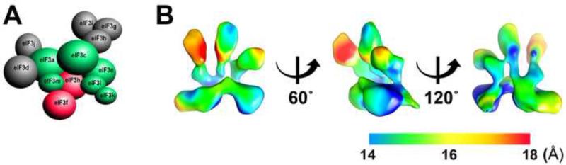
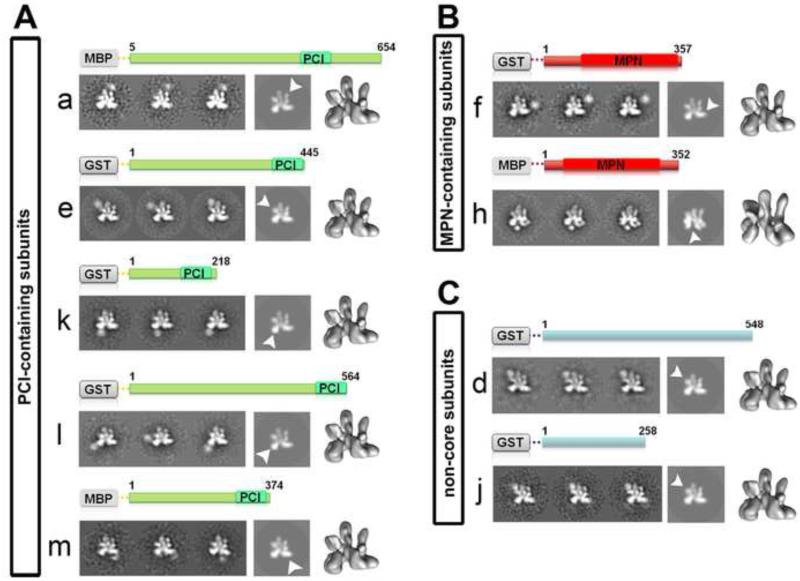
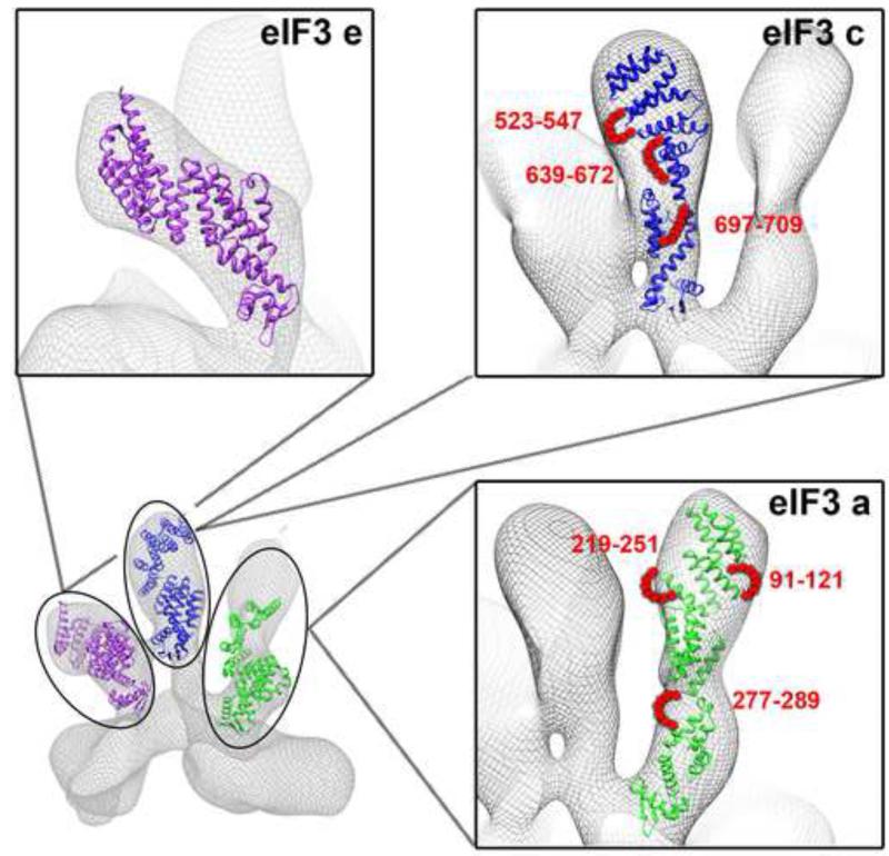
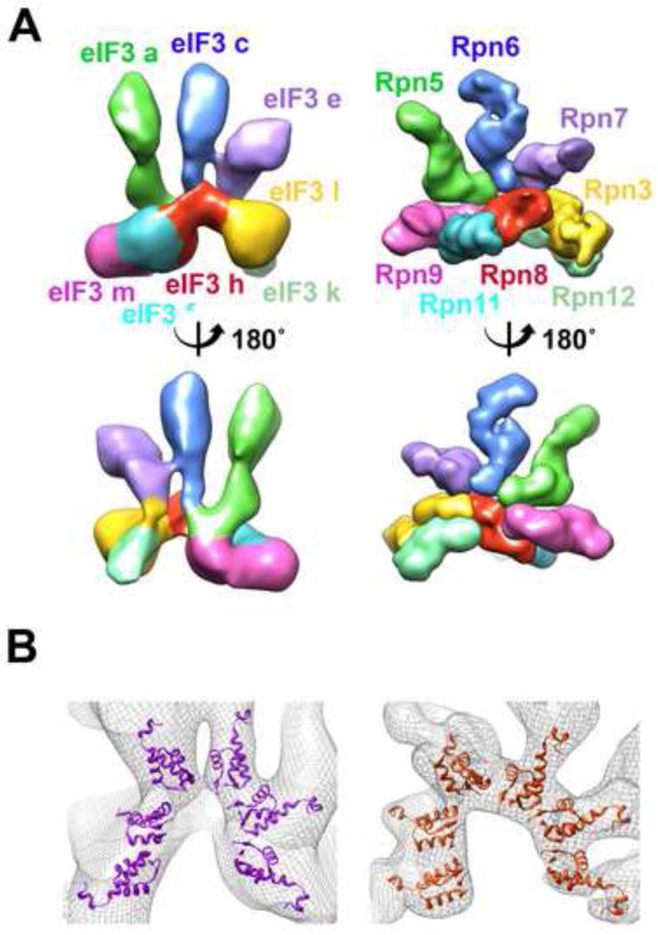
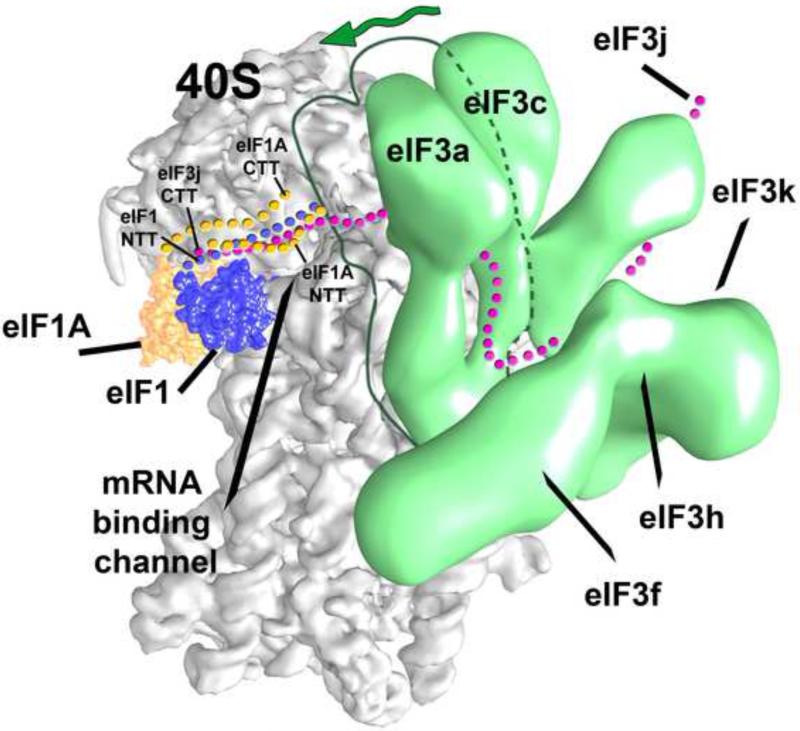
Eukaryotic translation initiation factor 3 (eIF3) plays a central role in protein synthesis by organizing the formation of the 43S preinitiation complex. Using genetic tag visualization by electron microscopy, we reveal the molecular organization of ten human eIF3 subunits, including an octameric core. The structure of eIF3 bears a close resemblance to that of the proteasome lid, with a conserved spatial organization of eight core subunits containing PCI and MPN domains that coordinate functional interactions in both complexes. We further show that eIF3 subunits a and c interact with initiation factors eIF1 and eIF1A, which control the stringency of start codon selection. Finally, we find that subunit j, which modulates messenger RNA interactions with the small ribosomal subunit, makes multiple independent interactions with the eIF3 octameric core. These results highlight the conserved architecture of eIF3 and how it scaffolds key factors that control translation initiation in higher eukaryotes, including humans.


Figures

Figure 1
Figure 1. 3D reconstruction of the human eIF3 core complex
(A) Subunit composition of human eIF3, arranged according prior mapping information (Sun et al., 2011). (B) Front, lateral and back views of the Cryo-EM reconstruction of the eIF3 8mer. Local resolution estimation is indicated by the color scheme (see also Supplementary Table 1 and Fig. S1).
Figure 2
Figure 2. Architecture of the core eIF3 octamer complex
Subunit localization for eIF3 8mer/9mer by visualization of MBP or GST tags on specific subunits. A set of three representative reference-free 2D class averages are shown for each labeled subunit containing a PCI domain (A) MPN domain (B) or to non-core subunits (C) compared with both a forward projection and a surface representation of the recombinant eIF3 8mer (shown to the right of each set of class averages). The position of each subunit based on the extra density due to the presence of the tag is indicated with an arrowhead. A schematic representation of each of the subunits showing the position of PCI and/or MPN domains is shown above each set (see also Supplementary Table 2 and Fig. S2).
Figure 3
Figure 3. Proposed location and structure of subunits a, c and e within the eIF3 cryo-EM reconstruction
The atomic models of subunits a (green), c (blue) and e (purple) have been fitted into the three-dimensional cryo-EM reconstruction of our reconstituted eIF3 core. The red strings indicate the position of insertions according to Phyre2 secondary structure predictions (see Supplemental Experimental Procedures and Fig. S3).
Figure 4
Figure 4. Conservation of the PCI/MPN core
(A) Conserved architecture between eIF3 8-mer and proteasome lid. The 3D reconstruction of the lid (colored by subunit) in its holoenzyme-bound state (right column) is compared with that of the eIF3 8-mer (left column). Subunit localization within the eIF core complex based on this comparison (and also on the tagging experiments shown in Figure 2) is indicated. The same color code for structurally equivalent subunits has been used for clarity. (B) Six copies of the crystal structure of a PCI domain (PDB ID: 1RZ4) are docked into the eIF3 octamer electron density (left), showing the same horseshoe-shaped arrangement of the winged-helix domains as in the 26S proteasome Lid (right).
Figure 5
Figure 5. Interaction of eIF3 octamer subunits with translation factors eIF1, eIF1A and subunit eIF3j
Coomassie blue-stained SDS gel showing eIF3a*c* dimer (A), a* monomer (B) and c* monomer (C) affinity-purified using MBP-tagged eIF1A, or using MBP-tagged eIF1. MW markers are shown in kilodaltons. Arrows indicate the position of MBP-eIF1A (lanes 1, 3 in panels A to C) and MBP-eIF1 (lanes 4, 6 in panels A to C). Binding and washing conditions prevent nonspecific binding of the eIF3a*c*, a* and c* to the MBP alone (lanes 7, 8 in panels A to C). (D) Schematic representations of GST fusions of full length eIF3j, and truncations GST-eIF3j3-50 GST-eIF3j103-258 (E) Native agarose (1.2%) gel shift assays showing interactions between Alexa-546 labeled GST fusions of the eIF3j proteins in (D) and the indicated concentrations of eIF3 octamer. (F) Native agarose gel shift with GST-eIF3j103-258 and 1uM of octamer, eIF3k or eIF3c*. The fusion protein does not enter the gel in the absence of eIF3.
Figure 6
Figure 6. Model for interactions of the eIF3 PCI/MPN octameric core with eIF3j and initiation factors eIF1 and eIF1A on the 40S ribosomal platform
The position of the PCI/MPN octamer relative to the 40S subunit is based on that in (Siridechadilok et al., 2005). The positions of eIF1 (blue surface) and eIF1A (orange surface) are based on the X-ray crystal structure of the eIF1/40S complex (Rabl et al., 2011), and directed hydroxyl radical probing experiments of eIF1A bound to the 40S subunit (Yu et al., 2009). The flexible N and C-terminal extensions of both factors reaching the decoding center are depicted using the same color code. eIF3j is represented by purple dots going along the eIF3 complex and reaching eIF1A. Subunits within the eIF3 core, which interact with eIF3j are explicitly indicated. Inferred flexibility of eIF3a and eIF3c subunits is indicated by the green line and green arrow.

Similar articles

Cited by

References

    1. Algire MA, Maag D, Lorsch JR. Pi release from eIF2, not GTP hydrolysis, is the step controlled by start-site selection during eukaryotic translation initiation. Mol Cell. 2005;20:251–262. - PubMed
    1. Asano K, Clayton J, Shalev A, Hinnebusch AG. A multifactor complex of eukaryotic initiation factors, eIF1, eIF2, eIF3, eIF5, and initiator tRNA(Met) is an important translation initiation intermediate in vivo. Genes Dev. 2000;14:2534–2546. - PMC - PubMed
    1. Ben-Shem A, Garreau de Loubresse N, Melnikov S, Jenner L, Yusupova G, Yusupov M. The structure of the eukaryotic ribosome at 3.0 A resolution. Science. 2011;334:1524–1529. - PubMed
    1. Bushell M, Wood W, Clemens MJ, Morley SJ. Changes in integrity and association of eukaryotic protein synthesis initiation factors during apoptosis. Eur J Biochem. 2000;267:1083–1091. - PubMed
    1. Cai Q, Todorovic A, Andaya A, Gao J, Leary JA, Cate JH. Distinct regions of human eIF3 are sufficient for binding to the HCV IRES and the 40S ribosomal subunit. J Mol Biol. 2010;403:185–196. - PMC - PubMed

LinkOut - more resources