

Wzi is an outer membrane lectin that underpins group 1 capsule assembly in Escherichia coli
- PMID: 23623732
- PMCID: PMC3791409
- DOI: 10.1016/j.str.2013.03.010
Wzi is an outer membrane lectin that underpins group 1 capsule assembly in Escherichia coli
Abstract
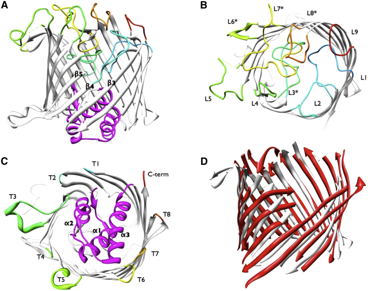
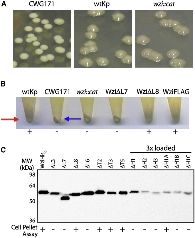
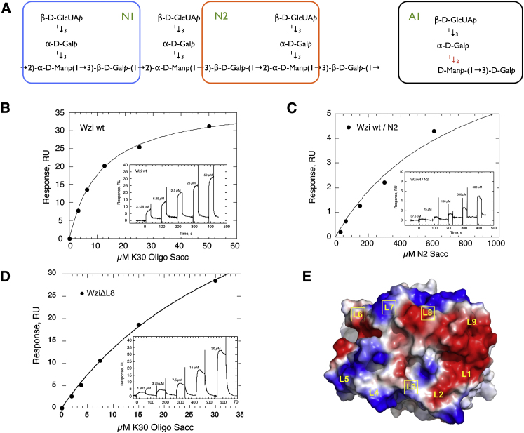
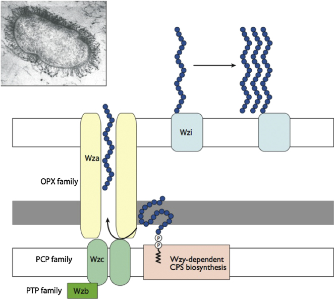
Many pathogenic bacteria encase themselves in a polysaccharide capsule that provides a barrier to the physical and immunological challenges of the host. The mechanism by which the capsule assembles around the bacterial cell is unknown. Wzi, an integral outer-membrane protein from Escherichia coli, has been implicated in the formation of group 1 capsules. The 2.6 Å resolution structure of Wzi reveals an 18-stranded β-barrel fold with a novel arrangement of long extracellular loops that blocks the extracellular entrance and a helical bundle that plugs the periplasmic end. Mutagenesis shows that specific extracellular loops are required for in vivo capsule assembly. The data show that Wzi binds the K30 carbohydrate polymer and, crucially, that mutants functionally deficient in vivo show no binding to K30 polymer in vitro. We conclude that Wzi is a novel outer-membrane lectin that assists in the formation of the bacterial capsule via direct interaction with capsular polysaccharides.
Copyright © 2013 Elsevier Ltd. All rights reserved.
Figures
Comment in
-
Encapsulating bacteria.Structure. 2013 May 7;21(5):692-3. doi: 10.1016/j.str.2013.04.011. Structure. 2013. PMID: 23664360
Similar articles
-
Key diffusion mechanisms involved in regulating bidirectional water permeation across E. coli outer membrane lectin.Sci Rep. 2016 Jun 20;6:28157. doi: 10.1038/srep28157. Sci Rep. 2016. PMID: 27320406 Free PMC article.
-
A novel outer membrane protein, Wzi, is involved in surface assembly of the Escherichia coli K30 group 1 capsule.J Bacteriol. 2003 Oct;185(19):5882-90. doi: 10.1128/JB.185.19.5882-5890.2003. J Bacteriol. 2003. PMID: 13129961 Free PMC article.
-
Crystallization and preliminary diffraction analysis of Wzi, a member of the capsule export and assembly pathway in Escherichia coli.Acta Crystallogr Sect F Struct Biol Cryst Commun. 2010 Dec 1;66(Pt 12):1621-5. doi: 10.1107/S1744309110040546. Epub 2010 Nov 26. Acta Crystallogr Sect F Struct Biol Cryst Commun. 2010. PMID: 21139210 Free PMC article.
-
Biosynthesis and assembly of capsular polysaccharides in Escherichia coli.Annu Rev Biochem. 2006;75:39-68. doi: 10.1146/annurev.biochem.75.103004.142545. Annu Rev Biochem. 2006. PMID: 16756484 Review.
-
Wza: a new structural paradigm for outer membrane secretory proteins?Trends Microbiol. 2007 Mar;15(3):96-100. doi: 10.1016/j.tim.2007.01.002. Epub 2007 Feb 1. Trends Microbiol. 2007. PMID: 17275308 Review.
Cited by
-
Key diffusion mechanisms involved in regulating bidirectional water permeation across E. coli outer membrane lectin.Sci Rep. 2016 Jun 20;6:28157. doi: 10.1038/srep28157. Sci Rep. 2016. PMID: 27320406 Free PMC article.
-
Evidence for a Widespread Third System for Bacterial Polysaccharide Export across the Outer Membrane Comprising a Composite OPX/β-Barrel Translocon.mBio. 2022 Oct 26;13(5):e0203222. doi: 10.1128/mbio.02032-22. Epub 2022 Aug 16. mBio. 2022. PMID: 35972145 Free PMC article.
-
Escherichia coli O127 group 4 capsule proteins assemble at the outer membrane.PLoS One. 2021 Nov 15;16(11):e0259900. doi: 10.1371/journal.pone.0259900. eCollection 2021. PLoS One. 2021. PMID: 34780538 Free PMC article.
-
Mechanisms governing bacterial capsular polysaccharide attachment and chain length.Ann N Y Acad Sci. 2025 Jun;1548(1):80-98. doi: 10.1111/nyas.15364. Epub 2025 May 14. Ann N Y Acad Sci. 2025. PMID: 40369709 Free PMC article. Review.
-
Capsules and Extracellular Polysaccharides in Escherichia coli and Salmonella.EcoSal Plus. 2021 Dec 15;9(2):eESP00332020. doi: 10.1128/ecosalplus.ESP-0033-2020. Epub 2021 Dec 1. EcoSal Plus. 2021. PMID: 34910576 Free PMC article.
References
-
- Amor P.A., Whitfield C. Molecular and functional analysis of genes required for expression of group IB K antigens in Escherichia coli: characterization of the his-region containing gene clusters for multiple cell-surface polysaccharides. Mol. Microbiol. 1997;26:145–161. - PubMed
-
- Baslé A., Rummel G., Storici P., Rosenbusch J.P., Schirmer T. Crystal structure of osmoporin OmpC from E. coli at 2.0 Å. J. Mol. Biol. 2006;362:933–942. - PubMed
-
- Biswas S., Mohammad M.M., Patel D.R., Movileanu L., van den Berg B. Structural insight into OprD substrate specificity. Nat. Struct. Mol. Biol. 2007;14:1108–1109. - PubMed
Publication types
MeSH terms
Substances
Grants and funding
LinkOut - more resources
Full Text Sources
Other Literature Sources
Molecular Biology Databases
