. 2013 May 7;17(5):709-18.
doi: 10.1016/j.cmet.2013.03.018. Epub 2013 Apr 25.

Tam41 is a CDP-diacylglycerol synthase required for cardiolipin biosynthesis in mitochondria

Affiliations

Tam41 is a CDP-diacylglycerol synthase required for cardiolipin biosynthesis in mitochondria

Yasushi Tamura et al. Cell Metab. .

Abstract

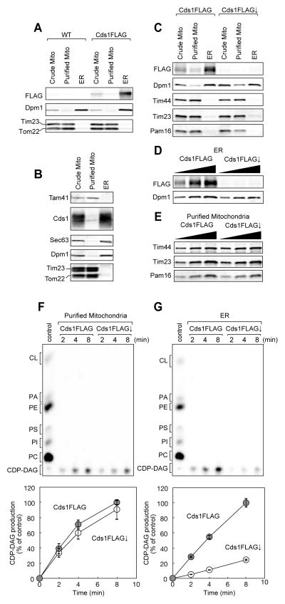
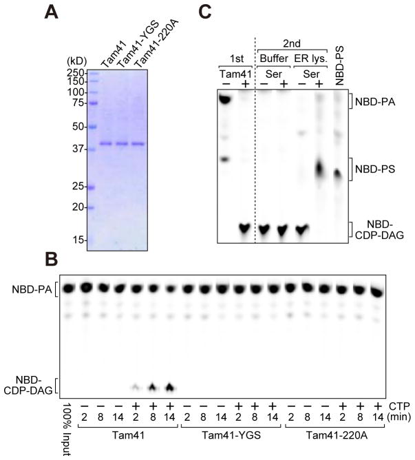
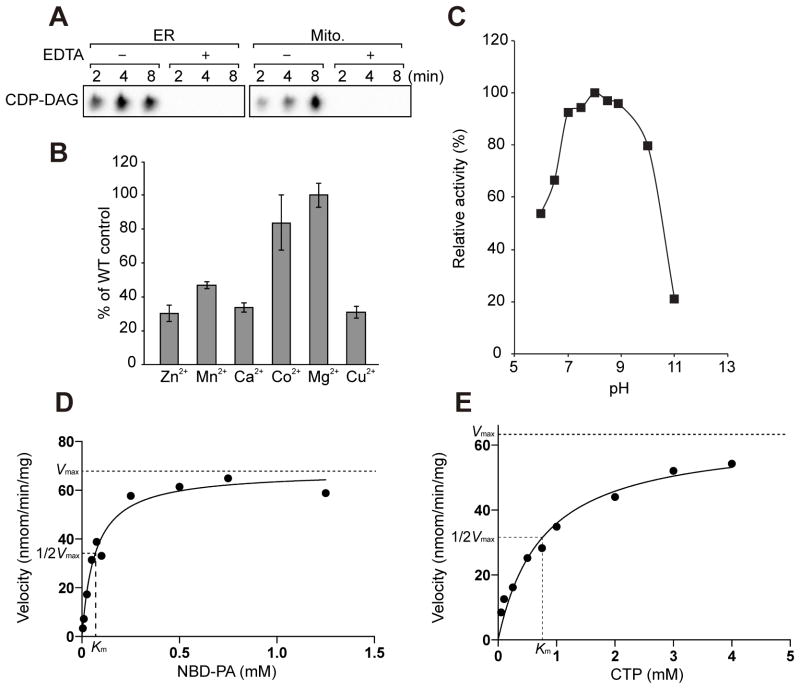
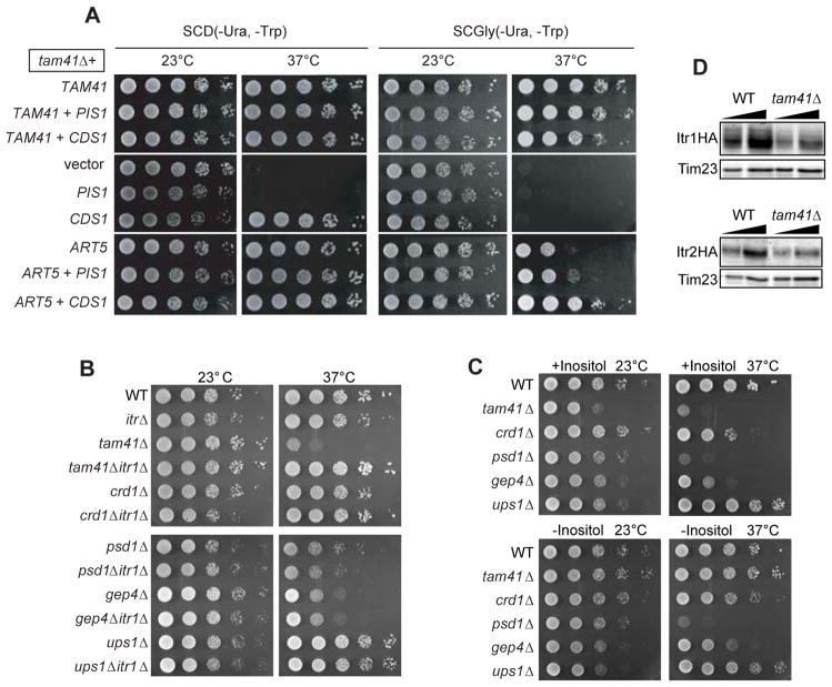
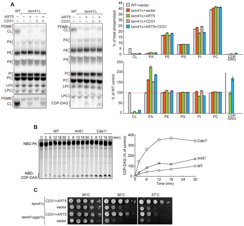
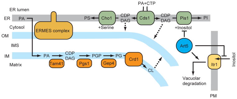
CDP-diacylglycerol (CDP-DAG) is central to the phospholipid biosynthesis pathways in cells. A prevailing view is that only one CDP-DAG synthase named Cds1 is present in both the endoplasmic reticulum (ER) and mitochondrial inner membrane (IM) and mediates generation of CDP-DAG from phosphatidic acid (PA) and CTP. However, we demonstrate here by using yeast Saccharomyces cerevisiae as a model organism that Cds1 resides in the ER but not in mitochondria, and that Tam41, a highly conserved mitochondrial maintenance protein, directly catalyzes the formation of CDP-DAG from PA in the mitochondrial IM. We also find that inositol depletion by overexpressing an arrestin-related protein Art5 partially restores the defects of cell growth and CL synthesis in the absence of Tam41. The present findings unveil the missing step of the cardiolipin synthesis pathway in mitochondria as well as the flexibile regulation of phospholipid biosynthesis to respond to compromised CDP-DAG synthesis in mitochondria.


Figures

Figure 1
Figure 1. Cds1 is an ER-resident protein
Crude and purified mitochondria and ER fractions were prepared from (A, B) wild-type and Cds1FLAG or (C) Cds1FLAG-expressing (Cds1FLAG) and Cds1FLAG-depleted (Cds1FLAG↓) cells and analyzed by immunoblotting with the indicated antibodies. (D, E) ER and purified mitochondria fractions isolated from Cds1FLAG and Cds1FLAG↓ cells were analyzed by immunoblotting with the indicated antibodies. (F, G) Phosphatidic acid (PA) and purified mitochondria or ER fractions solubilized with Triton X100 were incubated for the indicate time in the presence of 32P-CTP. Phospholipids were then extracted and analyzed by TLC. Amounts of CDP-DAG generated after 8 min incubation in wild-type cells is set to 100%. Values are mean ± SEM (n=3).
Figure 2
Figure 2. Tam41 is a mitochondrial CDP-DAG synthase
(A) Purified Tam41-His6 and its mutants were analyzed by SDS-PAGE followed by CBB-staining. (B) NBD-PA and purified Tam41 and Tam41 mutants (Tam41-YGS and Tam41-220A) were incubated for the indicated periods of time at 30°C in the presence or absence of CTP. Phospholipids were then extracted and analyzed by TLC. Note that Tam41 appears to belong to the superfamily of nucleotidetransferase (NTase) fold proteins containing conserved active site residues; hG[GS], [DE]h[DE]h and h[DE]h (h indicates a hydrophobic amino acid) (Kuchta et al., 2009). Tam41-YGS mutant has mutations in the hG[GS] motif, which may has a role in holding substrates within the active site, while it is not clear if Asp220 functions in the active site since intact [DE]h[DE]h and h[DE]h motifs are missing in Tam41. (C) CDP-DAG generated in vitro was further incubated with solubilized ER fractions (ER lys.) in the presence or absence of serine. See also Figure S1.
Figure 3
Figure 3. Enzymological analyses of Tam41
(A) CDP-DAG synthase activities of highly purified ER and mitochondria fractions were measured in the presence of 10 mM EDTA. (B) Purified Tam41 and NBD-PA were incubated in the presence of 3 mM divalent metal ions for 5 min. The CDP-DAG synthase activity detected in the presence of Mg2+ was set to 100%. Values are mean ± SEM (n=3). (C) pH profile of Tam41 CDP-DAG synthase. Tam41 CDP-DAG activity was determined at varying pH. The buffer used are 50 mM PIPES-NaOH pH 6.0 and 6.5; 50 mM Tris-HCl pH 7.0, 7.5, 8.0, 8.5 and 8.9; and 50 mM CAPS-NaOH pH 10 and 11. The CDP-DAG activity obtained at pH 8.0 was set to 100%. (D, E) Kinetic analyses of Tam41 CDP-DAG synthase on the concentrations of NBD-PA and CTP. NBD-PA and purified Tam41 were incubated for 10 min at 30°C. 2 mM CTP or 250 μM NBD-PA was used for (D) and (E) experiments, respectively. Curve fitting was performed with Prism 6.0 (GraphPad software).
Figure 4
Figure 4. Decreased inositol uptake restores the growth defects of tam41Δ cells
(A) Serial dilutions of tam41Δ cells with a multi-copy plasmid harboring the TAM41, PIS1, CDS1 or ART5 gene or empty vector were spotted onto glucose- or glycerol-containing media and cultivated for 2 or 4 days, respectively. (B, C) Serial dilutions of the indicated yeast strains were spotted on SCD plates at 23 or 37°C (B) or on Synthetic minimal plates with or without inositol at 23 or 37°C (C). (D) Whole cell extracts prepared from wild-type and tam41Δ cells expressing C-terminally HA-tagged Itr1 or Itr2 were analyzed by immunoblotting with the antibodies against HA-tag and Tim23.
Figure 5
Figure 5. Phospholipid profiles of tam41Δ cells upon overexpression of Art5 or Cds1
(A) WT and tam41Δ cells with a multi-copy plasmid harboring the indicated gene or empty vector were cultivated in YPD containing 32P at 30°C. Total phospholipids were extracted from whole cells and separated on TLC for 2h (left) or 4h (right). A part of TLC plate showing PDME and CL is shown with high contrast (left below). Amounts of each lipid relative to total phospholipids (above) and to those in WT cells (except for CDP-DAG) (below) were determined. In the latter representation, the amount of CDP-DAG relative to that in the “tam41Δ+ART5” strain (CDP-DAG) was shown. Values are mean ± SEM (n=3). (B) CDP-DAG synthetic activities in ER fractions. Triton X100-solubilized ER fractions prepared from wild-type cells with or without overexpression of Art5 or Cds1 were incubated with 100 μM NBD-PA in the presence of 2 mM CTP for the indicated times at 30°C. Amounts of CDP-DAG generated after 30 min incubation in wild-type ER fraction is set to 100%. (C) Overexpression of Cds1 and Art5 rescues growth defects of tam41Δpgs1Δ cells. Serial dilutions of tam41Δ and tam41Δpgs1Δ cells with multi-copy plasmids expressing both Cds1 and Art5 or with empty vectors were spotted onto YPD and cultivated for 3 days. See also Figure S2
Figure 6
Figure 6. Phospholipid biosynthesis leading to CL, PI, and PS in mitochondria and the ER
Broken lines indicate putative transport routes. OM, mitochondrial outer membrane; IM, mitochondrial inner membrane; PM, plasma membrane.
Figure 7
Figure 7. Taxonomic distribution of Tam41 and Cds1 homologs
Two sunburst charts adopted from Pfam show the distribution of PF09139 including Tam41 (A) and PF01148 including Cds1 (B) across species. Segments shown in purple represent the proteins in eukaryotes and green ones represent bacterial proteins. Detailed explanations of color assignments are given in a rectangle. Names of three bacterial species that have homologous proteins of PF09139 are shown. (C) Evolutionary relationships of yeast Tam41 and its homologs in bacteria. The tree was colored to depict subdivisions of proteobacteria. Confidence values from 1000 re-samplings are shown.

Similar articles

Cited by

References

    1. Beranek A, Rechberger G, Knauer H, Wolinski H, Kohlwein SD, Leber R. Identification of a cardiolipin-specific phospholipase encoded by the gene CLD1 (YGR110W) in yeast. J Biol Chem. 2009;284:11572–11578. - PMC - PubMed
    1. Chang SC, Heacock PN, Clancey CJ, Dowhan W. The PEL1 gene (renamed PGS1) encodes the phosphatidylglycero-phosphate synthase of Saccharomyces cerevisiae. J Biol Chem. 1998a;273:9829–9836. - PubMed
    1. Chang SC, Heacock PN, Mileykovskaya E, Voelker DR, Dowhan W. Isolation and characterization of the gene (CLS1) encoding cardiolipin synthase in Saccharomyces cerevisiae. J Biol Chem. 1998b;273:14933–14941. - PubMed
    1. Clancey CJ, Chang SC, Dowhan W. Cloning of a gene (PSD1) encoding phosphatidylserine decarboxylase from Saccharomyces cerevisiae by complementation of an Escherichia coli mutant. J Biol Chem. 1993;268:24580–24590. - PubMed
    1. Claypool SM. Cardiolipin, a critical determinant of mitochondrial carrier protein assembly and function. Biochim Biophys Acta. 2009;1788:2059–2068. - PMC - PubMed

Publication types

MeSH terms