. 2013 Jul 1;379(1):76-91.
doi: 10.1016/j.ydbio.2013.04.012. Epub 2013 Apr 23.

dusky-like is required to maintain the integrity and planar cell polarity of hairs during the development of the Drosophila wing

Affiliations

dusky-like is required to maintain the integrity and planar cell polarity of hairs during the development of the Drosophila wing

Paul N Adler et al. Dev Biol. .

Abstract

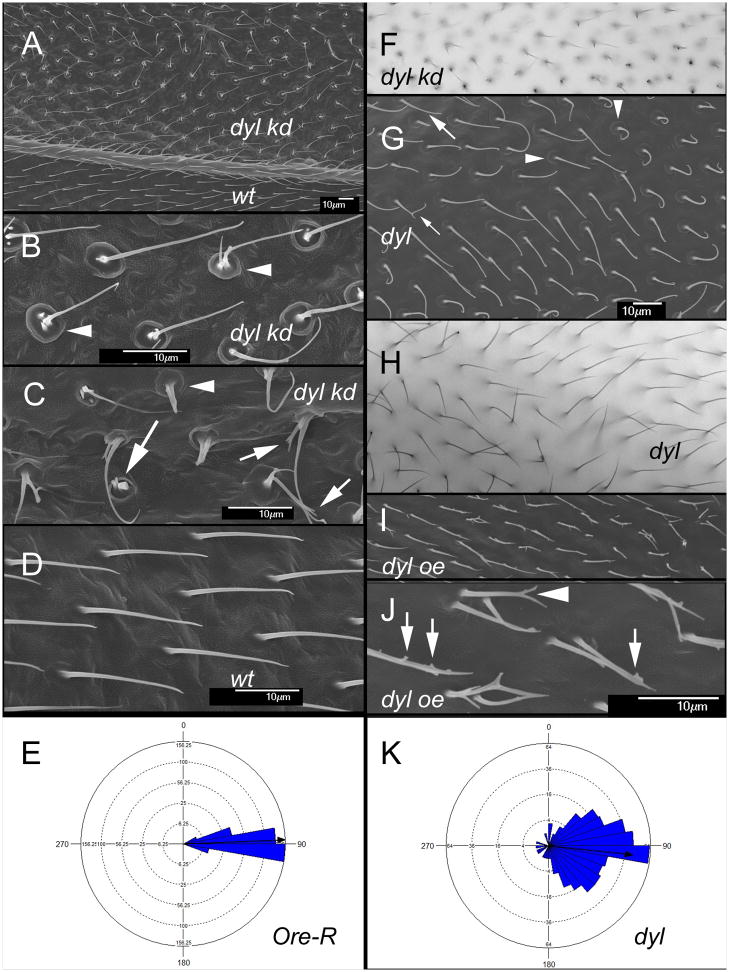
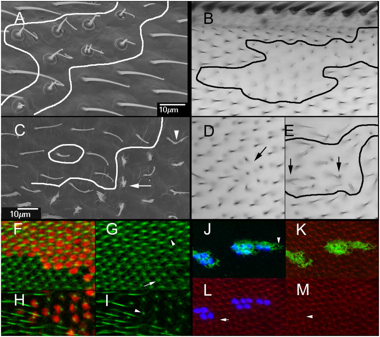
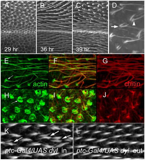
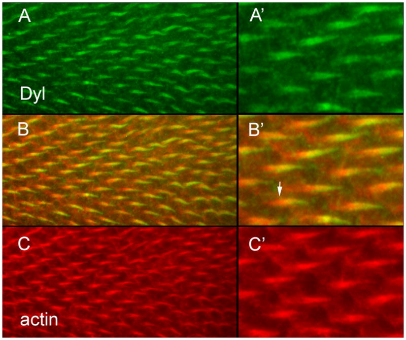
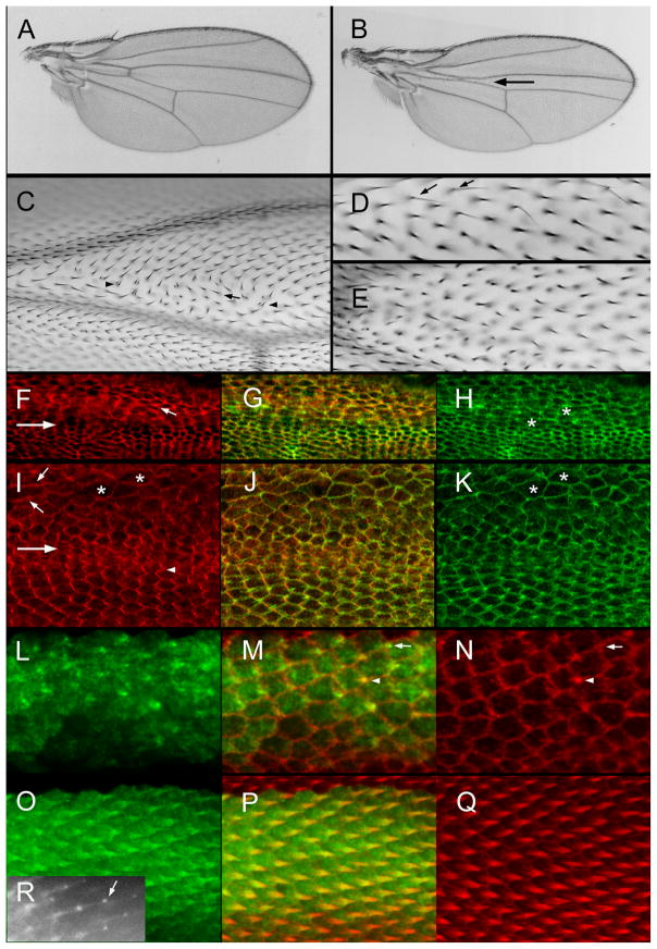
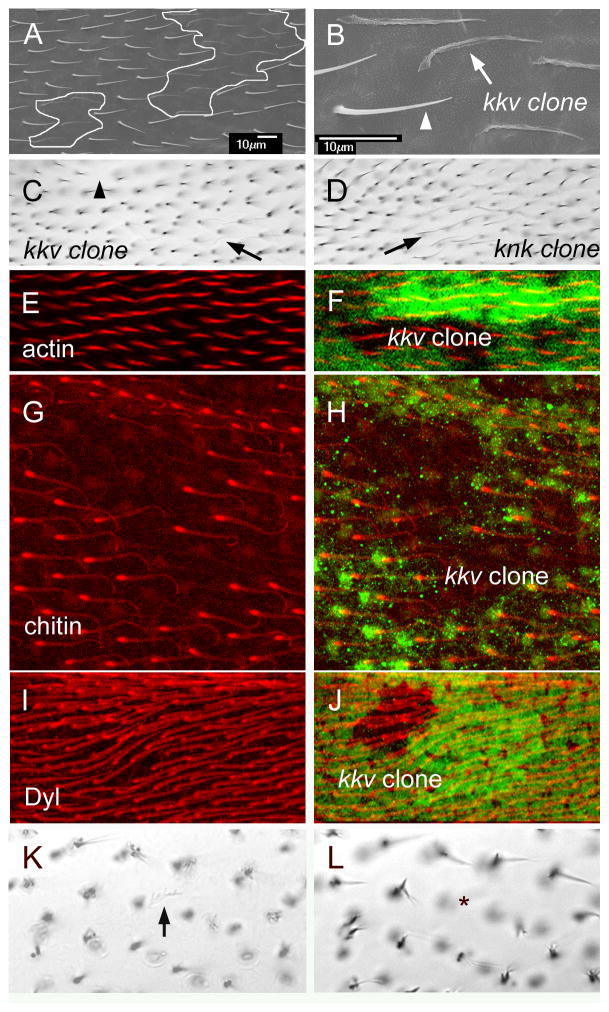
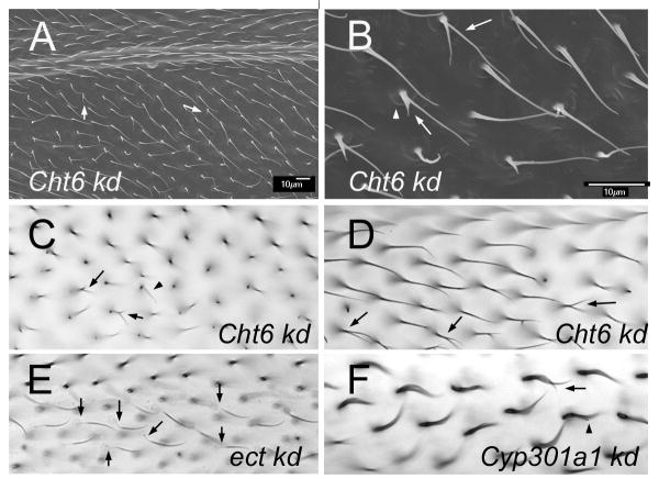
The cuticular hairs and sensory bristles that decorate the adult Drosophila epidermis and the denticles found on the embryo have been used in studies on planar cell polarity and as models for the cytoskeletal mediated morphogenesis of cellular extensions. ZP domain proteins have recently been found to be important for the morphogenesis of both denticles and bristles. Here we show that the ZP domain protein Dusky-like is a key player in hair morphogenesis. As is the case in bristles, in hairs dyl mutants display a dramatic phenotype that is the consequence of a failure to maintain the integrity of the extension after outgrowth. Hairs lacking dyl function are split, thinned, multipled and often very short. dyl is required for normal chitin deposition in hairs, but chitin is not required for the normal accumulation of Dyl, hence dyl acts upstream of chitin. A lack of chitin however, does not mimic the dyl hair phenotype, thus Dyl must have other targets in hair morphogenesis. One of these appears to be the actin cytoskeleton. Interestingly, dyl mutants also display a unique planar cell polarity phenotype that is distinct from that seen with mutations in the frizzled/starry night or dachsous/fat pathway genes. Rab11 was previously found to be essential for Dyl plasma membrane localization in bristles. Here we found that the expression of a dominant negative Rab11 can mimic the dyl hair morphology phenotype consistent with Rab11 also being required for Dyl function in hairs. We carried out a small directed screen to identify genes that might function with dyl and identified Chitinase 6 (Cht6) as a strong candidate, as knocking down Cht6 function led to weak versions of all of the dyl hair phenotypes.


Figures

Fig. 1
Fig. 1
Dyl functions in hair morphogenesis. (A) An SEM of an adult ptc-Gal4 UAS-dyl RNAi wing. The ptc domain includes the region above the vein shown in the image (marked by dyl kd). Note the hair polarity phenotype. (B) A high mag SEM of an adult ptc-Gal4 UAS-dyl wing. The arrowheads point to the hair “cups”. (C) A high mag SEM of an adult ptc-Gal4 UAS-dyl wing. The arrowhead points to a hair “cups”. The arrows point to thin and/or branched dyl kd hairs. (D) An SEM of phenotypically wild type hairs from outside of the ptc domain of the ptc-Gal4 UAS-dyl RNAi wing in (A). (E) A Rose diagram showing the distribution of hair orientation for Ore-R wings. The arrow shows the mean orientation. (F). A bright field micrograph of the phenotype associated with a dyl kd (ptc-Gal4 UAS-dyl RNAi). (G) An SEM of a dylMI02088/Df wing. Note the abnormal hair polarity. The larger arrow points to a thin hair, the smaller arrow to a split hair and the arrowheads to “cups” at the base of dyl mutant hairs. (H). A bright field micrograph of a dylMI02088/Df wing. Note the poor alignment of neighboring hairs. (I) An SEM of a ptc-Gal4 UAS-dyl wing. (J) A higher mag SEM of a ptc-Gal4 UAS-dyl wing where the small branches (arrows) and split (arrowheads) can be seen. (K). A Rose diagram showing the distribution of hair orientation for dylMI02088/Df wings. The arrow shows the mean orientation. Note how much broader the distribution is than in Ore-R.
Fig. 2
Fig. 2
Cell autonomy of dyl. (A) An SEM of a dyl kd flip out clone. The putative clone boundary is outlined. Note the strong dyl phenotype of the putative clone cells juxtaposed to wild type hairs. (B). A light micrograph showing of of a dyl kd flip out clone. Once again note the wild type cells that are juxtaposed next to cells that show a strong dyl mutant phenotype. (C). An SEM of a dyl oe flip out clone. Note some cells show a very strong phenotype (arrow), some a weak phenotype (arrowhead) and others appear wild type. (D) A bright field micrograph of a small dyl oe clone. A small group of hairs appear to point inward (arrow). (E) A bright field micrograph of a larger dyl oe clone. The putative clone is outlined. As was observed in the SEM such clones contain cells that display a range of phenotypes. The arrows point to abnormal hairs. (F) A dyl kd flip out clone in a 33 hr pupal wing marked by the expression of LacZ (red). F-actin (green) shows the growing hairs. (G) The same wing as in F but with only the green (F-actin) channel. Note the clone hairs (arrowhead) are of wild type morphology at this stage and appear longer on average than the wild type neighbors (arrow). (H) A dyl kd flip out clone in a 46 hr pupal wing marked by the expression of LacZ (red). F-actin (green) shows the growing hairs. (I) The same wing as in H but with only the green channel shown. The phenotype of the dyl hairs is dramatic while neighboring wild type hairs show no phenotype. At this late stage F-actin staining is less vigorous and consistent from hair to hair than in younger wings particularly when combined with antibody staining. (J) A flip out dyl oe clone marked by the expression of LacZ (blue). Note the accumulation of Dyl is fibrous and extends beyond the clone cells (arrowhead). Note that the endogenous Dyl found in hairs is not visible at this level of exposure. (K). The same cells as in J with F-actin staining shown. (L). The same cells as in J with actin and LacZ shown. Note that some wild type cells show abnormal hair F-actin (arrow). (M). The same cells showing only F-actin staining. NBote that some kd cells also display abnormal hair F-actin (arrowhead).
Fig. 3
Fig. 3
Dyl and hair morphogenesis. (A) A 29 hr ptc-Gal4 UAS-dyl RNAi pupal wing stained to show F-actin. The arrow shows the boundary between the ptc domain and the wild type wing posterior to it (below). Note the hairs forming inside the ptc domain and not outside it. (B) A 36 hr ptc-Gal4 UAS-dyl RNAi pupal wing stained for F-actin. Hairs are seen both in and outside of the ptc domain, but note the hairs inside the ptc domain appear longer, thicker and are stained more brightly. (C). A 39 hr ptc-Gal4 UAS-dyl RNAi pupal wing stained for F-actin. Note hairs inside the ptc domain are starting to appear abnormal. (D). A higher magnification view of abnormal ptc-Gal4 UAS-dyl RNAi hairs in a 39 hr wing. The arrow points to a multiple hair cell. The arrowhead points to a split hair. (E) A 43 hr ptc-Gal4 UAS-dyl RNAi pupal wing stained for F-actin. This image is for a region outside of the ptc domain. Note the F-actin (green) in the hair is central to chitin (red). Relatively weak staining of hair cups is visible (arrow). (F) A merged image of E and G. (G). The same wing region shown in E, but stained for chitin in red. (H) A 43 hr ptc-Gal4 UAS-dyl RNAi pupal wing stained for F-actin. This image is for a region inside of the ptc domain. The arrow points to the large accumulation of F-actin at the base of the hair and the abnormal structure of the hairs. (I) A merge of H and J. (J) The same wing region shown in H, but stained for chitin in red. Note the staining is far brighter in the proximal part of the hair. This is not seen in wt. (K). A 33 hr ptc-Gal4 Tub-Gal80ts UAS-dyl pupal wing inside of the ptc domain. The arrow points to a multiple/split hair cell. (L). A 33 hr ptc-Gal4 Tub-Gal80ts UAS-dyl pupal wing outside of the ptc domain. Note that the relative total hair F-actin staining is on average slightly stronger in K than L.
Fig. 4
Fig. 4
Dyl accumulates in growing hairs. (A) Dyl antibody staining shows the protein accumulates in growing hairs. (B) A merge of A and C. (C) F-actin staining. (A’) A higher magnification image of part of the field in A. (B’) A higher magnification image of part of the field in B. The arrow points to the proximal “root” of the hair that stains for actin but not the plasma membrane localized Dyl. (C’) A higher magnification image of part of the field in C.
Fig. 5
Fig. 5
Rab11 and wing development. (A) A ptcGal4 Gal80ts/UAS-DN-Rab11 wing from a fly grown at 21°C. (B) A ptcGal4 Gal80ts/UAS-DN-Rab11 wing from a fly grown at 27.5°C. The arrow points to the reduced size of the ptc domain. (C) A micrograph of a ptcGal4 Gal80ts/UAS-DN-Rab11 wing from a fly grown at 27.5°C. Just distal to the anterior cross vein (ACV) is a group of cells showing abnormal hair polarity (arrows) and multiple hair cells (arrowheads). (D). A micrograph of a ptcGal4 Gal80ts/UAS-DN-Rab11 wing from a fly grown at 21°C until wpp and then shifted to 27.5°C. Note the presence of several thin split hairs (arrows). (E). A micrograph of a ptcGal4 Gal80ts/UAS-DN-Rab11 wing from a fly grown at 21°C until wpp and then shifted to 27.5°C. Note the lack of precise alignment of neighboring hairs. (F). A micrograph of a ptcGal4 Gal80ts/UAS-DN-Rab11 pupal wing from a fly grown at 27.5°C. The large arrow shows the boundary of the ptc domain (above inside). This image shows immunolocalization of Stan. The small arrow shows a cell with an increased level of improperly localized Stan. (G). A merge of F and H. (H). The same wing shown in F but showing F-actin staining in green. The asterisks are on abnormally shaped cells. (I). A micrograph of a ptcGal4 Gal80ts/UAS-DN-Rab11 pupal wing from a fly grown at 27.5°C. The large arrow shows the boundary of the ptc domain (above inside). This image shows immunolocalization of In. Note the zigzag accumulation pattern for In outside of the ptc domain and the aberrant cell shape and In localization inside the ptc domain. (J). A merge of I and K. (K). The same wing as in I but showing F-actin staining. The asterisks are on abnormally shaped cells. (L). A 32 hr ptc-Gal/UAS-GFP-Rab11 pupal wing. (M) A merge of L and N. The arrow points to a cell where GFP-Rab11 is showing distal accumulation prior to F-actin accumulation. This shows that GFP-Rab11 is an earlier marker of hair outgrowth than F-actin. The arrowhead points to a cell where the hair is also marked by F-actin accumulation. (O). A 34 hr ptc-Gal/UAS-GFP-Rab11 pupal wing stained for GFP. (P) A merge of O and Q. (Q). A 34 hr ptc-Gal/UAS-GFP-Rab11 pupal wing stained for F-actin. (R) A 35 hr ap-Gal/UAS-GFP-Rab11 pupal wing imaged in vivo. The arrow points to the “blob” of GFP-Rab11 at the tip of the growing hair.
Fig. 6
Fig. 6
kkv and hair morphogenesis. (A) An SEM of an adult wing that contains a pair of small kkv1 clones. These are outlined in white. (B) A higher mag image of part of A. The arrow points to a kkv1 hair and the arrowhead to a neighboring wild type hair. Note how the kkv1 is flaccid and fainter. However, it is not branched and is neither thinner nor shorter than wild type. (C). Shown is a bright field image of a region of an adult wing bearing two small kkv1 clones. The arrow points to a faint kkv1 hair and the arrowhead to a location where the faint mutant hair cannot be seen due to being out of the plane of focus. (D) A bright field micrograph of a knk clone (arrow). Note the similarity to the kkv1 clone. (E). A region of a 34 hr pupal wing stained for F-actin (red) that contains a kkv1 clone. (F). The image from E showing the location of the clone as marked by the loss of GFP. (G). A region of a 44 hr pupal wing stained for chitin (red) that contains a kkv1 clone. (H) The image from G showing the location of the clone as marked by the loss of GFP. Note at this late stage GFP staining quality is lower than in younger wings. Note the bright spot of chitin staining at the base of the hairs. Note that chitin staining is lost in the clone cells showing the specificity of the chitin staining. (I) A region of a 34 hr pupal wing stained for Dyl (red) that contains a kkv1 clone. (J) The image from I showing the location of the clone as marked by the loss of GFP. Note that the hairs inside the clone do not show altered Dyl staining. (K) A kkv1 clone inside of the ptc domain where dyl has been knocked down by RNAi. The arrow points to a faint and flaccid hair (hence one that is mutant for kkv) that shows dramatic branching. (L). A different focal plane from the same region of the same wing shown in K. The asterisk marks the location of the kkv clone cell. No hair is seen due to it laying on the wing blade surface.
Fig. 7
Fig. 7
(A) An SEM of an adult ptc-Gal4 UAS-Cht6 RNAi wing. Arrows point to branched hairs. Note the poor alignment of neighboring hairs. (B). A higher magnification image of a region of A. The arro points to a split hair. The arrowhead points to a “cup” at the base of the hair. (C). A brightfield micrograph of an adult ptc-Gal4 UAS-Cht6 RNAi wing. The arrows point to branched and thin hairs. (D). A brightfield micrograph of an adult ptc-Gal4 UAS-Cht6 RNAi wing. The arrows point to branched hairs. (E). A brightfield micrograph of an adult ptc-Gal4 UAS-ect RNAi wing. The arrows point to hairs that show the “ect” phenotype of hairs with a faint and wimpy proximal region. (F). A brightfield micrograph of an adult ptc-Gal4 UAS-Cyp301a RNAi wing. The arrows point to split hairs. Note the curved shape of all hairs in this wing region.

References

    1. Adler PN. Planar signaling and morphogenesis in Drosophila. Dev Cell. 2002;2:525–535. - PubMed
    1. Adler PN. The frizzled/stan pathway and planar cell polarity in the Drosophila wing. Curr Top Dev Biol. 2012;101:1–31. - PMC - PubMed
    1. Adler PN, Charlton J, Liu J. Mutations in the cadherin superfamily member gene dachsous cause a tissue polarity phenotype by altering frizzled signaling. Development. 1998;125:959–968. - PubMed
    1. Adler PN, Zhu C, Stone D. Inturned Localizes to the Proximal Side of Wing Cells under the Instruction of Upstream Planar Polarity Proteins. Curr Biol. 2004;14:2046–2051. - PubMed
    1. Amonlirdviman K, Khare NA, Tree DR, Chen WS, Axelrod JD, Tomlin CJ. Mathematical modeling of planar cell polarity to understand domineering nonautonomy. Science. 2005;307:423–426. - PubMed

Publication types

MeSH terms

LinkOut - more resources