. 2013 Aug 1;319(13):1922-1931.
doi: 10.1016/j.yexcr.2013.04.013. Epub 2013 Apr 23.

Glycogen synthase kinase 3 regulates expression of nuclear factor-erythroid-2 related transcription factor-1 (Nrf1) and inhibits pro-survival function of Nrf1

Affiliations

Glycogen synthase kinase 3 regulates expression of nuclear factor-erythroid-2 related transcription factor-1 (Nrf1) and inhibits pro-survival function of Nrf1

Madhurima Biswas et al. Exp Cell Res. .

Abstract

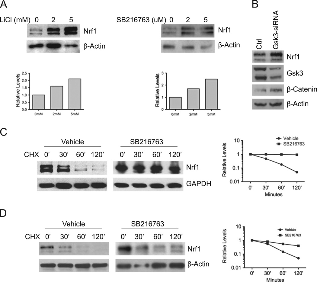
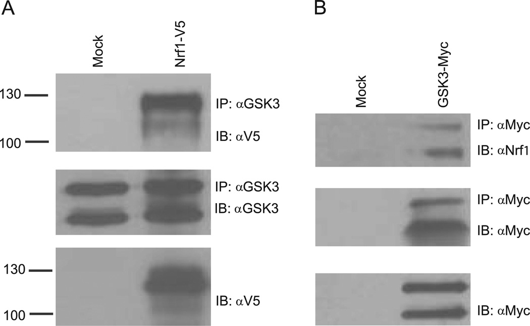
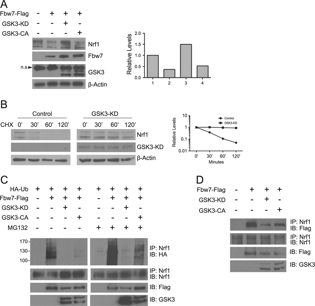
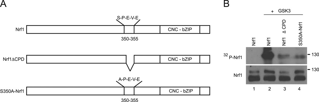
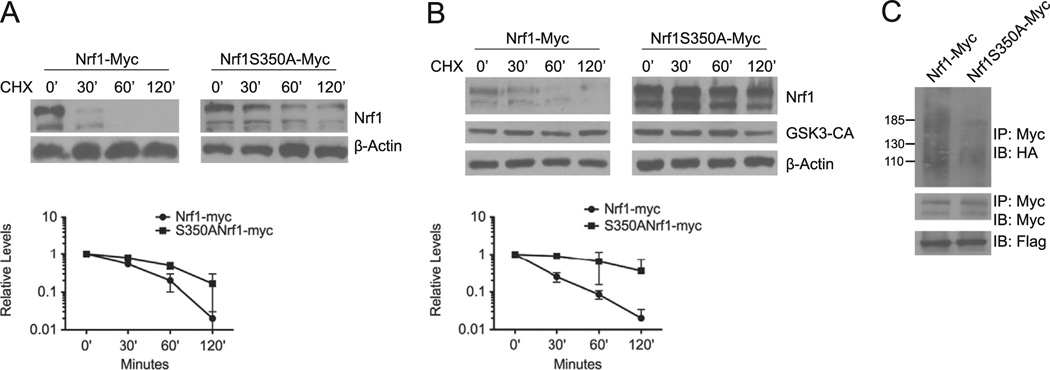
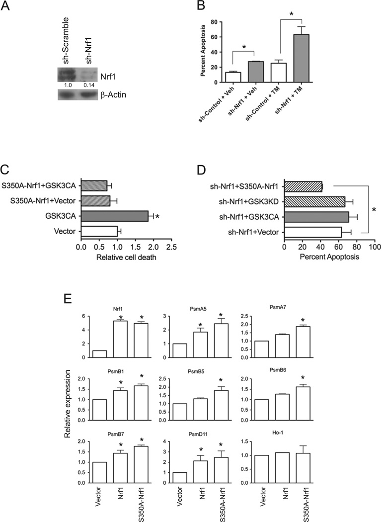
Nuclear factor E2-related factor-1 (Nrf1) is a basic leucine zipper transcription factor that is known to regulate antioxidant and cytoprotective gene expression. It was recently shown that Nrf1 is regulated by SCF-Fbw7 ubiquitin ligase. However our knowledge of upstream signals that targets Nrf1 for degradation by the UPS is not known. We report here that Nrf1 expression is negatively regulated by glycogen synthase kinase 3 (GSK3) in Fbw7-dependent manner. We show that GSK3 interacts with Nrf1 and phosphorylates the Cdc4 phosphodegron domain (CPD) in Nrf1. Mutation of serine residue in the CPD of Nrf1 to alanine (S350A), blocks Nrf1 from phosphorylation by GSK3, and stabilizes Nrf1. Knockdown of Nrf1 and expression of a constitutively active form of GSK3 results in increased apoptosis in neuronal cells in response to ER stress, while expression of the GSK3 phosphorylation resistant S350A-Nrf1 attenuates apoptotic cell death. Together these data suggest that GSK3 regulates Nrf1 expression and cell survival function in response to stress activation.

Keywords: ARE; CNC-BZIP; CNC-bZIP; CPD; Cap ‘n’ Collar-type basic leucine zipper; Cdc4 phosphodegron domain; ER; Fbw7; GSK3; Nrf1; Nuclear factor E2-related factor-1; SCF; Skp1–Cul1–F-box; Stress response; Transcription factor; UPS; antioxidant response element; endoplasmic reticulum; glycogen synthase kinase 3; nuclear factor E2-related factor-1; ubiquitin proteasome system.


Conflict of interest statement

Conflict of interest

The authors have no conflicts of interest to disclose.

Figures

Fig. 1
Fig. 1. GSK3 modulates Nrf1 expression
(A) HEK293 cells were treated with LiC1 or SB216763 at the indicated concentrations for 16 h, and whole cell extracts were immunoblotted with anti-Nrf1 antibody. In accord with previous findings, two major immunoreactive bands representing glycosylated and non-glycosylated forms of Nrf1 were detected [21]. Beta-actin levels were used as loading control. (B) HEK293 cells were transfected with a mixture of the two siRNAs against GSK3. After 48 h, lysates were Western blotted for Nrf1 levels. Western blotting was also done to verify the level of GSK3 knockdown and concomitant increase in β-Catenin protein as control. Beta-actin levels were used as loading control. (C) HEK293 cells were treated with 10 µM SB216763 for 16 h, followed with 50 µg/ml cycloheximide, and cells were harvested at the indicated time points. Whole cell extracts were immunoblotted with anti-Nrf1 antibody. Beta-actin levels were used as loading control. (D) Mouse embryonic fibroblasts were treated with SB216763 for 16 h, followed by 50 µg/ml cycloheximide, and cells were harvested at the indicated time points. Whole cell extracts were prepared and immunoblotted against Nrf1. Beta-actin levels were used as loading control. Graphs show quantitation of protein levels.
Fig. 2
Fig. 2. GSK3 binds Nrf1
(A) HEK293 cells were transfected with vector or Nrfl-V5. After 48 h, whole cell extracts were prepared, and immunoprecipitated with GSK3 antibody followed by immunblotting against V5 and GSK3. (B) HEK293 cells were transfected with vector or GSK3-myc and whole cell extracts were prepared 48 h after and immunoprecipitated with anti-Myc antibody. Immunoprecipitates were then immunoblotted with anti-Myc and anti-Nrf1 antibody.
Fig. 3
Fig. 3. GSK3 activity is required for Nrf1 turnover by Fbw7
(A) HEK293 cells were transfected with Fbw7-Flag alone, Fbw7-Flag and GSK3βKD-myc, or Fbw7 and GSK3βCA-myc. Whole cell extracts prepared 48 h after transfection and immunoblotted with Nrf1, Myc, or Flag antibodies. Beta-actin levels were used as loading control. The graph shows quantitation of the Nrf1 expression. Each point represents the mean±SEM of the remaining protein. (B) HEK293 cells were transfected with Nrf1-myc, or Nrf1-myc and GSK3β-KD. After 48 h, cells were treated with 50 µg/ml cycloheximide, and harvested at the indicated time points for Western blot analysis. Beta-actin levels were used to determine loading of each sample. Graph shows quantitation of the protein levels at each time points. Each point represents the mean±SEM of the remaining protein. (C) HEK293 cells were transfected with HA-Ub, HA-Ub and Flag-Fbw7, or HA-Ub, Flag-Fbw7 and GSK3βKD-myc or GSK3βCA-myc. After 48 h, whole cell extracts were immunoprecipitated with anti-Nrf1 antibody followed by immunoblotting with anti-HA antibody. As input control, cell extracts were also immunoblotted with anti-Flag and anti-Myc antibody for expression of Fbw7 and GSK3β, respectively. (D) HEK293 cells were transfected with Flag-Fbw7 alone, Flag-Fbw7 along with GSK3βKD-myc or GSK3βCA-myc. Cell extracts were prepared 48 h after, and immunoprecipitated with Nrf1 antibody. Immunoprecipitates were then Western blotted using anti-Flag and anti-Nrf1 antibodies. As input control, extracts were also probed with anti-Myc antibody for expression of GSK3β.
Fig. 4
Fig. 4. GSK3 Phosphorylates Nrf1 in-vitro
(A) Purified GST-Nrf1, GST-S350A–Nrf1, GST-Nrf1CPD were subjected to in-vitro ldnase assay with recombinant GSK3β in the presence of γ-32P ATP. Reaction products were separated on SDS-PAGE and subjected to autoradiography (upper panel). As input control, equal amounts of purified fusion products were immunoblotted against Nrf1 (lower panel).
Fig. 5
Fig. 5. GSK3 mediated degradation is attenuated by S350A substitution in Nrf1
(A) HEK293 cells were transfected with Nrf1-myc or S350A–Nrf1-myc. Cells were then treated with 50 µg/ml cycloheximide, and harvested at the indicated time points for Western blotting using anti-Myc antibody. Beta-actin levels were used to determine loading of each sample. The graph shows quantitation of protein levels. Each point represents the mean±SEM of remaining protein. (B) HEK293 cells were co-transfected with Nrf1-myc, or S350A–Nrf1-myc and GSK3β-CA After 48 h, cells were treated with 50 µg/ml cycloheximide, and harvested at the indicated time points for Western blot analysis. Beta-actin levels were used to determine loading of each sample. Graph shows quantitation of the protein levels at each time points. Each point represents the mean±SEM of the remaining protein. (C) HEK293 cells were transfected with Nrf1-Myc or S350A–Nrfl-myc along with HA-ubiquitin and Flag-Fbw7. 48 h after transfections, cell extracts were prepared and immunoprecipitated with anti-Myc antibody, followed by immunoblotting with anti-HA antibody. For input control, the filter was stripped and probed with anti-Myc antibody. Cell extracts were also immunoblotted with anti-Flag antibody to determine Fbw7 expression.
Fig. 6
Fig. 6. GSK3 modulates Nrf1 mediated stress response in neuronal cells
(A) Western blot analysis of Nrf1 in SH-SY5Y cells transduced with shScramble and shNrf1 virus. Densitometric quantitations of Nrf1 normalized against beta-Actin are shown. (B) shScramble- and shNrf1-transduced SH-SY5Y cells were treated with DMSO, or tunicamycin, and apoptosis was determined by Annexin V staining and flow cytometric analysis. Data represents means±SEM, n=3, *, P<0.05. (C) shScramble control cells co-transfected with EGFP and GSK3β-CA or EGFP, S350A–Nrf1 and GSK3β-CA were treated with tunicamycin, and cell death assessed by flow cytometric analysis of Annexin V staining in GFP-positive gated cells. Data represents means ± SEM, n=3, *, P<0.05. (D) shNrf1-transduced SH-SY5Y cells transfected with E-GFP and GSK3β-CA EGFP and GSK3β-KD, or EGFP and S350A–Nrfl were treated with tunicamycin. After 16 h, cell death assessed by flow cytometric analysis of Annexin V staining in GFP-positive gated cells. Data represents means±SEM, n=3, *, P<0.05. (E) Real time RTPCR analysis of gene expression in Nrf1 knockout MEF cells transfected with vector, Nrf1 or S350A–Nrf1. Expression levels of genes were quantitated relative to endogenous TBP levels as an internal reference, and calculated as 2(ct test gene-Ct TBP). Bars represent mean values±SEM of 3 experiments, and significance was assessed by Student’s t-test (*P<0.05).

Similar articles

Cited by

References

    1. Andrews NC, Erdjument-Bromage H, Davidson MB, Tempst P, Orkin SH, et al. Erythroid transcription factor NF-E2 is a haematopoietic-specific basic-leucine zipper protein. Nature. 1993;362:722–728. - PubMed
    1. Chan JY, Han XX, Kan YW, et al. Cloning of Nrf1, an NF-E2-related transcription factor, by genetic selection in yeast. Proc. Natl. Acad. Sci. USA. 1993;90:11371–11375. - PMC - PubMed
    1. Kobayashi A, Ito E, Told T, Kogame K, Takahashi S, Igarashi K, Hayashi N, Yamamoto M, et al. Molecular cloning and functional characterization of a new Cap ‘n’ collar family transcription factor Nrf3. J. Biol. Chem. 1999;274:6443–6452. - PubMed
    1. Moi P, Chan K, Asunis I, Cao A, Kan YW, et al. Isolation of NF-E2-related factor 2 (Nrf2), a NF-E2-like basic leucine zipper transcriptional activator that binds to the tandem NF-E2/AP1 repeat of the beta-globin locus control region. Proc. Natl. Acad. Sci. USA. 1994;91:9926–9930. - PMC - PubMed
    1. Oyake T, Itoh K, Motohashi H, Hayashi N, Hoshino H, Nishizawa M, Yamamoto M, Igarashi K, et al. Bach proteins belong to a novel family of BTB-basic leucine zipper transcription factors that interact with MafK and regulate transcription through the NF-E2 site. Mol. Cell Biol. 1996;16:6083–6095. - PMC - PubMed

Publication types

MeSH terms