. 2013 Aug 15;62(3):197-206.
doi: 10.1016/j.ymeth.2013.04.013. Epub 2013 Apr 23.

Multi-allelic phenotyping--a systematic approach for the simultaneous analysis of multiple induced mutations

Affiliations

Multi-allelic phenotyping--a systematic approach for the simultaneous analysis of multiple induced mutations

Christopher M Dooley et al. Methods. .

Abstract

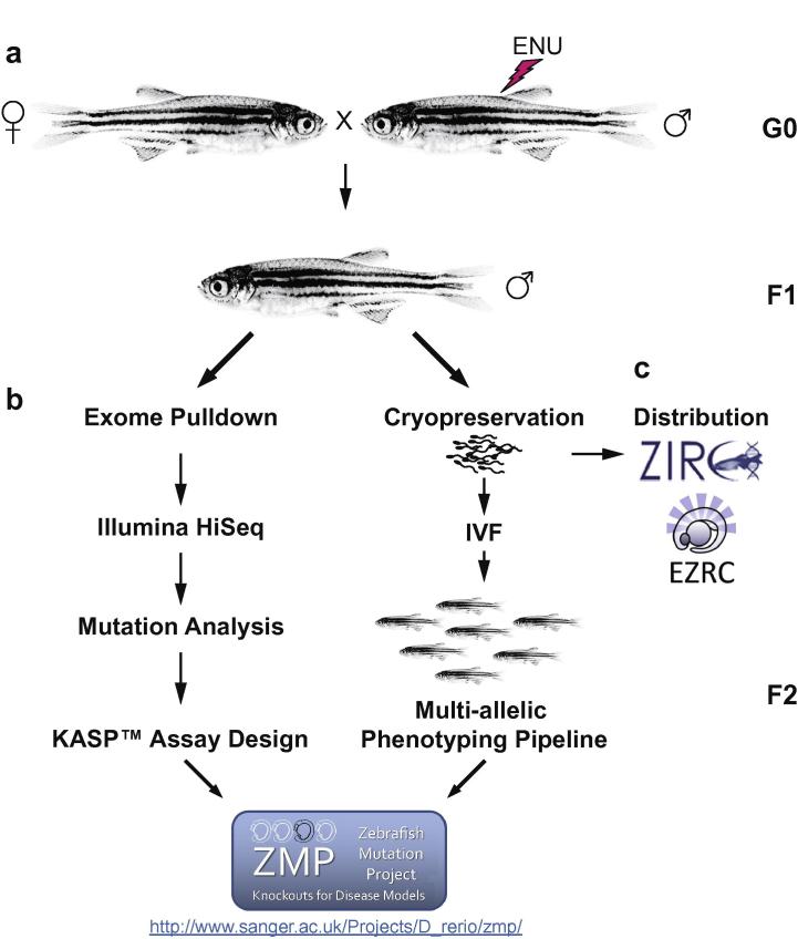
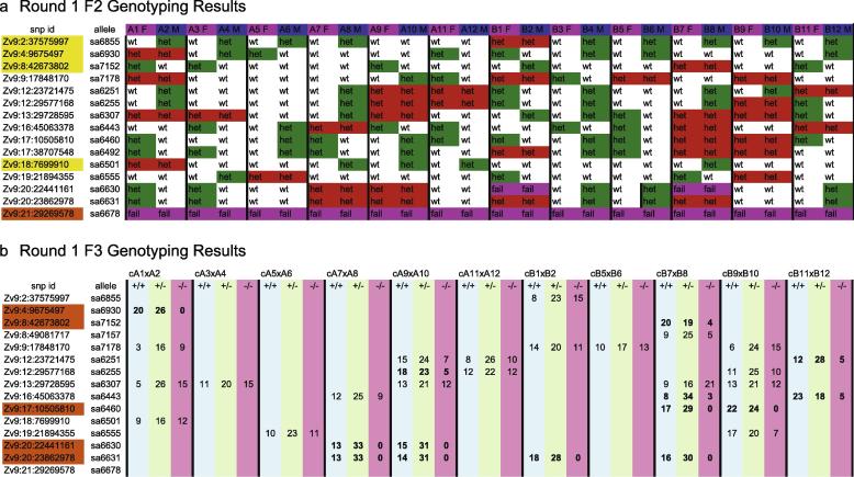
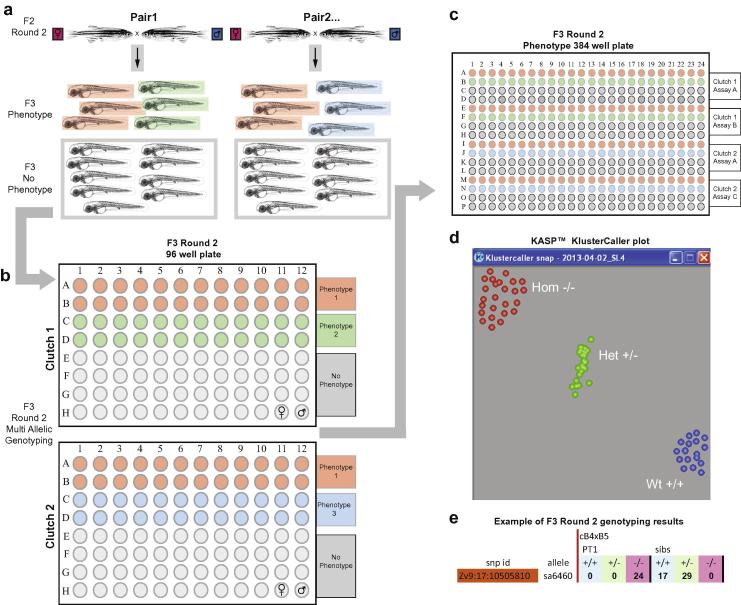
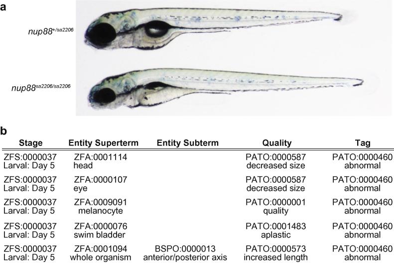
The zebrafish mutation project (ZMP) aims to generate a loss of function allele for every protein-coding gene, but importantly to also characterise the phenotypes of these alleles during the first five days of development. Such a large-scale screen requires a systematic approach both to identifying phenotypes, and also to linking those phenotypes to specific mutations. This phenotyping pipeline simultaneously assesses the consequences of multiple alleles in a two-step process. First, mutations that do not produce a visible phenotype during the first five days of development are identified, while a second round of phenotyping focuses on detailed analysis of those alleles that are suspected to cause a phenotype. Allele-specific PCR single nucleotide polymorphism (SNP) assays are used to genotype F2 parents and individual F3 fry for mutations known to be present in the F1 founder. With this method specific phenotypes can be linked to induced mutations. In addition a method is described for cryopreserving sperm samples of mutagenised males and their subsequent use for in vitro fertilisation to generate F2 families for phenotyping. Ultimately this approach will lead to the functional annotation of the zebrafish genome, which will deepen our understanding of gene function in development and disease.

Keywords: Cryopreservation; Knockout; Model organism; Phenomics; Screen; Zebrafish.


Figures

Fig. 1
Fig. 1
Mutation detection overview. Male TLF zebrafish (G0) are treated with ENU and then out-crossed (a) to produce F1 families heterozygously carrying induced mutations. F1 fish (b) are raised to an age of about 12 months and sperm is collected from the fish. Each individual is then sacrificed and tissue samples of the body are taken. The tails are used for genomic DNA preparation while the rest of the body is preserved for archival purposes. Genomic DNA is then subjected to exome pulldown (b), sequenced via Illumina paired end HiSeq and analysed to detect induced mutations. KASP™ assays are designed for each allele and then all information is made available on the ZMP website as the project proceeds. The cryopreserved sperm is made available to ZIRC and EZRC where alleles can be ordered. (c) An aliquot of sperm from each F1 male is retained at the Wellcome Trust Sanger Institute and used for the IVF of F2 families which are placed into the ZMP multi-allelic phenotyping pipeline. Phenotypic descriptions are published on the ZMP website.
Fig. 2
Fig. 2
Cryopreservation of zebrafish sperm. From left to right the cryopreservation station comprises (1) dry ice block with deep well block for F1 tissue samples, (2) tricaine for culling, (3) LM80 dissecting microscope fitted with ring light, 1000 and 200 μl pipettes, bin and pipette tips behind, (4) wet ice box with tube rack for cryoprotectant aliquots and cryovials, (5) sponge with slits to hold anaesthetised fish, (6) wet ice box for cryovial box, (7) plastic spoon to scoop fish out of tricaine, (8) tricaine for anaesthesia, (9) fish tank holding males, (10) recording sheet, (11) mallet to drive Falcon tubes into dry ice, (12) dry ice/ethanol box with Falcon tubes, (13) styrofoam box containing 3 cm of LN2 and cryoboxes. Not depicted are: timer, tissues and cotton buds to dry off fish, suction tube and capillaries. (a) Males are placed in sponge holder ventral side up. (b) Carefully dried males are squeezed and sperm is collected in 10 μl capillary. (c) Good quality sperm is visible inside the capillary (white arrow). (d) Sperm is expelled into cryoprotectant while holding tube such that the solution is not warmed between the fingers. (e) Aliquots are pipetted into cryovials and immediately dropped into Falcon tubes which are capped and then hammered into the dry ice (f).
Fig. 3
Fig. 3
In vitro fertilisation. The IVF station consists of (1) a LM80 dissecting microscope, (2) 9 cm Petri dishes, egg water without Methylene Blue in a (3) squirt bottle and (4) cell culture flask, (5) 6 cm glass dishes, (6) recovery tank for females, (7) lamp, (8) heating block at 28 °C for fructose aliquots, (9) 1 ml pipette with tips and disposable 2 ml pipette, (10) pieces of Parafilm, (11) water bath at 37 °C for BSMIS, (12) and (16) timer, (13) tricaine to anaesthetise females, (14) tissues to dry females, (15) box with LN2 and samples to use for IVF. (a) Anaesthetised females are transferred onto Parafilm where fins are moved out of the way using a pipette tip and urogenital opening is dried with a cotton bud. (b) Gentle abdominal pressure releases eggs. (c) Eggs are separated from the female using a pipette tip (d) and transferred into glass dish. (e) Activated sperm is pipetted directly onto eggs so that they are evenly dispersed (f, g).
Fig. 4
Fig. 4
Multi-allelic phenotyping – round 1. F2 families are randomly in-crossed and the 12 largest clutches are collected and enter the pipeline with their respective parents. 150–200 fertilized F3 embryos are raised till 5 dpf, all abnormal fry are removed and 46+ phenotypically normal fry are collected and frozen in MeOH. (a) Fin clips are taken from each F2 parent and placed into a 96 well plate. Genomic DNA is prepared from the tissue and transferred into 384 well plates allowing for up to 24 KASP™ genotyping assays per 384 well plates, with all disruptive mutations identified in the original F1 male being interrogated. (b) The 46 phenotypically normal embryos from each clutch are aliquoted into half of a 96 well plate, DNA is extracted, and aliquots of the corresponding parental DNA are added to the last two wells. (c) The 96 well plates are transferred into 384 well plates and each clutch is stamped out into an individual 384 well plate and assayed for all alleles that both parents were heterozygous for (d).
Fig. 5
Fig. 5
Genotyping results. An example of first round genotyping results from F2 parents and their non-phenotypic F3 progeny. The genomic position of each mutation is in the column labelled snp id followed by the allele designation directly to the right. The top row labels first the female of the pair followed by the male. Their IDs stem from their original positions in a 96 well plates. In total 12 pairs are shown. Red boxes indicate alleles where both parents are carriers, whereas green signifies alleles with only one heterozygous parent in a pair. The snp id which are highlighted in yellow indicate alleles where only one cross contains heterozygous parents. The orange highlighted snp id flags assays which require attention. In the case of sa6678 all samples have failed, which indicates that this is probably due to the assay not working either at the amplification or clustering step. (a) For each clutch, genotyping is carried out on 46 non-phenotypic 5 dpf fry for all alleles that both F2 parents are heterozygous for. Absolute numbers of genotypes per allele and clutch are shown. The alleles flagged in orange in the snp id column indicate alleles suspected of causing a phenotype and should be considered for second round phenotyping (b).
Fig. 6
Fig. 6
Multi-allelic phenotyping – round 2. Round 2 pairs are set up based on the genotyping results of the first round phenotyping. Clutches are collected and observed over the first five days of development. (a) When phenotypes arise in approximately 25% of embryos, they are collected along with non-phenotypic siblings and frozen in MeOH. Multiple phenotypes per clutch can be collected simultaneously but are archived in separate tubes. Phenotypic embryos are then aliquoted into the upper half of a 96 well plate. (b) Multiple phenotypes can be added to a single plate and multiple plates per clutch can be produced. Phenotypically normal siblings are then added to the lower half of the 96 well plates with wells H11 and H12 containing parental control DNA. The 96 well plates are then transferred into 384 well plates where all alleles flagged for each particular pair combination are assayed against all identified phenotypes per clutch. (c) A KASP Kluster Caller plot depicting the grouping of the three genotypes. (d) This is an example of a genotype-phenotype association that would be documented as likely causal (e).
Fig. 7
Fig. 7
An example of phenotype annotation. At 5 dpf nup88sa2206 mutants have smaller heads and eyes, abnormal melanocytes, increased body length and lack the swim bladder. (a) Entity quality annotations for nup88sa2206 phenotype (b).

Similar articles

Cited by

References

    1. Nusslein-Volhard C., Wieschaus E. Nature. 1980;287(5785):795–801. - PubMed
    1. Mullins M.C. Curr. Biol. 1994;4(3):189–202. - PubMed
    1. Haffter P. Development. 1996;123:1–36. - PubMed
    1. Driever W. Development. 1996;123:37–46. - PubMed
    1. Chalfie M., Horvitz H.R., Sulston J.E. Cell. 1981;24(1):59–69. - PubMed

Publication types

LinkOut - more resources