. 2013 May 9;497(7448):205-10.
doi: 10.1038/nature12076. Epub 2013 Apr 28.

Hierarchy of orofacial rhythms revealed through whisking and breathing

Affiliations

Hierarchy of orofacial rhythms revealed through whisking and breathing

Jeffrey D Moore et al. Nature. .

Abstract

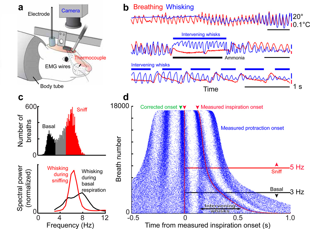
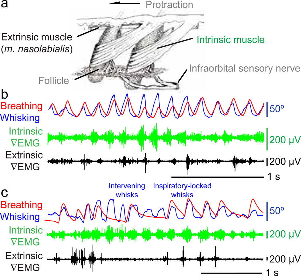
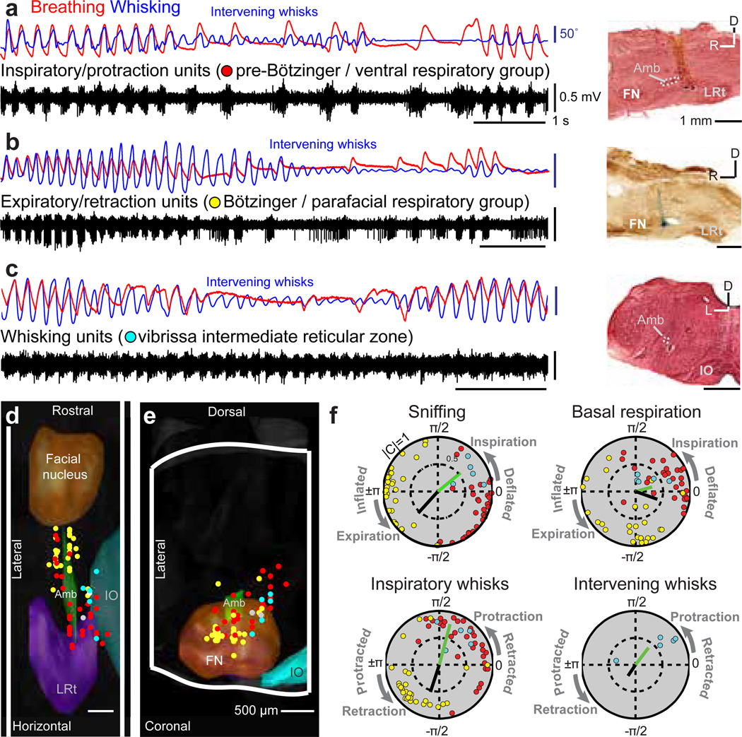
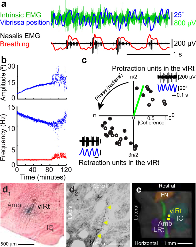
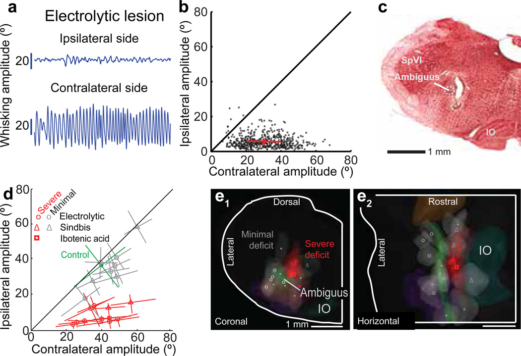
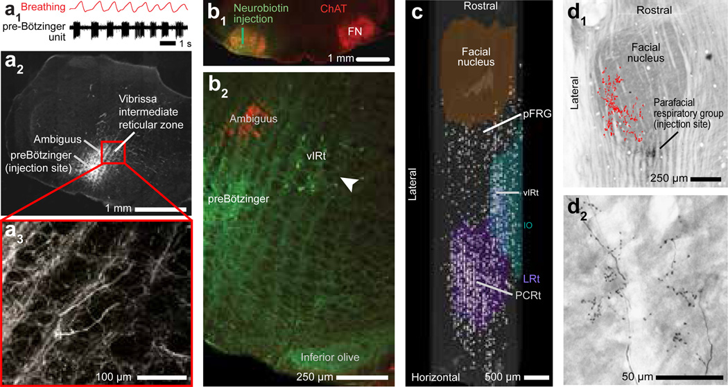
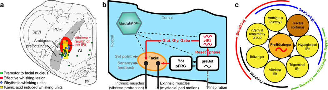
Whisking and sniffing are predominant aspects of exploratory behaviour in rodents. Yet the neural mechanisms that generate and coordinate these and other orofacial motor patterns remain largely uncharacterized. Here we use anatomical, behavioural, electrophysiological and pharmacological tools to show that whisking and sniffing are coordinated by respiratory centres in the ventral medulla. We delineate a distinct region in the ventral medulla that provides rhythmic input to the facial motor neurons that drive protraction of the vibrissae. Neuronal output from this region is reset at each inspiration by direct input from the pre-Bötzinger complex, such that high-frequency sniffing has a one-to-one relationship with whisking, whereas basal respiration is accompanied by intervening whisks that occur between breaths. We conjecture that the respiratory nuclei, which project to other premotor regions for oral and facial control, function as a master clock for behaviours that coordinate with breathing.


Figures

Figure 1
Figure 1. Coordination of whisking and breathing
(a) Procedures to measure whisking, breathing, and associated electrophysiology in headrestrained rats. (b) Simultaneous measurement of vibrissa position (blue) and breathing (red). Protraction and inspiration are upward. (c) Histogram of instantaneous breathing frequencies (top) delineates the classification of breaths below 3 Hz as basal respiration and those above 5 Hz as sniffs. The spectral power of whisking (bottom) is plotted during periods of basal respiration (black) as well as sniffing (red). (d) Rasters of inspiration onset times (red) and protraction onset times (blue) relative to the onset of inspiration for individual breaths are ordered by the duration of the breath; green arrow represents the 30 ms lead of inspiratory drive to facial muscles as opposed to the measured inspiration. Whisks and inspiration onset times are significantly correlated during both sniffing and basal respiration (p < 0.01).
Figure 2
Figure 2. Facial muscle activity during whisking and breathing
(a) The musculature responsible for vibrissa and mystacial pad motion; adapted from Dorfl. (b) Vibrissa motion (blue), breathing (red), and intrinsic (green) and extrinsic (black) ∇EMG activity during whisking and sniffing. (c) The same activity during whisking and mixed basal respiration and sniffing.
Figure 3
Figure 3. Activity in medullary respiratory centers during breathing and whisking
(a) Concurrent recordings of breathing (red), whisking (blue), and multiunit activity (black) in the preBötzinger complex. The location of the recording site is labeled with Chicago sky blue and is shown in a sagittal section counterstained with neutral red. LRt denotes the lateral reticular nucleus, FN the facial nucleus, Amb the ambiguus nucleus, and IO the inferior olive. (b) Multiunit spike activity in the Bötzinger complex. The section is counterstained for cytochrome oxidase. (c) Multiunit spike activity in the vibrissa zone of the intermediate reticular formation. The section is counterstained with neutral red. (d,e) The recording sites for all data imposed on a three dimensional reconstruction of the medulla. Whisking units are located dorsomedially to the preBötzinger complex in the IRt. Two units whose spiking had no relation to breathing or whisking are shown in white. (f) Polar plots of the magnitude (0 to 1 radial coordinate) and phase (angular coordinate) of the coherence between multiunit spiking activity and measured behaviors at the peak frequency for each behavior, i.e., 2 Hz for basal respiration, 6 Hz for sniffing and inspiratory whisks, and 8 Hz for intervening whisks (Fig. 1c). Only units with significant coherence (p < 0.01) are shown and correspond to the point in panels d to f. The coherence between the measured behavior and the ∇EMG of the intrinsic muscles (green bar) and Nasolabialis muscle ∇EMG (black bar) are shown.
Figure 4
Figure 4. Injection of kainic acid in the medullary reticular formation induces whisking
(a) Vibrissa motion (blue), breathing (red), intrinsic (green) and extrinsic (black) ∇EMG. (b) Time-course of kainic-acid induced whisking. Instantaneous peak-to-peak amplitude (top) and frequency (bottom) of vibrissa motion (blue) and frequency of breathing (red). The animal starts to wake by 100 minutes. (c) Polar plots of the coherence between spiking activity and vibrissa motion at the peak frequency of whisking (8.8 Hz median); only units with statistically significant coherence (32 of 33 units, p < 0.01) are shown. Open circles represent multiunit activity and closed circles represent single units. The green bar represents the coherence of the ∇EMG for the intrinsic muscle (panel b) with vibrissa motion. (Inserts) Spiking activity of neuronal units in the vIRt (black) in relation to vibrissa motion (blue). (d1) One of the locations that corresponded to a units in panel c, labeled via ionophoretic injection of Neurobiotin™ through the recording electrode. (d2) Axons (yellow arrows) and terminals in the ventral lateral division of the facial nucleus labeled after Neurobiotin™ injection at the recording site in panel d1. (e) Three dimensional reconstruction of the labeled recording locations for the units in panel d.
Figure 5
Figure 5. Lesion of the vIRt impairs ipsilateral whisking
(a) Example of whisking bout following an electrolytic lesion. (b) Scatter plot of ipsilateral versus contralateral whisk amplitudes reveals the functional completeness of the lesion; each dot represents one whisk, circle represents the mean, and lines represent the inter-quartile range. (c) Histological analysis confirms that the lesion is in the vIRt; coronal section stained with neutral red. (d) Composite results for a subset of lesions (19 rats) where vibrissa position was tracked; lines are central quartiles. Symbols correspond to the method of lesion. Results were scored by the severity of the ipsilateral whisking deficit: severe (red), for > 50 % reduction as in panels a and b, or minimal (gray), < 50 % reduction. Whisking of a non-lesioned control rat is shown in green. (e1–e2) Lesion sites were mapped onto a three dimensional reconstruction of the medulla and selected anatomical substructures, as in Figure 4f. The lesion centroids are denoted with the symbols in panel d and have a median volume of 0.2 µL. Sites marked with an asterisk (six rats) represent additional lesions not shown in panel d where animals were observed to have minimal whisking deficits by visual inspection.
Figure 6
Figure 6. Anatomical evidence for connections between respiratory and whisking zones
(a1–a3) Recording of a single inspiratory unit in the preBötzinger complex, together with breathing (panel a1). Injection of biotinylated dextran amine through the same pipette (panel a2) leads to anterograde labeling of axons and terminals in the vIRt (panel a3); panels a2 and a3 are coronal sections. (b1,b2) Injection of Neurobiotin™ (green) into the facial nucleus (FN) (panel b1) retrogradely labels neurons in the vIRt (panel b2; white arrow). Labeling with α-choline acetyl-transferase highlights motoneurons in the facial and ambiguus nuclei (red). (c) Compendium of the locations of cells that were retrogradely labeled from the facial nucleus with Neurobiotin™, superimposed on a three dimensional reconstruction of the medulla. Note labeled cells in the vIRt, located between coronal planes −12.5 and −13.0 mm relative to bregma, that span ~ 200 µm along the lateral-medial axis. pFRG denotes the parafacial respiratory group and PCRt the parvocellular reticular nucleus. (d1,d2) Injection of Neurobiotin™ into the parafacial region labels terminals in the dorsolateral aspect of the facial nucleus (panel e1). Individual axons and terminals are seen in panel e2, while a compendium across three consecutive sections is summarized in panel e1 (red dots). Horizontal sections stained for cytochrome oxidase.
Figure 7
Figure 7. The whisking rhythm generator circuit in the broader context of orofacial behaviors
(a) Summary of evidence for a rhythm generator in the vibrissa zone of the intermediate band of the reticular formation (IRt; gray). This region contains units that fire in phase with all whisking events in freely behaving animals as well as when whisking is induced by microinjection of kainic acid. This region also contains cells that project to the facial nucleus, and lesions of this area severely disable whisking on the ipsilateral side. (b) Model of the medullary circuitry that generates whisking in coordination with breathing. (c) Summary of all premotor nuclei (yellow) that are known to receive rhythmic drive from the preBötzinger complex (orange), or conjectured to receive input based on anatomical projections, along with a potential resetting circuit (brown). The nuclei subserve shared oral facial behaviors, as demonstrated here for whisking (black) and breathing (red).

References

    1. Smith JC, Abdala APL, Rybak IA, Paton JFR. Structural and functional architecture of respiratory networks in the mammalian brainstem. Philosophical Transactions of the Royal Society of London B. 2009;364:2577–2587. - PMC - PubMed
    1. Nakamura Y, Katakura N. Generation of masticatory rhythm in the brainstem. Neuroscience Research. 1995;23:1–19. - PubMed
    1. Travers JB, Dinardo LA, Karimnamazi H. Motor and premotor mechanisms of licking. Neuroscience and Biobehavioral Reviews. 1997;21:631–647. - PubMed
    1. Alheid GF, McCrimmon DR. The chemical neuroanatomy of breathing. Respiratory Physiology & Neurobiology. 2008;164:3–11. - PMC - PubMed
    1. Feldman JL, Del Negro CA, Gray PA. Understanding the rhythm of breathing: So near, yet so far. Annual Review of Physiology. 2013;75:423–452. - PMC - PubMed

Additional references for online methods

    1. Lewis TL, Jr, Mao T, Svoboda K. Myosin-dependent targeting of transmembrane proteins to neuronal dendrites. Nature Neuroscience. 2009;12:568–576. - PMC - PubMed
    1. Uchida N, Mainen ZF. Speed and accuracy of olfactory discrimination in the rat. Nature Neuroscience. 2003;6:1224–1229. - PubMed
    1. Ganguly K, Kleinfeld D. Goal-directed whisking behavior increases phase-locking between vibrissa movement and electrical activity in primary sensory cortex in rat. Proceedings of the National Academy of Sciences USA. 2004;101:12348–12353. - PMC - PubMed
    1. O'Connor DH, Clack NG, Huber D, Komiyama T, Myers EW, Svoboda K. Vibrissa-based object localization in head-fixed mice. Journal of Neuroscience. 2010;30:1947–1967. - PMC - PubMed
    1. Shusterman R, Smear MC, Koulakov AA, Rinberg D. Precise olfactory responses tile the sniff cycle. Nature Neuroscience. 2011;14:1039–1044. - PubMed

Publication types