. 2013 Jun;16(6):698-705.
doi: 10.1038/nn.3387. Epub 2013 Apr 28.

Circadian glucocorticoid oscillations promote learning-dependent synapse formation and maintenance

Affiliations

Circadian glucocorticoid oscillations promote learning-dependent synapse formation and maintenance

Conor Liston et al. Nat Neurosci. 2013 Jun.

Abstract

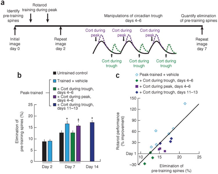
Excessive glucocorticoid exposure during chronic stress causes synapse loss and learning impairment. Under normal physiological conditions, glucocorticoid activity oscillates in synchrony with the circadian rhythm. Whether and how endogenous glucocorticoid oscillations modulate synaptic plasticity and learning is unknown. Here we show that circadian glucocorticoid peaks promote postsynaptic dendritic spine formation in the mouse cortex after motor skill learning, whereas troughs are required for stabilizing newly formed spines that are important for long-term memory retention. Conversely, chronic and excessive exposure to glucocorticoids eliminates learning-associated new spines and disrupts previously acquired memories. Furthermore, we show that glucocorticoids promote rapid spine formation through a non-transcriptional mechanism by means of the LIM kinase-cofilin pathway and increase spine elimination through transcriptional mechanisms involving mineralocorticoid receptor activation. Together, these findings indicate that tightly regulated circadian glucocorticoid oscillations are important for learning-dependent synaptic formation and maintenance. They also delineate a new signaling mechanism underlying these effects.


Figures

Figure 1
Figure 1
Circadian glucocorticoid peaks promote spine formation after learning. (a) Experimental procedure. Blue arrows: formed on day 2, persisting on day 7. Red arrows: formed on day 2, eliminated on day 7. White arrows: eliminated on day 2. Scale bar, 2 μm. (b) ELISA quantification of plasma corticosterone (cort) (F4,34 = 41.0, P < 0.001). *P < 0.05 versus circadian peak–trained group after Holm-Bonferroni correction. (c) Effects of circadian cort on training-induced spine formation (F6,33 = 42.2, P < 0.001). Spine formation rates represent the number of spines formed during the 2-d training period, as a percentage of the total number quantified at day 0 baseline. *P < 0.05 (corrected) versus untrained control. **P < 0.001 versus untrained control and peak-trained group. (d) Effects of circadian cort on spine survival on day 7 (F6,28 = 9.40, P < 0.001). Spine survival represents the number of spines formed during the 2-d training period and persisting on day 7, as a percentage of the total number quantified at baseline. *P < 0.05 (corrected) versus untrained control. (e) Corresponding cort effects on rotarod performance on day 7 (F5,55 = 2.84, P = 0.02). Day 7 performance is expressed as percent change in each subject's baseline performance on day 1. *P < 0.05 (corrected) versus day 1 baseline. (f) Survival of spines that formed during the training period predicted memory retention on day 7 (r = 0.86, P < 0.001). Error bars, s.e.m. See Supplementary Table 1 for statistics and details.
Figure 2
Figure 2
Circadian glucocorticoid troughs preserve newly formed spines. (a) Schematic of experimental procedure. (b) Disruption of the circadian trough reduced the survival of spines that formed during the training period (F4,18 = 5.02, P = 0.007). Corticosterone (cort) that was administered during the second week (days 11–13) had no effect on spine survival, indicating that new spines require circadian troughs only during a critical period after their formation. New spine survival represents the number of spines formed during the 2-d training period and persisting on day 7, as a percentage of the total number of spines at baseline. *P < 0.05 (corrected) versus untrained control. (c) Manipulations of the circadian trough had corresponding effects on motor skill memory retention (F3,35 = 4.24, P = 0.01). Rotarod performance is depicted as a percent change in each group's baseline performance on day 1. *P < 0.05 (corrected) versus day 1 baseline. (d) Survival of spines that formed during the 2-d training period predicted retention of the learned skill on day 7 (r = 0.89, P < 0.001). Error bars, s.e.m. See Supplementary Table 2 for statistics and details.
Figure 3
Figure 3
Disruption of glucocorticoid troughs reduces learning-dependent spine pruning. (a) Schematic of experimental procedure. (b) Learning caused a delayed increase in the elimination of pre-existing spines, which represents the number of spines that were present before training (day 0) and eliminated on day 2 or 7, expressed as a percentage of the total number of spines at baseline. Whereas training had no immediate impact on spine elimination on day 2 (F1,12 = 0.03, P = 0.87), spine elimination was significantly elevated on day 7 (F4,15 = 3.56, P = 0.03). Learning-induced spine pruning required an intact glucocorticoid trough 4–6 d after training. *P < 0.05 versus untrained control after Holm-Bonferroni correction. P < 0.05 uncorrected, P < 0.10 corrected. (c) Elimination of pre-training spines was correlated with retention of the learned motor skill on day 7 (r = 0.83, P < 0.001). Cort, corticosterone. Error bars, s.e.m. See Supplementary Table 3 for statistics and details.
Figure 4
Figure 4
Chronic glucocorticoid exposure causes spine loss and memory impairment. (a) Experimental procedure. (b) Prolonged corticosterone (cort) exposure disrupted the survival of learning-related spines that were present for at least 1 week (F1,5 = 54.2, P < 0.001). New spine survival represents the number of spines formed during the 2-d training period and persisting on day 20, expressed as a percentage of the total number of spines at baseline. (c) Prolonged cort exposure was associated with corresponding deficits in retention of the motor skill (F1,16 = 42.2, P < 0.001). (d) Elimination rates for spines present before training also increased (F1,5 = 66.6, P < 0.001). Elimination rates describe the number of spines that were present before training (day 0) and eliminated on day 20, expressed as a percentage of the total number of spines at baseline. (e) Chronic cort (F1,5 = 125.9, P < 0.001) but not transient cort or learning (F4,19 = 2.07, P = 0.13), caused significant spine loss. Net change in spine number represents the combined effects of elimination and formation on total spine number, relative to the small (4–5%) rate of net spine loss observed in untrained controls (ctrl). (f) The survival of new spines formed during training was strongly correlated with the elimination of pre-existing spines (r = 0.91, P < 0.001), but this balance was disrupted after chronic cort exposure (red). *P < 0.05 (corrected) versus vehicle-treated control. Error bars, s.e.m. See Supplementary Table 4 for statistics and details.
Figure 5
Figure 5
Glucocorticoids promote spine formation and pruning through distinct signaling pathways. (a) Corticosterone (cort) increased spine formation rapidly (F1,40 = 124.0, P < 0.001). (b) Cort caused a delayed increase in spine elimination (F1,40 = 102.3, P < 0.001). (c) Glucocorticoids promote spine formation rapidly through a GR-dependent, non-transcriptional mechanism. Direct cortical application of cort caused rapid increases in spine formation (t = 14.6, P < 0.001), with comparable effects evident after cortical application of cort plus actinomycin D (50 μg ml−1; t = 18.8, P < 0.001) or a membrane-impermeant cort-BSA conjugate (t = 4.83, P < 0.001). Co-administration of the GR antagonist (antag) mifepristone (100 μM) blocked this effect. (d) Mifepristone (20 mg kg−1 i.p.) administered immediately after training reduced learning-dependent spine formation (t = 8.46, P < 0.001). (e) Glucocorticoids cause a delayed increase in spine elimination through a MR-dependent, transcriptional mechanism. A selective MR antagonist (spironolactone, 10 μM) reduced 24-h spine elimination (t = 6.19, P < 0.001), whereas a selective MR agonist (aldosterone, 10 μM) had the opposite effect (t = 18.3, P < 0.001). This effect was blocked by cotreatment with either actinomycin D or anisomycin, consistent with a transcriptional mechanism of action. (f) Spironolactone administered during the circadian trough on days 4, 5 and 6 after training interfered with learning-induced spine elimination (t = 4.84, P = 0.002). *P < 0.05 versus corresponding control. Error bars, s.e.m. See Supplementary Table 5 for statistics and details.
Figure 6
Figure 6
Glucocorticoids promote spine formation rapidly through non-transcriptional regulation of LIMK1-cofilin activity. (a) Expression of phospho-LIMK1 (pLIMK1; upper panels) and phospho-cofilin (pCofilin; lower panels) in cortical lysates 20 min after corticosterone (cort). (b) Cort rapidly increased the expression of phospho-LIMK1 in vivo (F4,68 = 2.74, P = 0.036). This effect was blocked by mifepristone but not by actinomycin D. (c) Similar effects of cort on pCofilin (F4,68 = 6.95, P < 0.001). (d) Cortical pyramidal cells in culture were transfected with a construct encoding GFP plus an interfering RNA (shRNA) specific for GR (lower panels) or a scrambled control construct (upper panels). Phospho-GR (pGR; left), pLIMK1 (middle) and pCofilin (right) immunofluorescence (white) and GFP expression in the same frame (green insets), which identifies pyramidal cells, are depicted after treatment with cort (1 μM) or vehicle for 20 min. White insets show 3× magnifications of boxed representative dendritic segments. Scale bar, 10 μm. (e) Cort increased dendritic and somatic pGR, pLIMK, and pCofilin immunofluorescence after transfection with a scrambled RNA construct (P < 0.001) but not the GR-specific shRNA construct (P > 0.21). A.u., arbitrary units. (f) Direct cortical application of cort (10 μM) increased spine formation within 20 min in wild-type (WT) controls, but not in Limk1 knockout mice (main effect of cort: F1,11 = 58.7, P < 0.001; interaction between genotype and cort: F1,11 = 71.1, P < 0.001). Spine formation at 24 h in vehicle-treated Limk1 knockouts was also reduced (t = 5.34, P < 0.001 versus wild-type). *P < 0.05 versus corresponding control; NS, not significant. Error bars, s.e.m. See Supplementary Table 6 for statistics and details.

Comment in

Similar articles

Cited by

References

    1. de Kloet ER, Vreugdenhil E, Oitzl MS, Jöels M. Brain corticosteroid receptor balance in health and disease. Endocr Rev. 1998;19:269–301. - PubMed
    1. McEwen BS. Protective and damaging effects of stress mediators. N Engl J Med. 1998;338:171–179. - PubMed
    1. Lupien SJ, et al. Cortisol levels during human aging predict hippocampal atrophy and memory defcits. Nat Neurosci. 1998;1:69–73. - PubMed
    1. de Quervain DJF, Roozendaal B, McGaugh JL. Stress and glucocorticoids impair retrieval of long-term spatial memory. Nature. 1998;394:787–790. - PubMed
    1. Bangasser DA, Shors TJ. The hippocampus is necessary for enhancements and impairments of learning following stress. Nat Neurosci. 2007;10:1401–1403. - PMC - PubMed

Publication types

MeSH terms

Substances