. 2013 Apr;25(4):1416-29.
doi: 10.1105/tpc.113.110684. Epub 2013 Apr 26.

BINDING PROTEIN is a master regulator of the endoplasmic reticulum stress sensor/transducer bZIP28 in Arabidopsis

Affiliations

BINDING PROTEIN is a master regulator of the endoplasmic reticulum stress sensor/transducer bZIP28 in Arabidopsis

Renu Srivastava et al. Plant Cell. 2013 Apr.

Abstract

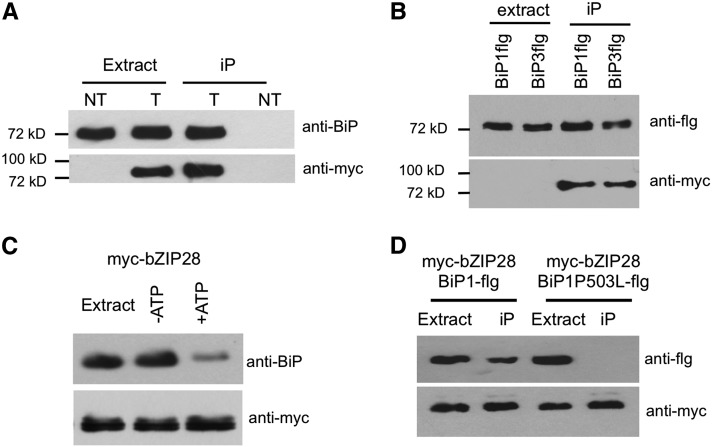
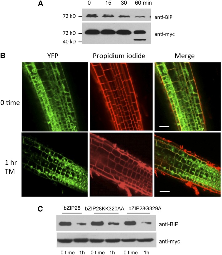
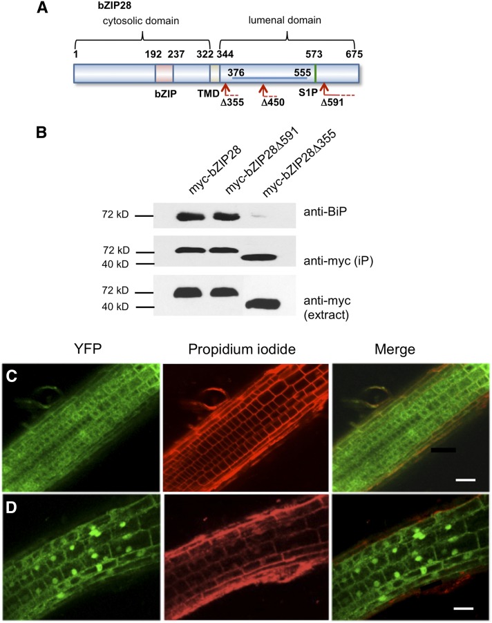
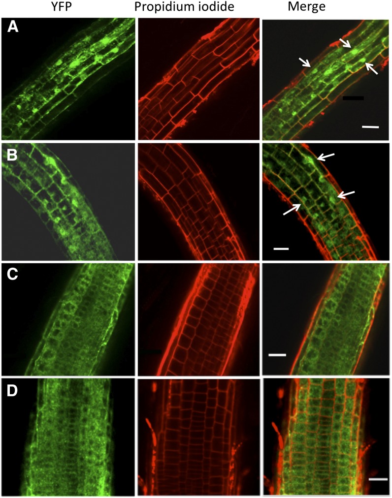
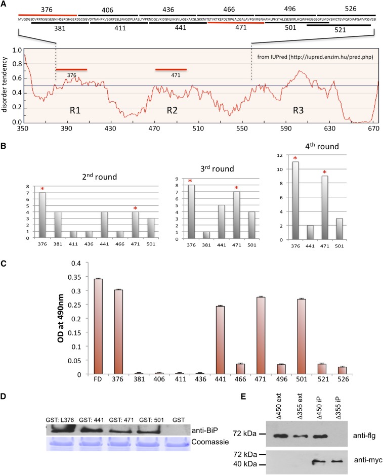
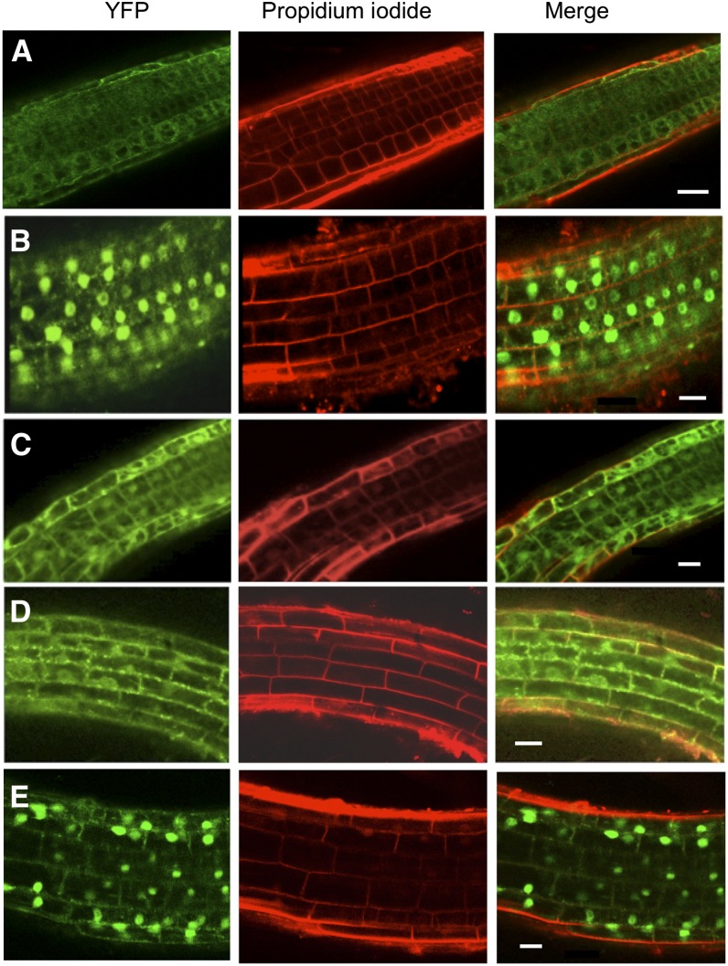
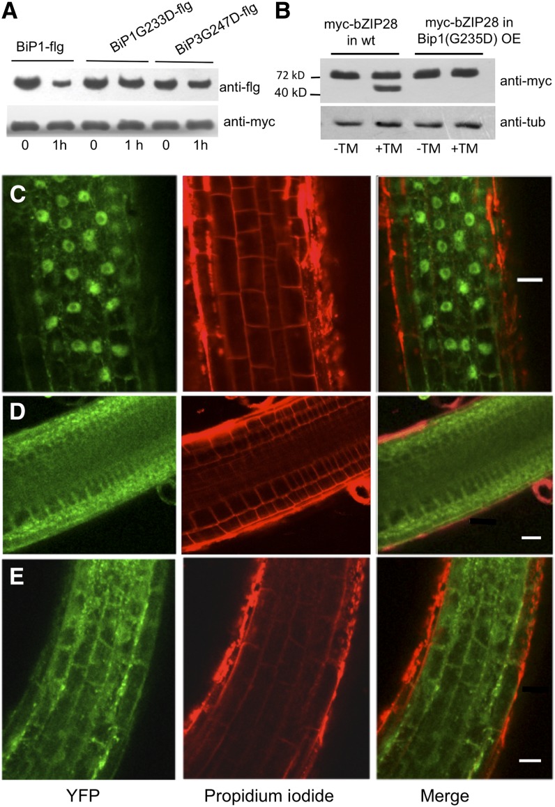
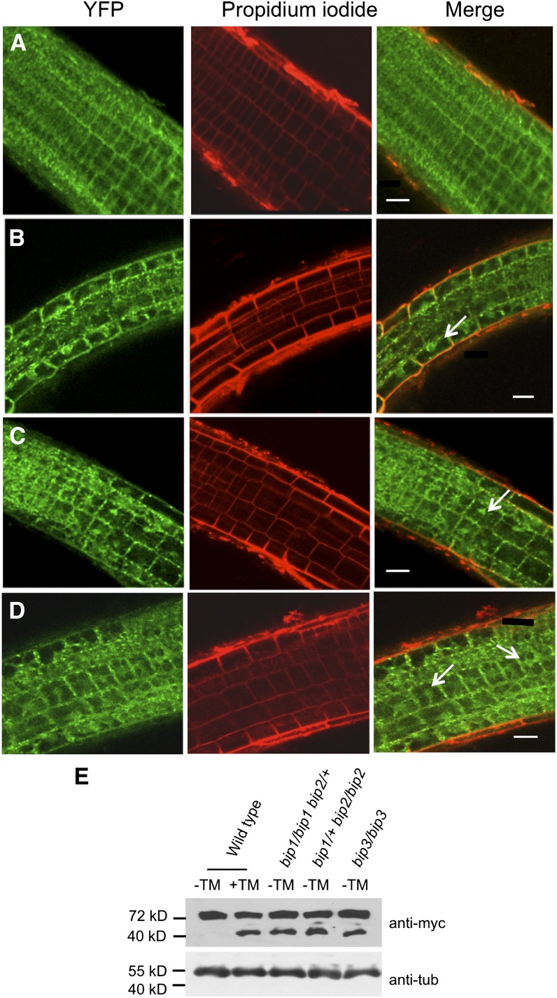
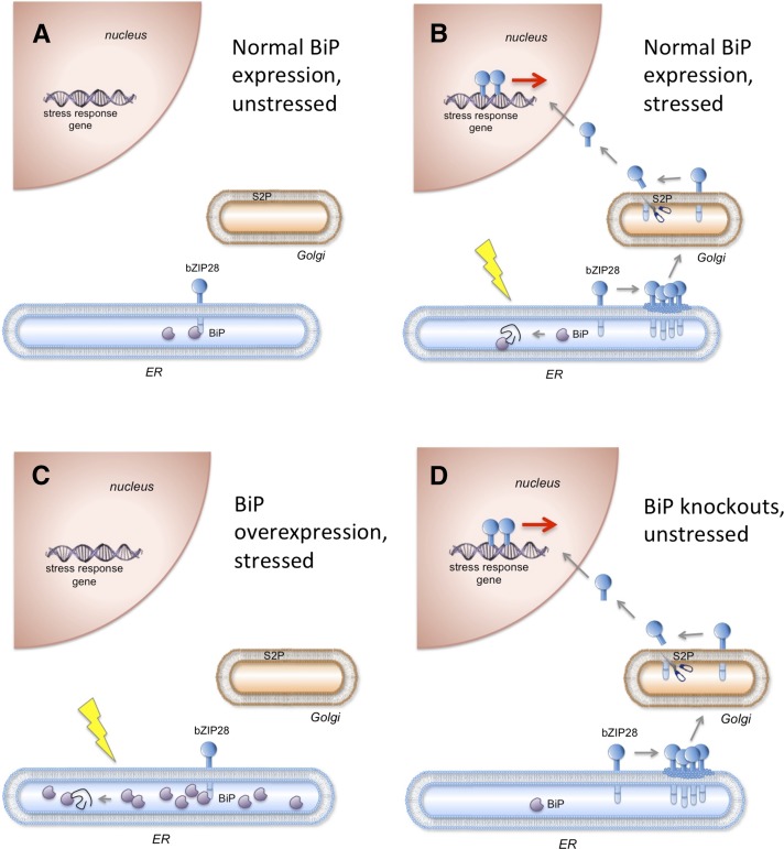
BINDING PROTEIN (BiP) is a major chaperone in the endoplasmic reticulum (ER) lumen, and this study shows that BiP binds to the C-terminal tail of the stress sensor/transducer bZIP28, a membrane-associated transcription factor, retaining it in the ER under unstressed conditions. In response to ER stress, BiP dissociates from bZIP28, allowing it to be mobilized from the ER to the Golgi where it is proteolytically processed and released to enter the nucleus. Under unstressed conditions, BiP binds to bZIP28 as it binds to other client proteins, through its substrate binding domain. BiP dissociates from bZIP28 even when bZIP28's exit from the ER or its release from the Golgi is blocked. Both BiP1 and BiP3 bind bZIP28, and overexpression of either BiP detains bZIP28 in the ER under stress conditions. A C-terminally truncated mutant of bZIP28 eliminating most of the lumenal domain does not bind BiP and is not retained in the ER under unstressed conditions. BiP binding sites in the C-terminal tail of bZIP28 were identified in a phage display system. BiP was found to bind to intrinsically disordered regions on bZIP28's lumen-facing tail. Thus, the dissociation of BiP from the C-terminal tail of bZIP28 is a major switch that activates one arm of the unfolded protein response signaling pathway in plants.


Figures

Figure 1.
Figure 1.
BiP Binds to bZIP28. (A) myc-bZIP28 was detected in extracts and immunoprecipitates (iP) from of roots of unstressed 7-d-old transgenic (T) and nontransgenic control (NT) Arabidopsis seedlings. Immunoblots were probed with anti-BiP and anti-myc antibodies. The anti-BiP antibody that was used for immunoprecipitations did not bind to agarose beads alone. (B) Both BiP1-flg and BiP3-flg bind to myc-bZIP28. myc-bZIP28 was immunoprecipitated from extracts of N. benthamiana leaves transiently expressing BiP1-flg and BiP3-flg. Immunoblots were probed with anti-flg and anti-myc antibodies. (C) BiP bound to myc-bZIP28 extracted from myc-bZIP28 expression lines is released by ATP. myc-bZIP28 was immunoprecipitated with anti-myc antibodies and incubated for 30 min with or without 2 mM ATP and 2 mM MgCl2. Immunoblot was probed with anti-BiP and anti-myc antibodies. (D) Mutant BiP with a defect in substrate binding does not bind bZIP28. myc-bZIP28 coexpressed with BiP1-flg or BiP1P503L-flg in the transient expression system was immunoprecipitated with anti-myc antibodies. Immunoblot was probed with anti-flg and anti-myc antibodies.
Figure 2.
Figure 2.
BiP Dissociates from bZIP28 in Response to ER Stress. (A) Seven-day-old Arabidopsis seedlings expressing myc-bZIP28 were treated with 2 μg/mL TM, and proteins were extracted (from whole seedlings) at times indicated following TM treatment. Extracts were immunoprecipitated with anti-myc antibodies, and immunoblot was probed with anti-BiP and anti-myc antibodies. The fast migrating band visualized by the anti-myc antibody is the proteolytically processed form of myc-bZIP28. (B) Confocal images of roots of Arabidopsis seedlings expressing YFP-bZIP28. Seedlings were treated for 1 h with 2 μg/mL TM. Roots were stained with propidium iodide to show cell outlines. Bars = 50µm. (C) BiP dissociates in response to stress from mutant forms of bZIP28 that are prevented from exiting the ER or from being released from Golgi bodies. Seven-day-old Arabidopsis expressing various forms of myc-bZIP28 were treated with TM for 1 h, and proteins were extracted, immunoprecipitated with anti-myc, subjected to immunoblot analysis, and probed with anti-BiP and anti-myc antibodies. bZIP28KK320AA is blocked in exiting the ER, and bZIP28G329A is not proteolytically cleaved by S2P and therefore is not released from Golgi bodies.
Figure 3.
Figure 3.
The Effect of Truncations on bZIP28 Mobilization. (A) Map shows truncations in myc-bZIP28 and the region of the protein (blue line) that was used in the phage display analysis. (B) Coimmunoprecipitation experiments of BiP with truncated forms of bZIP28. Proteins were extracted from 7-d-old Arabidopsis seedlings expressing myc-tagged forms of the truncated constructs myc-bZIP28Δ591 and myc-bZIP28Δ355 and subjected to immunoblot analysis probed with anti-BiP and anti-myc antibodies. The expression levels of truncated forms of the myc-bZIP28 constructs in the lines used are shown in the crude protein extracts [labeled anti-myc (extract)]. (C) Subcellular localization of YFP-bZIP28Δ591 in root cells under unstressed conditions. (D) Relocation of YFP-bZIP28Δ591 to nuclei in root cells of seedlings treated with 2 μg/mL TM for 1 h. Confocal images of Arabidopsis roots counterstained with propidium iodide to show cell outlines. Bars = 50 µm.
Figure 4.
Figure 4.
Translocation of bZIP28Δ355 to the Nucleus Requires S2P. YFP-bZIP28Δ355 was imaged by confocal microscopy in a wild-type background at zero time (A) and after 1 h treatment (B) with 2 μg/mL TM. Arrows point out the nuclear localization of some of the YFP-bZIP28Δ355. YFP-bZIP28Δ355 was imaged in an s2p background at zero time (C) and after 1 h treatment (D) with TM. In each case the root cells were counterstained with propidium iodide. Bars = 50 µm.
Figure 5.
Figure 5.
bZIP28 Peptides Used in Phage Display Library. (A) Overlapping peptides from residues 376 to 555 in the lumenal domain of bZIP28 were displayed in M13 phage. Tendency for intrinsic disorder in the lumenal domain of bZIP28 is plotted against the map of the lumenal domain for bZIP28. The tendency for disorder was determined by IUPred (http://iupred.enzim.hu/pred.php). (B) Recombinant phages were pooled and panned against immobilized BiP1-His in four rounds of panning. At each round, bound phages were released and the inserts encoding the bZIP28 peptides were sequenced. The frequency in recovering phage expressing the various peptides in progressive rounds of screening is shown. Red asterisks indicate the peptides in phage recovered with the highest frequency in the fourth round of panning. (C) Separate recombinant phage lines were incubated with immobilized BiP1-His, and bound phage were quantified in an ELISA assay. Error bars indicate se. (D) Overlay immunoblot demonstrating that soluble BiP1-His binds to GST-tagged bZIP28 peptides. The four peptides (441, 471, 376, and 501) enriched in panning were tagged with GST, purified by binding to glutathione beads, eluted, subjected to SDS-PAGE, and transferred to a nitrocellulose filter. The filter was incubated with purified BiP1-His, washed, and incubated with a primary anti-BiP antibody and then a secondary antibody was used to detect BiP binding. The GST-tagged peptides pulled down with glutathione beads and stained with Coomassie blue were used as a loading control. (E) bZIP28 construct containing only the R1 region of lumenal domain (as shown in [A]) binds BiP1-flg in vivo. bZIP28 truncation constructs myc-bZIP28Δ450 (containing region R1) and myc-bZIP28Δ355 (lacking region R1) were each coexpressed with BiP1-flg in a tobacco leaf transient expression assay. Leaf extracts were subjected to immunoblotting and probed with anti-flg and anti-myc antibodies. [See online article for color version of this figure.]
Figure 6.
Figure 6.
The Mobilization of bZIP28 in Lines Overexpressing BiP. (A) YFP-bZIP28 localization in untreated Arabidopsis seedlings in a wild-type background. (B) YFP-bZIP28 localization in wild-type Arabidopsis seedlings lines treated with 2 μg/mL TM for 2 h. (C) YFP-bZIP28 localization in a line overexpressing BiP1 from a 35Spromoter:BiP transgene treated with 2 μg/mL TM for 2 h. (D) YFP-bZIP28 localization in lines overexpressing BiP3 treated with 2 μg/mL TM for 2 h. (E) YFP-bZIP28 localization in lines overexpressing BiP1P503L-flg (a form of BiP1 with a defect in substrate binding as shown in Figure 1D) treated with 2 μg/mL TM for 2 h. YFP fluorescence was imaged by confocal microscopy in roots counterstained with propidium iodide. Bars = 50 µm.
Figure 7.
Figure 7.
The Mobilization of bZIP28 in Lines Overexpressing Mutant Forms of BiP. (A) Immunoprecipitation of myc-bZIP28 in extracts from Arabidopsis lines expressing flag-tagged mutated forms of BiP (BiP1G233D-flg and BipG247D-flg) that are predicted to be defective in binding ATP and releasing client proteins. Seedlings were treated with 2 μg/mL TM for 2 h to determine whether the BiP constructs dissociated from myc-bZIP28. Immunoblot of extracted proteins was probed with anti-flg and anti-myc. (B) Processing of YFP-bZIP28 in wild-type (wt) and BiP1G233D-flg overexpressing (OE) backgrounds. Seedlings were either untreated or treated with 2 mM DTT for 2 h. Immunoblot of extracted proteins was probed with anti-myc and antitubulin as a loading control. (C) YFP-bZIP28 localization in wild-type Arabidopsis seedlings lines treated with TM for 2 h. (D) and (E) YFP-bZIP28 localization in lines overexpressing BiP1G233D (D) or BiP3G247D (E), mutants with defects in ATP binding and release of client proteins. Lines were treated with TM for 2 h. YFP fluorescence was imaged by confocal microscopy in roots counterstained with propidium iodide. Bars = 50 µm.
Figure 8.
Figure 8.
Mobilization of bZIP28 in BiP Knockout Lines. (A) YFP-bZIP28 in wild-type unstressed Arabidopsis seedlings. (B) to (D) YFP-bZIP28 in the bip1/bip1 bip2/+ (B), bip1/+ bip2/bip2 (C), and bip3/bip3 (D) mutant lines. YFP fluorescence was imaged by confocal microscopy in roots stained with propidium iodide. Arrows point out nuclear localization. Bars = 50 µm. (E) Proteolytic processing of mycbZIP28 in wild-type seedlings in response to 2 μg/mL TM treatment for 2 h. Processing of myc-bZIP28 in untreated BiP knockout lines as indicated.
Figure 9.
Figure 9.
The Effect of BiP Expression on the Mobilization of bZIP28. (A) BiP normally associates with bZIP28 under unstressed conditions and detains bZIP28 in the ER. (B) In response to stress, BiP is competed away by the accumulation of misfolded proteins, releasing bZIP28 to relocate to the nucleus via the Golgi. (C) When BiP is overexpressed, the accumulation of misfolded proteins fails to compete BiP away from bZIP28 under stress. As a result bZIP28 is detained in the ER even under stress conditions. (D) When BiP is underexpressed, bZIP28 escapes to the nucleus under unstressed conditions. [See online article for color version of this figure.]

Similar articles

Cited by

References

    1. Alvim F.C., Carolino S.M., Cascardo J.C., Nunes C.C., Martinez C.A., Otoni W.C., Fontes E.P. (2001). Enhanced accumulation of BiP in transgenic plants confers tolerance to water stress. Plant Physiol. 126: 1042–1054 - PMC - PubMed
    1. Anelli T., Sitia R. (2008). Protein quality control in the early secretory pathway. EMBO J. 27: 315–327 - PMC - PubMed
    1. Batoko H., Zheng H.Q., Hawes C., Moore I. (2000). A rab1 GTPase is required for transport between the endoplasmic reticulum and golgi apparatus and for normal Golgi movement in plants. Plant Cell 12: 2201–2218 - PMC - PubMed
    1. Bechtold N., Ellis J., Pelletier G. (1993). In planta Agrobacterium-mediated gene transfer by infiltration of adult Arabidopsis thaliana plants. C. R. Acad. Sci. Paris 316: 1194–1199
    1. Bertolotti A., Zhang Y., Hendershot L.M., Harding H.P., Ron D. (2000). Dynamic interaction of BiP and ER stress transducers in the unfolded-protein response. Nat. Cell Biol. 2: 326–332 - PubMed

Publication types

MeSH terms