. 2013 Jul;38(7):1454-66.
doi: 10.1007/s11064-013-1044-x. Epub 2013 Apr 27.

Inhibition of ROS-activated p38MAPK pathway is involved in the protective effect of H2S against chemical hypoxia-induced inflammation in PC12 cells

Affiliations

Inhibition of ROS-activated p38MAPK pathway is involved in the protective effect of H2S against chemical hypoxia-induced inflammation in PC12 cells

Aiping Lan et al. Neurochem Res. 2013 Jul.

Abstract

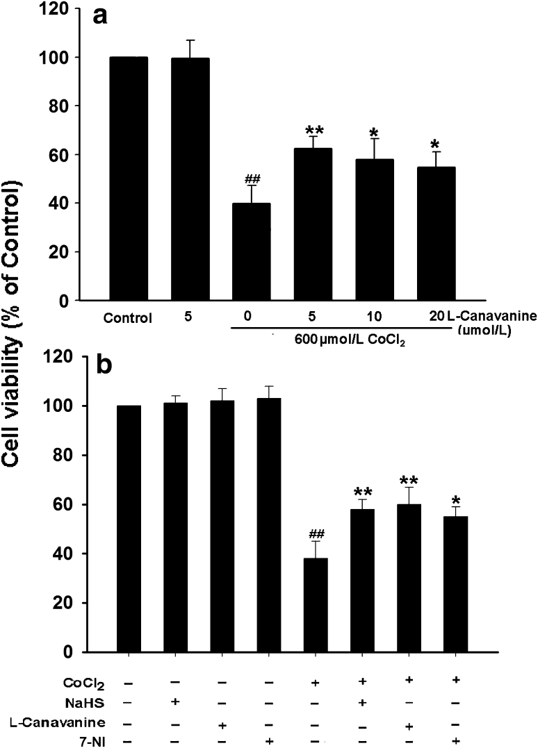
We have demonstrated the neuroprotection of hydrogen sulfide (H2S) against chemical hypoxia-induced injury by inhibiting p38MAPK pathway. The present study attempts to evaluate the effect of H2S on chemical hypoxia-induced inflammation responses and its mechanisms in PC12 cells. We found that treatment of PC12 cells with cobalt chloride (CoCl2, a hypoxia mimetic agent) enhanced IL-6 secretion, nitric oxide (NO) generation and expression levels of inducible nitric oxide synthase (iNOS) and neuronal nitric oxide synthase (nNOS). L-canavanine, a selective iNOS inhibitor, partly blocked CoCl2-induced cytotoxicity, apoptosis and mitochondrial insult. In addition, 7-Nitroindazole (7-NI), an inhibitor of nNOS, also partly attenuated the CoCl2-induced cytotoxicity. The inhibition of p38MAPK by SB203580 (a selective p38MAPK inhibitor) or genetic silencing of p38MAPK by RNAi (Si-p38) depressed not only CoCl2-induced iNOS expression, NO production, but also IL-6 secretion. In addition, N-acetyl-L-cysteine, a reactive oxygen species (ROS) scavenger, conferred a similar protective effect of SB203580 or Si-p38 against CoCl2-induced inflammatory responses. Importantly, pretreatment of PC12 cells with exogenous application of sodium hydrosulfide (a H2S donor, 400 μmol/l) for 30 min before exposure to CoCl2 markedly attenuated chemical hypoxia-stimulated iNOS and nNOS expression, NO generation and IL-6 secretion as well as p38MAPK phosphorylation in PC12 cells. Taken together, we demonstrated that p38MAPK-iNOS pathway contributes to chemical hypoxia-induced inflammation and that H2S produces an anti-inflammatory effect in chemical hypoxia-stimulated PC12 cells, which may be partly due to inhibition of ROS-activated p38MAPK-iNOS pathway.


Figures

Fig. 1
Fig. 1
CoCl2 increases expression of iNOS and production of NO in PC12 cells. a PC12 cells were treated with 600 μmol/l CoCl2 for indicated times. The expression of iNOS was tested by Western blot assay. b Densitometric analysis of the results from (a). c PC12 cells were treated with 600 μmol/l CoCl2 for 24 or 48 h. Nitrite in the culture supernatant was determined using the Griess reagent as described in “Materials and Methods”. Data were presented as mean ± SEM (n = 3). ## P < 0.01 versus control group
Fig. 2
Fig. 2
NaHS, L-canavanine or 7-nitroindazole inhibits CoCl2-induced cytotoxicity in PC12 cells. a PC12 cells were pretreated with L-canavanine, an inhibitor of iNOS, at indicated concentrations for 60 min before exposure to 600 μmol/l CoCl2 for 24 h. b PC12 cells were pretreated with 400 μmol/l NaHS for 30 min or 5 μmol/l L-canavanine for 60 min or 250 μmol/l 7-nitroindazole (7-NI), an inhibitor of nNOS, for 30 min prior to exposure to 600 μmol/l CoCl2 for 24 h. Cell viability was measured by the CCK-8 assay. Data were presented as mean ± SEM (n = 3). ## P < 0.01 versus control group; *P < 0.05, **P < 0.01 versus CoCl2 group
Fig. 3
Fig. 3
NaHS and L-canavanine block CoCl2-induced apoptosis in PC12 cells. a Morphological changes in apoptotic cells were assessed by Hoechst33258 staining. b Apoptosis rate was determined by FCM as described in “Materials and Methods”. Control group: Normal PC12 cells; NaHS group: cells were treated with 400 μmol/l NaHS for 30 min alone; L-canavanine group: cells were exposed to 5 μmol/l L-canavanine for 60 min alone; CoCl2 group: cells were treated with 600 μmol/l CoCl2 for 48 h; NaHS + CoCl2: cells were pretreated with 400 μmol/l NaHS for 30 min followed by 600 μmol/l CoCl2 treatment for 48 h. L-canavanine + CoCl2: cell were pretreated with 5 μmol/l L-canavanine for 60 min followed by 600 μmol/l CoCl2 treatment for 48 h. Data were presented as mean ± SEM (n = 5). ## P < 0.01 versus control group; **P < 0.01 versus CoCl2 group
Fig. 4
Fig. 4
NaHS and L-canavanine attenuate CoCl2-induced mitochondrial insult. MMP was measured by JC-1 staining followed by photofluorography. Dual emission images (527 and 590 nm) represent the signals from monomeric (green) and J-aggregate (red) JC-1 fluorescence in PC12 cells. a Random micrographs of fluorescence in PC12 cells. Control group: normal PC12 cells; NaHS group: cells were exposed to 400 μmol/l NaHS for 30 min alone; L-canavanine group: cells were incubated with 5 μmol/l L-canavanine for 60 min alone; CoCl2 group: cells were treated with 600 μmol/l CoCl2 for 24 h; NaHS + CoCl2: cells were pretreated with 400 μmol/l NaHS for 30 min followed by exposure to 600 μmol/l CoCl2 for 24 h. L-canavanine + CoCl2: cells were pretreated with 5 μmol/l L-canavanine for 60 min followed by exposure to 600 μmol/l CoCl2 for 24 h. b Quantitative analysis of the ratio of red/green fluorescence in the indicated groups. Data were presented as mean ± SEM (n = 5). ## P < 0.01 versus control group; **P < 0.01 versus CoCl2 group (Color figure online)
Fig. 5
Fig. 5
p38MAPK activation participates in CoCl2-induced iNOS expression and NO production in PC12 cells. The expression of iNOS was tested by Western blot assay. a PC12 cells were preconditioned with 20 μmol/l SB203580 for 60 min before exposure to 600 μmol/l CoCl2 for 24 h. c PC12 cells were co-cultured with small interfering RNA (Si-p38) or random non-coding RNA(Si-NC) for 6 h. e PC12 cells were co-incubated with Si-p38 for 6 h followed by treated with 600 μmol/l CoCl2 for 24 h. b, d, and f densitometric analysis of the results from a, c, and e, respectively. g PC12 cells were treated with 600 μmol/l CoCl2 for 48 h in the presence or absence of pretreatment with 20 μmol/l SB203580 for 60 min or co-cultured with Si-p38 for 6 h. Nitrite in the culture supernatant was determined using the Griess reagent as described in “Materials and Methods”. Data were presented as mean ± SEM (n = 5). ## P < 0.01 versus Control group; **P < 0.01 versus CoCl2 group
Fig. 6
Fig. 6
ROS contribute to CoCl2-induced iNOS expression and NO generation in PC12 cells. The expression of iNOS was detected by Western blot assay. a PC12 cells were preconditioned with 500 μmol/l NAC for 60 min before exposure to 600 μmol/l CoCl2 for 24 h. b Densitometric analysis of the results from A. c PC12 cells were treated with 600 μmol/l CoCl2 for 48 h in the presence or absence of pretreatment with 500 μmol/l NAC for 60 min. Nitrite in the culture supernatant was determined using the Griess reagent as described in “Materials and Methods”. Data were presented as the mean ± SEM (n = 5). ## P < 0.01 versus control group; *P < 0.05, **P < 0.01 versus CoCl2 group
Fig. 7
Fig. 7
Effects of indicated treatments on secretions of IL-6 induced by CoCl2 from PC12 cells. a PC12 cells were treated with 600 μmol/l CoCl2 for indicated times. b PC12 cells were treated with 600 μmol/l CoCl2 for 48 h in the absence or presence of pretreatment with 20 μmol/l SB203580 for 60 min or 500 μmol/l NAC for 60 min or co-incubation with Si-p38 for 6 h. ELISA was performed to detect the levels of IL-6 in cell supernatants. Data were presented as the mean ± SEM (n = 3). # P < 0.05, ## P < 0.01 versus control group; **P < 0.01 versus CoCl2 group
Fig. 8
Fig. 8
NaHS attenuates CoCl2-induced expression levels of iNOS and nNOS as well as NO production in PC12 cells. PC12 cells were treated with 600 μmol/l CoCl2 for 24 h in the absence or presence of the preconditioning with 400 μmol/l NaHS for 30 min. The expression levels of iNOS (a) and nNOS (c) were determined by Western blot assay. b and d Densitometric analysis of the results from a and c, respectively. e PC12 cells were treated with 600 μmol/l CoCl2 for 48 h in the presence or absence of pretreatment with 400 μmol/l NaHS for 30 min. Nitrite in the culture supernatant was determined using the Griess reagent as described in “Materials and Methods”. Data were presented as the mean ± SEM (n = 5). ## P < 0.01 versus Control group; **P < 0.01 versus CoCl2 group
Fig. 9
Fig. 9
NaHS attenuates CoCl2-stimulated IL-6 secretion from PC12 cells. PC12 cells were treated with 600 μmol/l CoCl2 for 48 h in the presence or absence of pretreatment with 400 μmol/l NaHS for 30 min. ELISA was performed to detect the levels of IL-6 in cell supernatants. Data were presented as the mean ± SEM (n = 3). ## P < 0.01 versus Control group; **P < 0.01 versus CoCl2 group
Fig. 10
Fig. 10
NaHS blocks CoCl2-induced activation of p38MAPK in PC12 cells. The expression of p-p38MAPK was determined by Western blot assay. a PC12 cells were treated with 600 μmol/l CoCl2 for 120 min in the presence or absence of pretreatment with 400 μmol/l NaHS for 30 min. b Densitometric analysis of the results from a. Data were presented as the mean ± SEM (n = 3). ## P < 0.01 versus Control group; **P < 0.01 versus CoCl2 group

Similar articles

Cited by

References

    1. Wang R. The gasotransmitter role of hydrogen sulfide. Antioxid Redox Signal. 2003;5:493–501. doi: 10.1089/152308603768295249. - DOI - PubMed
    1. Chahl LA. Hydrogen sulphide: an endogenous stimulant of capsaicin-sensitive primary afferent neurons? Br J Pharmacol. 2004;142:1–2. doi: 10.1038/sj.bjp.0705765. - DOI - PMC - PubMed
    1. Kimura H, Nagai Y, Umemura K, Kimura Y. Physiological roles of hydrogen sulfide: synaptic modulation, neuroprotection, and smooth muscle relaxation. Antioxid Redox Signal. 2005;7:795–803. doi: 10.1089/ars.2005.7.795. - DOI - PubMed
    1. Li L, Bhatia M, Moore PK. Hydrogen sulphide–a novel mediator of inflammation? Curr Opin Pharmacol. 2006;6:125–129. doi: 10.1016/j.coph.2005.10.007. - DOI - PubMed
    1. Zhang H, Zhi L, Moore PK, Bhatia M. Role of hydrogen sulfide in cecal ligation and puncture-induced sepsis in the mouse. Am J Physiol Lung Cell Mol Physiol. 2006;290:L1193–L1201. doi: 10.1152/ajplung.00489.2005. - DOI - PubMed

Publication types

LinkOut - more resources