. 2014 Apr 3;33(14):1828-39.
doi: 10.1038/onc.2013.122. Epub 2013 Apr 29.

ERK and AKT signaling cooperate to translationally regulate survivin expression for metastatic progression of colorectal cancer

Affiliations

ERK and AKT signaling cooperate to translationally regulate survivin expression for metastatic progression of colorectal cancer

Q Ye et al. Oncogene. .

Abstract

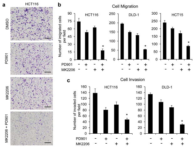
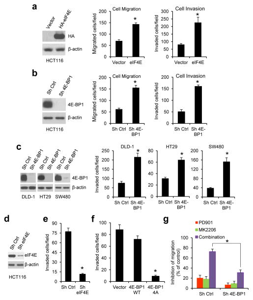
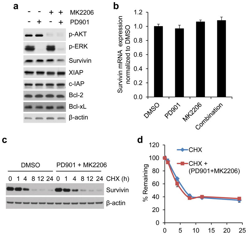
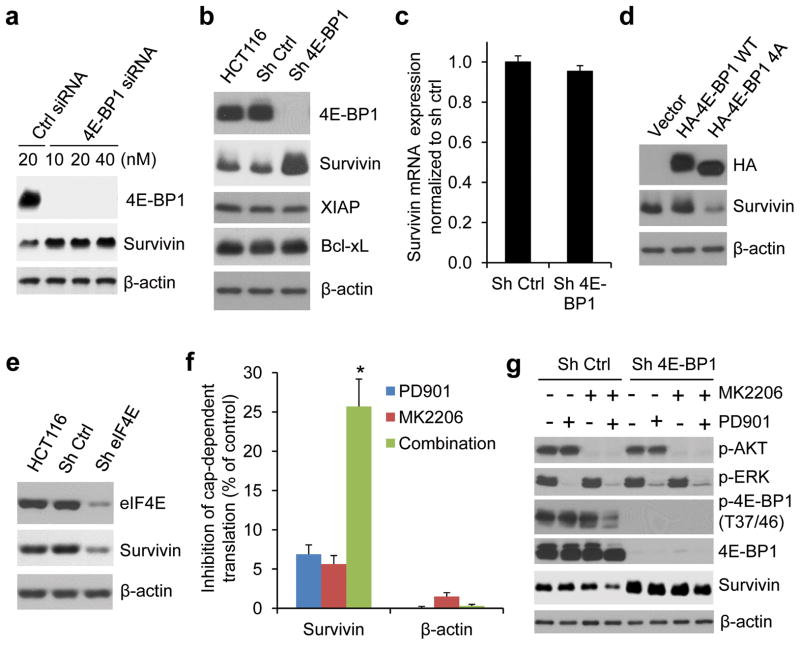
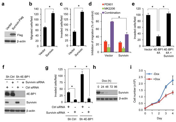
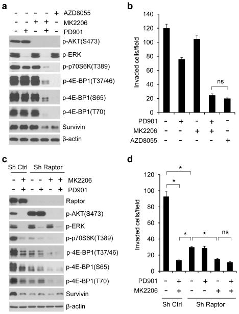
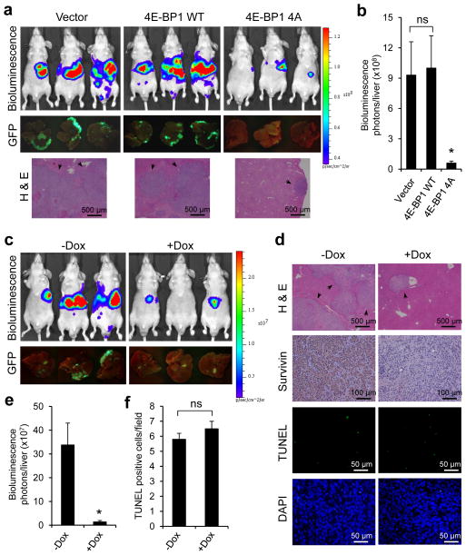
The mitogen-activated extracellular signal-regulated kinase/extracellular signal-regulated kinase (MEK/ERK) and phosphatidylinositol 3-kinase (PI3K)/AKT pathways are often concurrently activated by separate genetic alterations in colorectal cancer (CRC), which is associated with CRC progression and poor survival. However, how activating both pathways is required for CRC metastatic progression remains unclear. Our recent study showed that both ERK and AKT signaling are required to activate eukaryotic translation initiation factor 4E (eIF4E)-initiated cap-dependent translation via convergent regulation of the translational repressor 4E-binding protein 1 (4E-BP1) for maintaining CRC transformation. Here, we identified that the activation of cap-dependent translation by cooperative ERK and AKT signaling is critical for promotion of CRC motility and metastasis. In CRC cells with coexistent mutational activation of ERK and AKT pathways, inhibition of either MEK or AKT alone showed limited activity in inhibiting cell migration and invasion, but combined inhibition resulted in profound effects. Genetic blockade of the translation initiation complex by eIF4E knockdown or expression of a dominant active 4E-BP1 mutant effectively inhibited migration, invasion and metastasis of CRC cells, whereas overexpression of eIF4E or knockdown of 4E-BP1 had the opposite effect and markedly reduced their dependence on ERK and AKT signaling for cell motility. Mechanistically, we found that these effects were largely dependent on the increase in mammalian target of rapamycin complex 1 (mTORC1)-mediated survivin translation by ERK and AKT signaling. Despite the modest effect of survivin knockdown on tumor growth, reduction of the translationally regulated survivin profoundly inhibited motility and metastasis of CRC. These findings reveal a critical mechanism underlying the translational regulation of CRC metastatic progression, and suggest that targeting cap-dependent translation may provide a promising treatment strategy for advanced CRC.


Conflict of interest statement

CONFLICT OF INTEREST

The authors declare no conflict of interest.

Figures

Figure 1
Figure 1
Combined inhibition of MEK and AKT is required for effective inhibition of migration and invasion of CRC cells with coexistent KRAS and PIK3CA mutations. (a and b) Transwell migration analysis of HCT116, DLD-1 and HCT15 cells in the presence of 50 nM PD0325901 (PD901) and 1 μM MK2206, alone or in combination or DMSO as control for 6 hours. The results represent the mean number of migrated cells per field ± s.e.m. (n=3). Scale bar = 500 μm. (c) Transwell invasion analysis of HCT116 and DLD-1 cells in the presence of the drugs as indicated in (a and b) for 30 hours. The results represent the mean number of invaded cells per field ± s.e.m. (n=3). * P < 0.02 for combination of PD901 and MK2206 versus DMSO control, PD901 or MK2206.
Figure 2
Figure 2
Cap-dependent translation apparatus mediates the effect of ERK and AKT activation on CRC cell migration and invasion. (a–c) Overexpression of eIF4E (a) or silencing 4E-BP1 expression (b and c) promoted CRC cell migration and invasion. Exogenous eIF4E expression (a, left) and 4E-BP1 knockdown efficiency by shRNA (b and c, left) in the indicated cell lines were verified by immunoblot analysis. (d) HCT116 cells with stable expression of control (ctrl) shRNA or eIF4E shRNA were immunoblotted with the indicated antibodies. (e and f) Silencing eIF4E expression (e) or expression of 4E-BP1 4A but not 4E-BP1 WT (f) markedly inhibited invasion of HCT116 cells. (g) Silencing 4E-BP1 expression significantly reduced the inhibitory effect on cell migration induced by combined inhibition of MEK and AKT. Migration analysis of HCT116 cells with stable expression of control shRNA or 4E-BP1 shRNA was performed in the presence of 50 nM PD901 and 1 μM MK2206, alone or in combination or DMSO as control for 6 hours. The results are expressed as the inhibition of migration relative to the DMSO-treated controls. Data shown in graphs represent the mean ± s.e.m. (n=3). * P < 0.02 for eIF4E versus vector; 4E-BP1 4A versus 4E-BP1 WT or vector; sh eIF4E or sh 4E-BP1 versus sh Ctrl; and combination of PD901 and MK2206 in sh 4E-BP1 cells versus that in sh ctrl cells.
Figure 3
Figure 3
Combined inhibition of MEK and AKT downregulates survivin expression at the level of protein but not total mRNA or protein stability. (a) HCT116 cells were treated with 50 nM PD901 and 1 μM MK2206, alone or in combination or DMSO as control for 12 hours. Cell lysates were immunoblotted with the indicated antibodies. (b) Quantitative RT-PCR analysis of mRNA expression of survivin relative to β-actin in HCT116 cells that were treated with the drugs as indicated in (a) for 12 hours (n=3). (c) HCT116 cells were treated with combination of 50 nM PD901 and 1 μM MK2206 or DMSO as control for 30 min, followed by addition of 20 μg/ml cycloheximide (CHX) for the indicated times. Cell lysates were immunoblotted with survivin and β-actin antibodies. (d) Immunoblots of survivin as shown in (c) were quantified using the FluoChem digital imaging system. The level of survivin remaining was obtained by normalizing β-actin level at each time, and the results are presented as mean ± s.e.m. (n=3).
Figure 4
Figure 4
Survivin is translationally regulated by ERK and AKT signaling in CRC cells with coexistent pathway activation. (a) HCT116 cells were transfected with the indicated concentrations of 4E-BP1 siRNAs or control non-targeting siRNAs for 48 hours. Cell lysates were immunoblotted with the indicated antibodies. (b) Immunoblot analysis of HCT116 cells alone or with stable expression of control shRNA or 4E-BP1 shRNA with different targeting sequences used in (a). (c) Quantitative RT-PCR analysis of mRNA expression of survivin relative to β-actin in HCT116 cells with stable expression of control shRNA or 4E-BP1 shRNA (n=3). (d) Immunoblot analysis of HCT116 cells with stable expression of vector, HA-4E-BP1 WT or HA-4E-BP1 4A. (e) Immunoblot analysis of HCT116 cells alone or with stable expression of control shRNA or eIF4E shRNA. (f) HCT116 cells were transfected with a bicistronic luciferase reporter that detects cap-dependent translation of the Renilla luciferase gene linked to the 5′-UTR of either survivin or β-actin and cap-independent Polio IRES-mediated translation of the firefly luciferase gene. The transfected cells were treated with 50 nM PD0325901 and 1 μM MK2206, alone or in combination for 12 hours. Luciferase activities were measured by a dual-luciferase assay, and the Renilla/firefly luciferase luminescence ratio was calculated for cap-dependent translational activity. The results are expressed as the inhibition of cap-dependent translation relative to the DMSO-treated controls and presented as means ± s.e.m. (n=3). * P < 0.01 for combination of PD901 and MK2206 versus PD901 or MK2206. (g) Immunoblot analysis of HCT116 cells with stable expression of control shRNA or 4E-BP1 shRNA that were treated with 50 nM PD901 and 1 μM MK2206, alone or in combination for 12 hours.
Figure 5
Figure 5
Survivin is an important effector of ERK and AKT signaling responsible for translational control of CRC cell migration and invasion. (a) Immunoblot analysis of HCT116 cells with stable expression of vector or flag-tagged survivin. (b and c) Migration (b) or invasion (c) analysis of HCT116 cells with stable expression of vector or survivin. (d) Migration analysis of HCT116 cells with stable expression of vector or survivin in the presence of 50 nM PD901 and 1 μM MK2206, alone or in combination or DMSO as control for 6 hours. The results are expressed as the inhibition of migration relative to the DMSO-treated controls. (e) Invasion analysis of HCT116 cells with stable expression of vector, 4E-BP1 4A or 4E-BP1 4A with co-expressing survivin over 30 hours. (f and g) HCT116 cells with stable expression of control shRNA or 4E-BP1 shRNA were transfected with control siRNA or survivin siRNA for 48 hours, followed by immunoblot analysis with the indicated antibodies (f) and invasion analysis over 30 hours (g). (h and i) HCT116 cells with stable expression of tet-inducible survivin shRNA were treated with or without 50 ng/ml doxycycline (dox) for the indicated times, followed by immunoblot analysis with the indicated antibodies (h) and cell proliferation analysis (i). Data shown in graphs represent the mean ± s.e.m. (n=3). * P < 0.01.
Figure 6
Figure 6
The effects of ERK and AKT activation on translational regulation of survivin and cell invasion are largely mediated by mTORC1. (a and b) Immunoblot (a) and invasion (b) analyses of HCT116 cells that were treated with 50 nM PD901, 1 μM MK2206 and 500 nM AZD8055, alone or in combination for 12 hours (a) and 30 hours (b) respectively. (c and d) Immunoblot (c) and invasion (d) analyses of HCT116 cells with stable expression of control shRNA or raptor shRNA that were treated with 50 nM PD901 and 1 μM MK2206, alone or in combination for 12 hours (c) and 30 hours (d) respectively. Data shown in graphs represent the mean ± s.e.m. (n=3). *P < 0.02; ns, not significant.
Figure 7
Figure 7
Genetic blockade of cap-dependent translation or silencing survivin expression suppresses lung metastasis of CRC. (a) Bioluminescence, GFP images and H & E staining of lung metastasis in athymic nude mice that were injected intravenously with HCT116-Luc/GFP cells expressing vector, 4E-BP1 WT or 4E-BP1 4A at week 8 post-injection. (b) Quantitative analysis of bioluminescence in lung metastasis as shown in (a) (n=6 mice/group). (c) Bioluminescence and GFP images of lung metastasis in athymic nude mice that were injected intravenously with HCT116-Luc/GFP cells expressing tet-inucible survivin shRNA followed by maintenance with or without Dox (0.5 mg/ml) in the drinking water for 8 weeks. (d) Representative sections of lung metastasis as shown in (c) that are evaluated histologically for H & E, survivin, TUNEL and DAPI. (e) Quantitative analysis of bioluminescence in lung metastasis as shown in (c) (n=6 mice/group). (f) Scoring of TUNEL staining as shown in (d). The results represent the mean number of TUNEL positive tumor cells per field ± s.e.m. (n=4). (g) Mice (n=2) bearing established HCT116 xenografts expressing tet-inducible survivin shRNA were treated with Dox (0.5 mg/ml) in the drinking water for the indicated times. Tumor lysates were immunoblotted with the indicated antibodies. (h) Mice bearing established HCT116 xenografts expressing tet-inducible survivin shRNA were maintained with or without Dox (0.5 mg/ml) in the drinking water. The results represent the mean tumor volume ± s.e.m. (n=6 mice/group). * P < 0.03 for 4E-BP1 4A versus 4E-BP1 WT or vector, and + Dox versus − Dox; # P < 0.05; ns, not significant.
Figure 8
Figure 8
Targeted inhibition of cap-dependent translation or silencing survivin expression suppresses liver metastasis of CRC. (a) Bioluminescence, GFP images and H & E staining of liver metastasis in athymic nude mice that were injected intrasplenically with HCT116-Luc/GFP cells expressing vector, 4E-BP1 WT or 4E-BP1 4A at week 3 post-injection. (b) Quantitative analysis of bioluminescence in liver metastasis as shown in (a) (n=5 mice/group). (c) Bioluminescence and GFP images of liver metastasis in athymic nude mice that were injected intrasplenically with HCT116-Luc/GFP cells expressing tet-inucible survivin shRNA followed by maintenance with or without Dox (0.5 mg/ml) in the drinking water for 3 weeks. (d) Representative sections of liver metastasis as shown in (c) that are evaluated histologically for H & E, survivin, TUNEL and DAPI. (e) Quantitative analysis of bioluminescence in liver metastasis as shown in (c) (n=5 mice/group). (f) Scoring of TUNEL staining as shown in (d). The results represent the mean number of TUNEL positive tumor cells per field ± s.e.m. (n=4). * P < 0.03 for 4E-BP1 4A versus 4E-BP1 WT or vector, and + Dox versus − Dox; ns, not significant.

Similar articles

Cited by

References

    1. Jemal A, Bray F, Center MM, Ferlay J, Ward E, Forman D. Global cancer statistics. CA Cancer J Clin. 2011;61:69–90. - PubMed
    1. Chu E. An update on the current and emerging targeted agents in metastatic colorectal cancer. Clin Colorectal Cancer. 2012;11:1–13. - PubMed
    1. Sebolt-Leopold JS, Herrera R. Targeting the mitogen-activated protein kinase cascade to treat cancer. Nat Rev Cancer. 2004;4:937–947. - PubMed
    1. Cancer Genome Atlas Research Network. Comprehensive molecular characterization of human colon and rectal cancer. Nature. 2012;487:330–337. - PMC - PubMed
    1. Parsons DW, Wang TL, Samuels Y, Bardelli A, Cummins JM, DeLong L, et al. Colorectal cancer: mutations in a signalling pathway. Nature. 2005;436:792. - PubMed

Publication types

MeSH terms

Substances