. 2013 Jun;69(6):1534-40.
doi: 10.1002/mrm.24736. Epub 2013 Apr 26.

Design of multishell sampling schemes with uniform coverage in diffusion MRI

Affiliations

Design of multishell sampling schemes with uniform coverage in diffusion MRI

Emmanuel Caruyer et al. Magn Reson Med. 2013 Jun.

Abstract

Purpose: In diffusion MRI, a technique known as diffusion spectrum imaging reconstructs the propagator with a discrete Fourier transform, from a Cartesian sampling of the diffusion signal. Alternatively, it is possible to directly reconstruct the orientation distribution function in q-ball imaging, providing so-called high angular resolution diffusion imaging. In between these two techniques, acquisitions on several spheres in q-space offer an interesting trade-off between the angular resolution and the radial information gathered in diffusion MRI. A careful design is central in the success of multishell acquisition and reconstruction techniques.

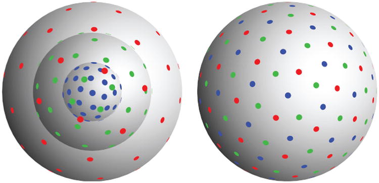
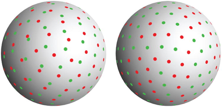
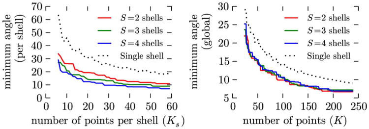
Methods: The design of acquisition in multishell is still an open and active field of research, however. In this work, we provide a general method to design multishell acquisition with uniform angular coverage. This method is based on a generalization of electrostatic repulsion to multishell.

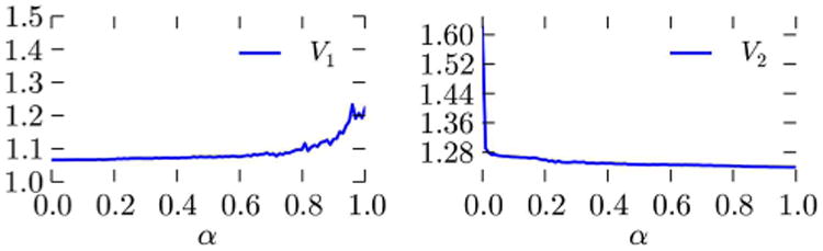
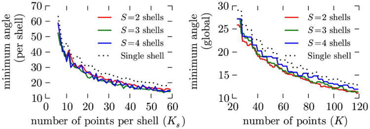
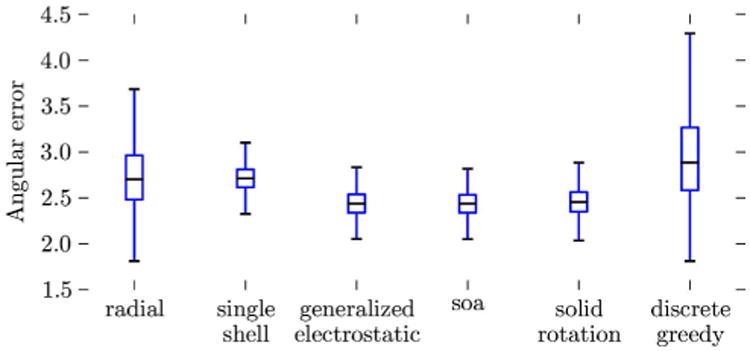
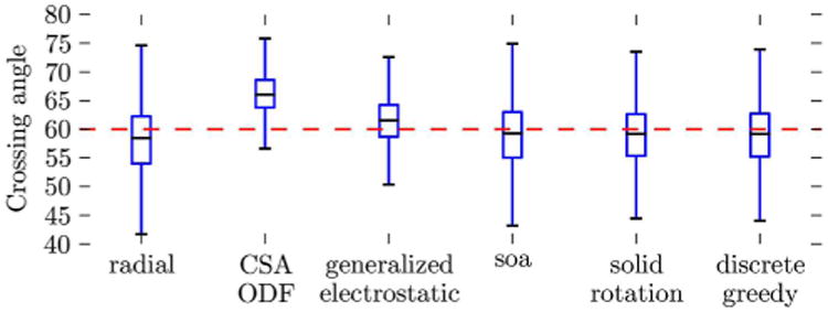
Results: We evaluate the impact of our method using simulations, on the angular resolution in one and two bundles of fiber configurations. Compared to more commonly used radial sampling, we show that our method improves the angular resolution, as well as fiber crossing discrimination.

Discussion: We propose a novel method to design sampling schemes with optimal angular coverage and show the positive impact on angular resolution in diffusion MRI.


Figures

Fig. 1
Fig. 1
Optimal point set on three shells, 30 points per shell. The weighting parameter is set to α = 0.5. (Left) sample points in the q-space, (right) sampling directions, i.e., the sampling points reprojected onto the unit sphere.
Fig. 2
Fig. 2
Extremal values of the weighting factor α: minimum cost configurations for S = 2 shells, K1 = K2 = 50 points per shell, points reprojected on the unit sphere. (Left) α = 0: all the importance is given to the uniformity of the configuration on each shell, independently of the other. As a result, the global coverage is nonuniform, and so we get almost “aligned” sampling on both shells. (Right) α = 1: the global angular coverage is uniform, but if we consider the repartition of points on shell 1 (green dots) or shell 2 (red dots) separately, the angular coverage is not uniform.
Fig. 3
Fig. 3
(Left) Cost function V1, measuring uniformity on each shell, and (right) V2, measuring uniformity of the set of directions as a whole. Cost function evaluated for the optimum configurations, for various α. Except near 0 and 1, the solution does not depend strongly on the choice of α. [Color figure can be viewed in the online issue, which is available at wileyonlinelibrary.com.]
Fig. 4
Fig. 4
Minimum angular distance (◦) between any two points (left) within the same shell and (right) globally. We compare the sets generated using generalized electrostatic analogy, for S = 2–4 shells, to the optimum electrostatic point sets in one shell. [Color figure can be viewed in the online issue, which is available at wileyonlinelibrary.com.]
Fig. 5
Fig. 5
Geometrical properties of the incrementally constructed point sets on S shells. Minimum angular distance (◦) between any two points (left) within the same shell and (right) globally. We compare the incremental sets generated using generalized electrostatic analogy, for S = 2–4 shells, to the optimum electrostatic point sets in one shell. [Color figure can be viewed in the online issue, which is available at wileyonlinelibrary.com.]
Fig. 6
Fig. 6
Average angular error between the principal direction of the estimated tensor and the axis of the cylinder. Signal simulation and reconstruction were repeated with 2000 different cylinder orientations following a random uniform distribution on the sphere, and 100 repetitions with random, Rician noise for each orientation. We plot here the median, first and third quartiles, and extremal values of the angular error (in degrees). The height of each boxplot represents how rotationally invariant each method performs. See Table 2 for quantitative results. [Color figure can be viewed in the online issue, which is available at wileyonlinelibrary.com.]
Fig. 7
Fig. 7
Crossing angle for a reconstructed fiber crossing. The q-space signal was reconstruction in the mSPF basis, with Laplace regularization (see Ref. 23), fixed regularization weight λ = 0.5. The ODF was further estimated using the SPFI method in Ref. 38. The boxplot represents min and max values, as well as the first and third quartiles. We also compare to the ODF reconstructed in constant solid angle (19), for a single shell experiment with the same number of points K = 100. See also Table 3. [Color figure can be viewed in the online issue, which is available at wileyonlinelibrary.com.]

Similar articles

Cited by

References

    1. Le Bihan D, Breton E. Imagerie De diffusion it in vivo par rsonnance magntique nuclaire. CR Acad Sci. 1985;301:1109–1112.
    1. Taylor D, Bushell M. The spatial mapping of translational diffusion coefficients by the NMR imaging technique. Phys Med Biol. 1985;30:345–349. - PubMed
    1. Merboldt K, Hanicke W, Frahm J. Self-diffusion NMR imaging using stimulated echoes. J Magn Reson. 1985;64:479–486.
    1. Jones DK, editor. Diffusion MRI: theory, methods, and applications. New York: Oxford University Press; 2010.
    1. Johansen-Berg H, Behrens TE, editors. Diffusion MRI: from quantitative measurement to in-vivo neuroanatomy. London: Academic Press; 2009.

Publication types

MeSH terms