

Relationships between regional cerebellar volume and sensorimotor and cognitive function in young and older adults
- PMID: 23625382
- PMCID: PMC3820158
- DOI: 10.1007/s12311-013-0481-z
Relationships between regional cerebellar volume and sensorimotor and cognitive function in young and older adults
Abstract
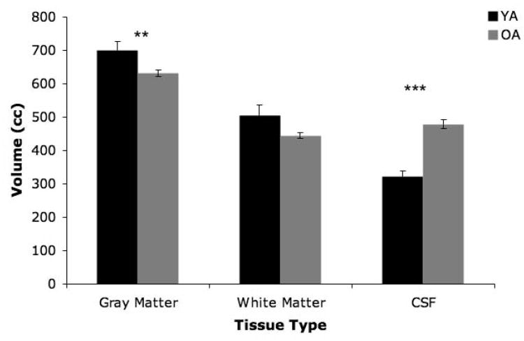
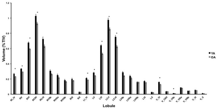
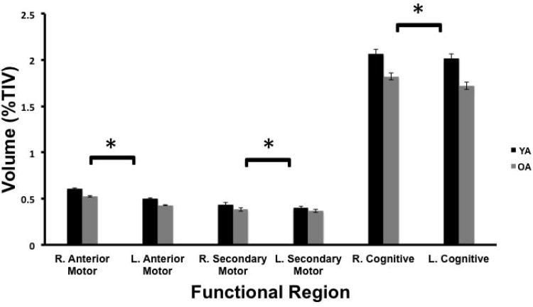
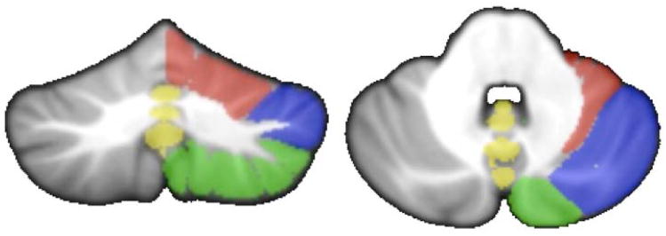
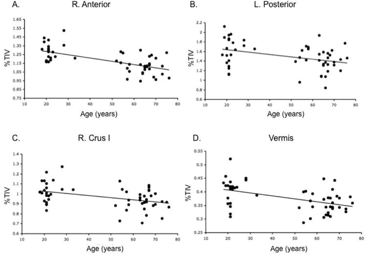
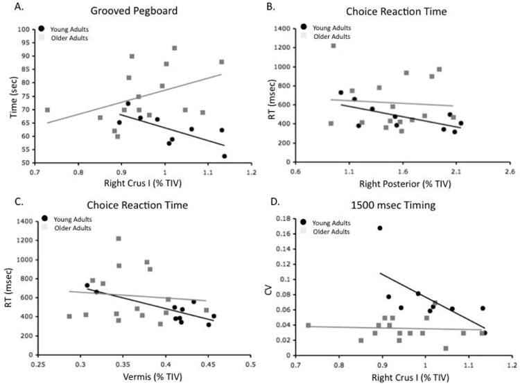
The cerebellum has been implicated in both sensorimotor and cognitive function, but is known to undergo volumetric declines with advanced age. Individual differences in regional cerebellar volume may therefore provide insight into performance variability across the lifespan, as has been shown with other brain structures and behaviors. Here, we investigated whether there are regional age differences in cerebellar volume in young and older adults, and whether these volumes explain, in part, individual differences in sensorimotor and cognitive task performance. We found that older adults had smaller cerebellar volume than young adults; specifically, lobules in the anterior cerebellum were more impacted by age. Multiple regression analyses for both age groups revealed associations between sensorimotor task performance in several domains (balance, choice reaction time, and timing) and regional cerebellar volume. There were also relationships with working memory, but none with measures of general cognitive or executive function. Follow-up analyses revealed several differential relationships with age between regional volume and sensorimotor performance. These relationships were predominantly selective to cerebellar regions that have been implicated in cognitive functions. Therefore, it may be the cognitive aspects of sensorimotor task performance that are best explained by individual differences in regional cerebellar volumes. In sum, our results demonstrate the importance of regional cerebellar volume with respect to both sensorimotor and cognitive performance, and we provide additional insight into the role of the cerebellum in age-related performance declines.
Conflict of interest statement
Figures
Similar articles
-
Regional cerebellar volume and cognitive function from adolescence to late middle age.Hum Brain Mapp. 2015 Mar;36(3):1102-20. doi: 10.1002/hbm.22690. Epub 2014 Nov 13. Hum Brain Mapp. 2015. PMID: 25395058 Free PMC article.
-
Altered cortical-cerebellar circuits during verbal working memory in essential tremor.Brain. 2011 Aug;134(Pt 8):2274-86. doi: 10.1093/brain/awr164. Epub 2011 Jul 11. Brain. 2011. PMID: 21747127
-
Volumetric variation in subregions of the cerebellum correlates with working memory performance.Neurosci Lett. 2012 Feb 2;508(1):47-51. doi: 10.1016/j.neulet.2011.12.016. Epub 2011 Dec 22. Neurosci Lett. 2012. PMID: 22206834
-
The cerebellum and cognition: evidence from functional imaging studies.Cerebellum. 2012 Jun;11(2):352-65. doi: 10.1007/s12311-011-0260-7. Cerebellum. 2012. PMID: 21373864 Review.
-
Cerebellar-Subcortical-Cortical Systems as Modulators of Cognitive Functions.Neuropsychol Rev. 2021 Sep;31(3):422-446. doi: 10.1007/s11065-020-09465-1. Epub 2021 Jan 29. Neuropsychol Rev. 2021. PMID: 33515170 Review.
Cited by
-
The Impact of 6 and 12 Months in Space on Human Brain Structure and Intracranial Fluid Shifts.Cereb Cortex Commun. 2020;1(1):tgaa023. doi: 10.1093/texcom/tgaa023. Epub 2020 Jun 15. Cereb Cortex Commun. 2020. PMID: 32864615 Free PMC article.
-
Regional cerebellar volume and cognitive function from adolescence to late middle age.Hum Brain Mapp. 2015 Mar;36(3):1102-20. doi: 10.1002/hbm.22690. Epub 2014 Nov 13. Hum Brain Mapp. 2015. PMID: 25395058 Free PMC article.
-
Cerebellar-motor dysfunction in schizophrenia and psychosis-risk: the importance of regional cerebellar analysis approaches.Front Psychiatry. 2014 Nov 25;5:160. doi: 10.3389/fpsyt.2014.00160. eCollection 2014. Front Psychiatry. 2014. PMID: 25505424 Free PMC article. Review.
-
Bimanual coordinated motor skill learning in patients with a chronic cerebellar stroke.Exp Brain Res. 2024 Jun;242(6):1517-1531. doi: 10.1007/s00221-024-06830-x. Epub 2024 May 9. Exp Brain Res. 2024. PMID: 38722346
-
Sex Differences and Behavioral Associations with Typically Developing Pediatric Regional Cerebellar Gray Matter Volume.Cerebellum. 2024 Apr;23(2):589-600. doi: 10.1007/s12311-023-01569-7. Epub 2023 Jun 29. Cerebellum. 2024. PMID: 37382829 Free PMC article.
References
-
- Kanai R, Rees G. The structural basis of inter-individual differences in human behaviour and cognition. Nat Rev Neurosci. 2011;12:231–242. - PubMed
-
- Morton SM, Bastian AJ. Relative contributions of balance and voluntary leg-coordination deficits to cerebellar gait ataxia. J Neurophysiol. 2003;89:1844–1856. - PubMed
-
- Morton SM, Bastian AJ. Cerebellar control of balance and locomotion. Neuroscientist. 2004;10:247–259. - PubMed
-
- Schmahmann JD, Sherman JC. The cerebellar cognitive affective syndrome. Brain. 1998;121:561–579. - PubMed
Publication types
MeSH terms
Grants and funding
LinkOut - more resources
Full Text Sources
Other Literature Sources
