. 2013 Jul;195(13):3022-34.
doi: 10.1128/JB.00287-13. Epub 2013 Apr 26.

A putative transmembrane leucine zipper of agrobacterium VirB10 is essential for t-pilus biogenesis but not type IV secretion

Affiliations

A putative transmembrane leucine zipper of agrobacterium VirB10 is essential for t-pilus biogenesis but not type IV secretion

Isaac Garza et al. J Bacteriol. 2013 Jul.

Abstract

The Agrobacterium tumefaciens VirB/VirD4 type IV secretion system is composed of a translocation channel and an extracellular T pilus. Bitopic VirB10, the VirB7 lipoprotein, and VirB9 interact to form a cell envelope-spanning structural scaffold termed the "core complex" that is required for the assembly of both structures. The related pKM101-encoded core complex is composed of 14 copies each of these VirB homologs, and the transmembrane (TM) α helices of VirB10-like TraF form a 55-Å-diameter ring at the inner membrane. Here, we report that the VirB10 TM helix possesses two types of putative dimerization motifs, a GxxxA (GA4) motif and two leucine (Leu1, Leu2) zippers. Mutations in the Leu1 motif disrupted T-pilus biogenesis, but these or other mutations in the GA4 or Leu2 motif did not abolish substrate transfer. Replacement of the VirB10 TM domain with a nondimerizing poly-Leu/Ala TM domain sequence also blocked pilus production but not substrate transfer or formation of immunoprecipitable complexes with the core subunits VirB7 and VirB9 and the substrate receptor VirD4. The VirB10 TM helix formed weak homodimers in Escherichia coli, as determined with the TOXCAT assay, whereas replacement of the VirB10 TM helix with the strongly dimerizing TM helix from glycophorin A blocked T-pilus biogenesis in A. tumefaciens. Our findings support a model in which VirB10's TM helix contributes to the assembly or activity of the translocation channel as a weakly self-interacting membrane anchor but establishes a heteromeric TM-TM helix interaction via its Leu1 motif that is critical for T-pilus biogenesis.


Figures

Fig 1
Fig 1
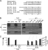
Effects of GA4 and Leu motif mutations on VirB10 function. (A) Schematics showing VirB10 domains, the TM domain sequence, and putative GA4 and Leu dimerization motifs. Domains: Cyto, cytoplasmic; PRR, proline rich region; Linker, region between the PRR and the β-barrel domain; Barrel/AP, C-terminal β-barrel domain positioned near the OM/AP and extending across the OM. Numbers in the schematic and above the TM domain sequence correspond to residues relative to VirB10's N terminus. The extended GA4 motif is depicted with the GA4 residues in bold and underlined. The Leu1 motif is in bold and underlined, and the Leu2 motif is in plain letters. The helical-wheel diagram at the right depicts the VirB10 TM domain sequence and the putative Leu1 and Leu2 zipper motifs. (B) T-pilus production by nonpolar ΔvirB10 (ΔB10) mutant strain PC1010 lacking or producing native (B10) or the GA4 or Leu motif mutant proteins shown. Top to bottom: B2 surface, colony immunoblot assays developed with anti-VirB2 antibodies; B2 shear, VirB2 comprising the T pilus detected in extracellular shear fractions; B2 cellular, VirB2 pilin detected in total cell lysates; B10, VirB10 detected in total cell lysates by immunostaining with anti-VirB10 antibodies. (C) Effects of mutations on T-DNA transfer as monitored by virulence on wounded Kalanchoe leaves (black bars; −, avirulent; +++, WT virulence) and transfer of the mobilizable IncQ plasmid pML122 to A. tumefaciens recipients (gray bars; Tc's/D, number of transconjugants per donor cell). For IncQ plasmid transfer, results of a single experiment with standard deviations from triplicate matings are presented.
Fig 2
Fig 2
Effects of TM domain swaps on VirB10 function. (A) Alignment of TM domain sequences of native VirB10 (B10) and mutant TM domains bearing replacements of FtsN′s TM domain sequence and synthetic pLA sequences. Substituted residues are in bold, residues at positions corresponding to the Leu1 motif are gray shaded, and the position of the Leu1 motif is underlined at the bottom. (B) T-pilus production by the ΔvirB10 (ΔB10) mutant producing native (B10) or mutant proteins with the TM domain residue substitutions indicated. Two chimeric proteins with substitutions of the FtsN TM domain or cytoplasmic and TM domains (Cyto/TM) were analyzed. Top to bottom: B2 surface, surface-exposed VirB2 pilin protein; B2 shear, pilin in shear fraction; B2 cellular, total cellular levels of pilin; B10, total cellular levels of VirB10. (C) Effects of mutations on T-DNA transfer as monitored by plant tumor production (black bars; −, avirulent; +++, WT virulence) and mobilization of IncQ plasmid pML122 to A. tumefaciens recipients (gray bars; Tc's/D, number of transconjugants per donor cell).
Fig 3
Fig 3
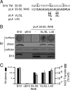
Effects of pLA TM domain sequences bearing substitutions of residues conserved among close VirB10 homologs on VirB10 function. (A) Alignment of the VirB10 TM domain and pLA (33-50, W48) sequences. Substituted residues are in bold, residues at positions corresponding to the Leu1 motif are gray shaded, and the position of the Leu1 motif is highlighted at the top. The two variant pLA sequences bearing the conserved VL/SL and Leu40 residues are indicated below. (B) T-pilus production by the ΔvirB10 (ΔB10) mutant producing native (B10) or pLA mutant proteins with the VL/SL or Leu40 (L40) substitutions indicated. Top to bottom: B2 surface, surface-exposed VirB2 pilin protein; B2 shear, pilin in shear fraction; B2 cellular, total cellular levels of pilin; B10, total cellular levels of VirB10. (C) Effects of mutations on T-DNA transfer as monitored by plant tumor production (black bars; −, avirulent; +++, WT virulence) and mobilization of IncQ plasmid pML122 to A. tumefaciens recipients (gray bars; Tc's/D, number of transconjugants per donor cell).
Fig 4
Fig 4
Effects of TM domain mutations on VirB10 partner interactions. Extracts from DSP-treated and detergent-solubilized cells were immunoprecipitated with anti-VirB10 antibodies, and precipitates were analyzed by immunoblot development with antibodies specific to VirB10, VirD4, VirB9, and VirB7. Strains: PC1010 lacking (ΔB10) or producing native VirB10 (B10) or the TM domain mutants listed; Leu1, L33A L40A I47A variant (see Fig. 1); FtsN and pLA 33-50, W48 TM domain swaps (see Fig. 3); ΔN30 and ΔN46, VirB10 N-terminal truncations; FtsN Cyto/TM, VirB10 chimera with cytoplasmic and TM domains from FtsN.
Fig 5
Fig 5
Effects of TM domain mutations on release of VirE2 to the cell surface. Surface: extracellular FLAG-VirE2 (FL-E2) was assessed by colony immunoblotting with anti-FLAG antibodies. Cellular: total cellular levels of FLAG-VirE2 and native and mutant forms of VirB10 (B10) were monitored by immunoblot development with anti-FLAG or anti-VirB10 antibodies. Strains: PC1010 (ΔB10) producing native VirB10 (B10), the substrate-leaky mutant (G272R), or the VirB10 TM domain mutant proteins listed.
Fig 6
Fig 6
Oxidative cross-linking of Cys-containing VirB10 derivatives. PC1010 lacking (ΔB10) or producing native VirB10 (B10) or derivatives with the mutations indicated were left untreated (−) or treated with Cu-OP (+). Total cell extracts were prepared and electrophoresed through gels in the absence of reductant, and VirB10 species were detected by immunostaining with anti-VirB10 antibodies. The i2 mutants carry Ala-Cys insertions immediately after the residue number indicated. The positions of VirB10 monomer (B10) and putative dimer (B10-B10) species are indicated at the right. MW, molecular mass markers with sizes (in kilodaltons) listed at the left.
Fig 7
Fig 7
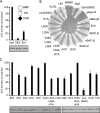
Self-association of native and mutant VirB10 TM domains as quantitated by TOXCAT assay. (A) Schematic of the MBP-TM-ToxR fusion protein. TM-mediated dimerization activates the ToxR-dependent ctx promoter controlling cat gene expression; total cellular CAT activity reflects the strength of TM domain self-association. Histogram depicts CAT activities normalized to that of the reporter strain for the strongly dimerizing GpA TM domain sequence. (B) Growth of MM39 cells expressing ToxR-TM-MBP proteins on maltose minimal medium. pMal-c2 producing a cytoplasmic form of MBP and pMal-p2 producing a periplasmic form of MBP served as negative and positive controls, respectively. (C) Histogram depicting CAT activities of reporter strains for the mutant TM domain sequences normalized to the CAT activity of the reporter strain for the native VirB10 TM domain. For panels A and C, the steady-state abundance of the fusion proteins was assessed by immunoblot development with anti-MBP antibodies.
Fig 8
Fig 8
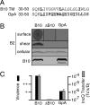
Effect of a GpA TM domain swap on VirB10 function (see Fig. 3). (A) Alignment of TM domain sequences of native VirB10 (B10) and the GpA TM domain variant. Substituted residues are in bold, residues at positions corresponding to the Leu1 motif are gray shaded, and the position of the extended GA4 motif is underlined at the bottom. (B) T-pilus production by the ΔvirB10 (ΔB10) mutant producing the native (B10) or VirB10-GpATM mutant protein. Top to bottom: B2 surface, surface-exposed VirB2 pilin protein; B2 shear, pilin in shear fraction; B2 cellular, total cellular levels of pilin; B10, total cellular levels of VirB10. (C) Effects of the GpATM swap on substrate transfer as monitored by plant tumor production (black bars; −, avirulent; +++, WT virulence) and mobilization of IncQ plasmid pML122 to A. tumefaciens recipients (gray bars; Tc's/D, number of transconjugants per donor cell).

Similar articles

Cited by

References

    1. Alvarez-Martinez CE, Christie PJ. 2009. Biological diversity of prokaryotic type IV secretion systems. Microbiol. Mol. Biol. Rev. 73:775–808 - PMC - PubMed
    1. Fronzes R, Schafer E, Wang L, Saibil HR, Orlova EV, Waksman G. 2009. Structure of a type IV secretion system core complex. Science 323:266–268 - PMC - PubMed
    1. Chandran V, Fronzes R, Duquerroy S, Cronin N, Navaza J, Waksman G. 2009. Structure of the outer membrane complex of a type IV secretion system. Nature 462:1011–1015 - PMC - PubMed
    1. Walldén K, Williams R, Yan J, Lian PW, Wang L, Thalassinos K, Orlova EV, Waksman G. 2012. Structure of the VirB4 ATPase, alone and bound to the core complex of a type IV secretion system. Proc. Natl. Acad. Sci. U. S. A. 109:11348–11353 - PMC - PubMed
    1. Christie PJ. 2009. Structural biology: translocation chamber's secrets. Nature 462:992–994 - PMC - PubMed

Publication types

MeSH terms

LinkOut - more resources