. 2013 Jun 7;288(23):17030-17041.
doi: 10.1074/jbc.M112.411272. Epub 2013 Apr 26.

Store-operated Ca2+ entry (SOCE) induced by protease-activated receptor-1 mediates STIM1 protein phosphorylation to inhibit SOCE in endothelial cells through AMP-activated protein kinase and p38β mitogen-activated protein kinase

Affiliations

Store-operated Ca2+ entry (SOCE) induced by protease-activated receptor-1 mediates STIM1 protein phosphorylation to inhibit SOCE in endothelial cells through AMP-activated protein kinase and p38β mitogen-activated protein kinase

Premanand C Sundivakkam et al. J Biol Chem. .

Abstract

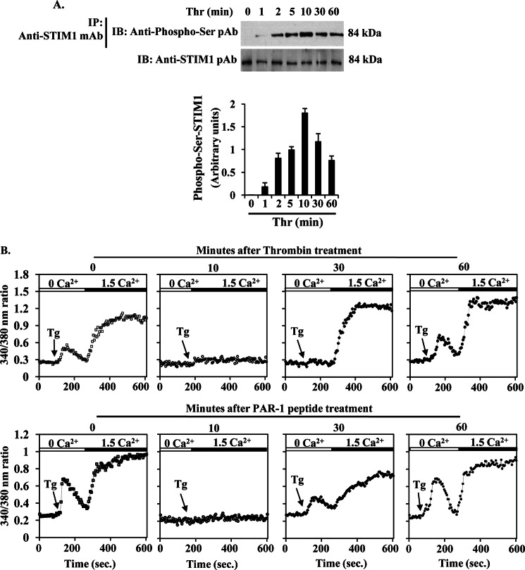
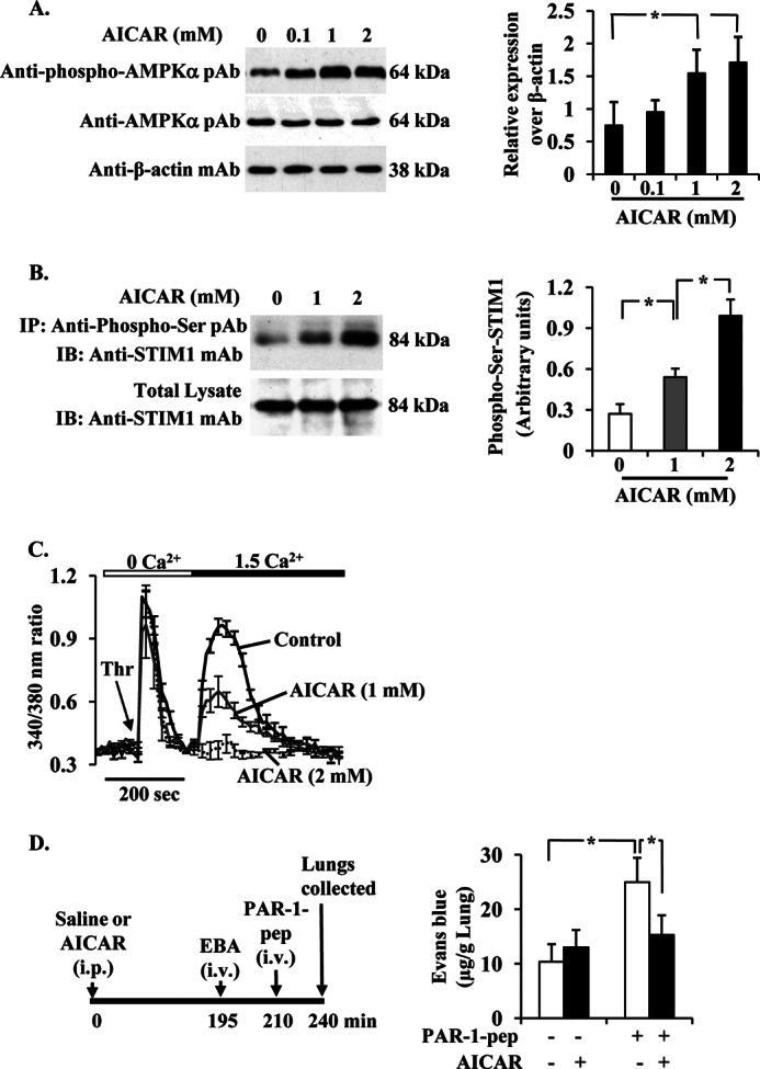
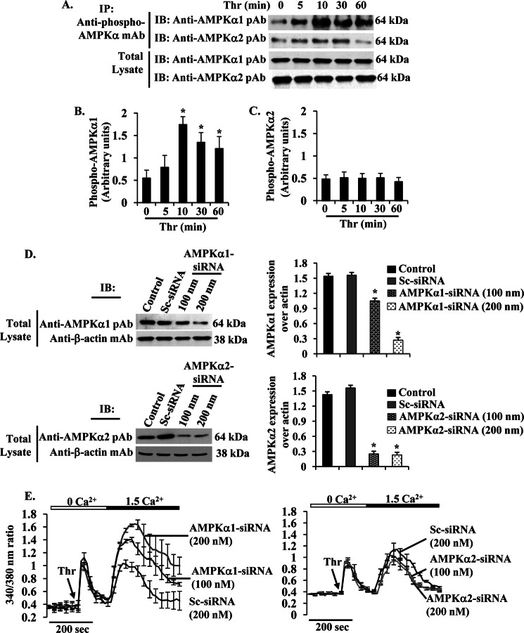
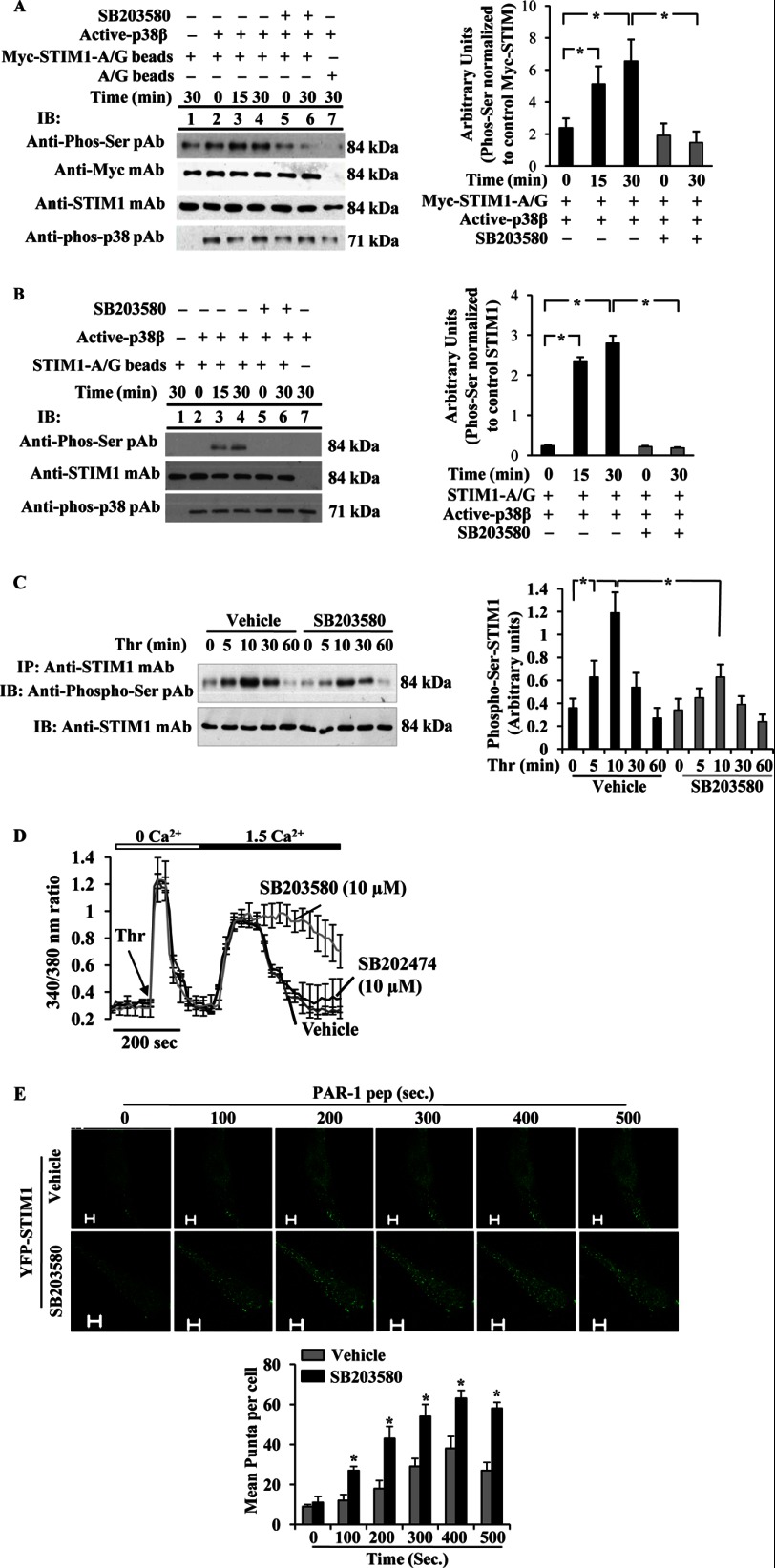
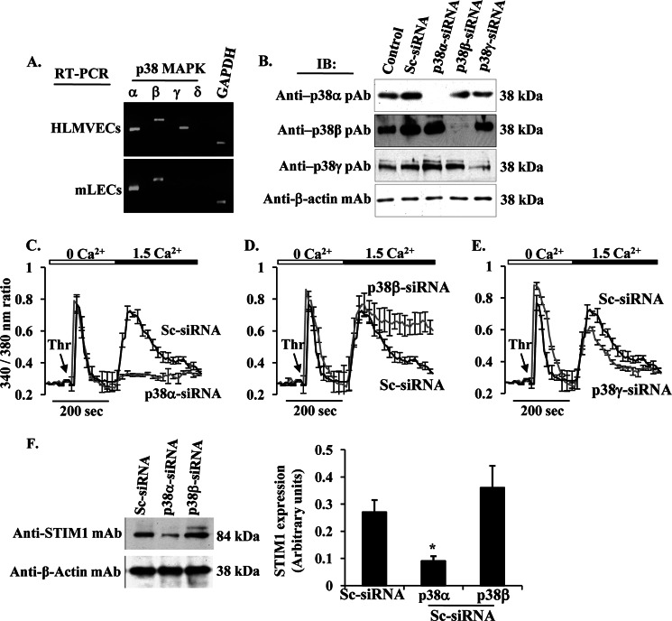
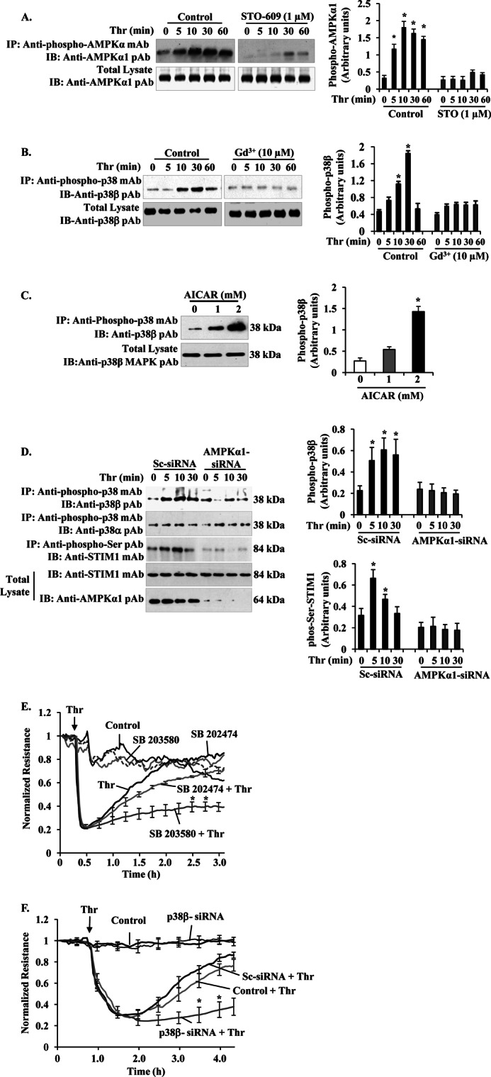
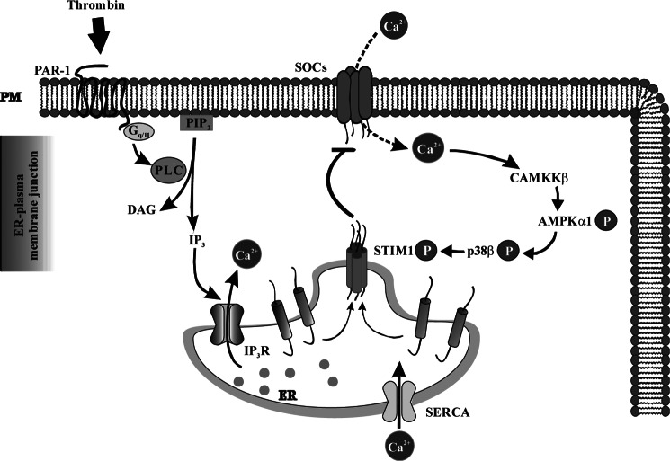
The Ca(2+) sensor STIM1 is crucial for activation of store-operated Ca(2+) entry (SOCE) through transient receptor potential canonical and Orai channels. STIM1 phosphorylation serves as an "off switch" for SOCE. However, the signaling pathway for STIM1 phosphorylation is unknown. Here, we show that SOCE activates AMP-activated protein kinase (AMPK); its effector p38β mitogen-activated protein kinase (p38β MAPK) phosphorylates STIM1, thus inhibiting SOCE in human lung microvascular endothelial cells. Activation of AMPK using 5-aminoimidazole-4-carboxamide-1-β-d-ribofuranoside (AICAR) resulted in STIM1 phosphorylation on serine residues and prevented protease-activated receptor-1 (PAR-1)-induced Ca(2+) entry. Furthermore, AICAR pretreatment blocked PAR-1-induced increase in the permeability of mouse lung microvessels. Activation of SOCE with thrombin caused phosphorylation of isoform α1 but not α2 of the AMPK catalytic subunit. Moreover, knockdown of AMPKα1 augmented SOCE induced by thrombin. Interestingly, SB203580, a selective inhibitor of p38 MAPK, blocked STIM1 phosphorylation and led to sustained STIM1-puncta formation and Ca(2+) entry. Of the three p38 MAPK isoforms expressed in endothelial cells, p38β knockdown prevented PAR-1-mediated STIM1 phosphorylation and potentiated SOCE. In addition, inhibition of the SOCE downstream target CaM kinase kinase β (CaMKKβ) or knockdown of AMPKα1 suppressed PAR-1-mediated phosphorylation of p38β and hence STIM1. Thus, our findings demonstrate that SOCE activates CaMKKβ-AMPKα1-p38β MAPK signaling to phosphorylate STIM1, thereby suppressing endothelial SOCE and permeability responses.

Keywords: AMP-activated Kinase (AMPK); Calcium Channels; Endothelial Dysfunction; Phosphorylation; Protease-activated Receptor-1; Store-operated Calcium Entry; Stromal Interaction Molecule-1; Thrombin; Vascular Permeability; p38 MAPK.


Figures

FIGURE 1.
FIGURE 1.
STIM1 phosphorylation inhibits SOCE in endothelial cells. A, thrombin-induced STIM1 phosphorylation was measured. HLMVECs exposed to thrombin (50 nm) for different time periods were lysed; lysates were immunoprecipitated (IP) with STIM1 mAb, and the immunoprecipitate was immunoblotted (IB) with anti-phospho-Ser pAb (top row). Total cell lysates were immunoblotted with anti-STIM1 mAb (bottom row). Phosphoprotein bands were quantified by densitometry and expressed as relative to control (bottom panel). Results shown are mean ± S.E. of four experiments. B, HLMVECs were preincubated either with thrombin (50 nm) (top panel) or PAR-1 peptide (40 μm) (bottom panel) for different time periods. After the specified time points, cells were used to measure TG (1 μm)-induced store Ca2+ release and Ca2+ entry (see details under “Experimental Procedures”). The experiment was repeated three times with similar results.
FIGURE 2.
FIGURE 2.
AMPK activation abrogates PAR-1-induced Ca2+ entry in ECs and lung microvessel permeability. A, AICAR induces phosphorylation of AMPKα. HLMVECs grown to 80% confluence were pretreated with AICAR (0, 0.1, 1, and 2 mm) for 2 h in 1% serum-containing medium. Cells were then lysed and immunoblotted with anti-phospho-AMPKα mAb (top), anti-AMPKα pAb (middle), and anti-β-actin mAb. A representative blot is shown from four independent experiments. The protein bands were quantified by densitometry relative to β-actin (right panel). *, significantly different from cells not treated with AICAR. B, AICAR induces STIM1 phosphorylation. HLMVECs exposed to the indicated concentrations of AICAR for 2 h as above. Cells were lysed; lysates were immunoprecipitated (IP) with anti-phospho-Ser pAb, and then the precipitate was immunoblotted (IB) with anti-STIM1 mAb. Results shown are from representative of four experiments. The quantified results are shown in the right panel. *, significantly different from control. C, AICAR inhibits thrombin-induced Ca2+ entry. HLMVECs grown on coverslips and pretreated with AICAR (1 or 2 mm) for 2 h were used to measure Ca2+ entry. Fura-2-loaded cells placed in Ca2+- and Mg2+-free HBSS were stimulated with thrombin (50 nm). After return of [Ca2+]i to base-line levels, CaCl2 (1.5 mm) was added to extracellular medium to induce Ca2+ entry. Arrow indicates time at which cells were stimulated with thrombin (Thr). Results shown are mean ± S.E. of four experiments. D, AICAR pretreatment abrogates PAR-1-induced lung vascular permeability increase. C57BL/6 mice either injected with AICAR (500 mg/kg, intraperitoneally) or saline were used to measure PAR-1-peptide-induced EBA uptake in lungs. Top panel shows experimental design. Results are mean ± S.E. of changes in lung EBA after PAR-1 agonist peptide administration (n = 6; in each group). * indicates the significance between the treatment groups and the respective control groups (p < 0.05).
FIGURE 3.
FIGURE 3.
PAR-1-activated AMPKα1 regulates SOCE in HLMVECs. A, HLMVECs were challenged with thrombin (50 nm) for different time intervals at 37 °C. After thrombin stimulation, cells were lysed and immunoprecipitated (IP) with anti-phospho-AMPKα mAb. The precipitated proteins were immunoblotted (IB) with anti-AMPKα1 pAb (1st lane) or AMPKα2 pAb (2nd lane). Total cell lysates were immunoblotted with anti-AMPKα1 pAb (3rd lane) or AMPKα2 pAb (4th lane). Results are shown as the mean ± S.E. of four independent experiments for AMPKα1 (B) or AMPKα2 (C). * indicates the significance compared with cells not treated with thrombin. D, HLMVECs transfected with Sc-siRNA, AMPKα1-siRNA, or AMPKα1-siRNA (see details under “Experimental Procedures”) were lysed and immunoblotted with anti-AMPKα1 pAb (left, top panel) or anti-AMPKα2 pAb (left, bottom panel). The membrane was stripped and probed with anti-β-actin mAb as loading control. In right panels, AMPKα1 and AMPKα2 proteins were quantified by densitometry relative to β-actin. Results shown are mean ± S.E. of four experiments. *, significantly different compared with control or Sc-siRNA transfected cells. E, HLMVECs transfected with Sc-siRNA, AMPKα1-siRNA (100 or 200 nm), or AMPKα2-siRNA (100 or 200 nm) were used to measure thrombin (Thr)-induced Ca2+ store release and Ca2+ entry as described in Fig. 2C. Arrow indicates the time when the cells are challenged with thrombin (Thr). Results shown are mean ± S.E. of four experiments.
FIGURE 4.
FIGURE 4.
p38 MAPK downstream of AMPK signaling controls SOCE via phosphorylation of STIM1. A and B, phosphorylation of STIM1 by active p38β2 was determined using in vitro kinase assay (see details under “Experimental Procedures”). A, Myc-STIM1 was ectopically expressed in HEK293 cells and immunoprecipitated (IP) using anti-Myc; mAb was used as substrate for active p38β2. The assay was performed in the presence (+) and absence (−) of p38 inhibitor, SB203580 (10 μm). Lane 1, active p38β2 was not included in the kinase mixture; lane 7, control A/G beads were incubated with Myc-STIM1 expressing HEK cell lysates included in the kinase assay mixture loaded. Equal volume of assay mixture was immunoblotted (IB) with anti-phospho-Ser pAb, anti-Myc pAb, anti-STIM1 mAb, or anti-phospho-p38 pAb (left panel). Phosphoprotein bands were quantified by densitometry and expressed as relative to Myc-STIM1 (right panel). Results shown are mean ± S.E. of three independent experiments. *, significantly different from SB203580 treatment. B, unstimulated HLMVECs were immunoprecipitated using anti-STIM1 pAb, and the immunoprecipitate was used as substrate for active p38β2. The assay was performed as above. Equal volume of assay mixture was immunoblotted with anti-phospho-Ser pAb, anti-STIM1 mAb, or anti-phospho-p38 pAb (left panel). Results shown are mean ± S.E. of four independent experiments (right panel). *, significantly different from SB203580 treatment. Note that active p38β2-mediated STIM1 phosphorylation was detectable by anti-phospho-Ser pAb. C, HLMVECs pretreated with vehicle (DMSO, 0.01%) or SB203580 (10 μm) for 30 min were used to measure thrombin-induced phosphorylation of STIM1 as above in Fig. 1A. Phosphoprotein bands were quantified by densitometry and expressed as relative to control (right panel). Results shown are mean ± S.E. of three experiments. *, significantly different from cells not stimulated with thrombin or significant difference between control and SB203580-treated cells. D, HLMVECs pretreated with SB203580 (10 μm) or SB202474 (10 μm) were used to measure thrombin-induced Ca2+ entry as described above. Arrow indicates time of addition of thrombin (Thr). Results shown are mean ± S.E. of four independent experiments. E, HLMVECs grown to ∼70% confluence on glass-bottomed 35-mm dishes were transfected with YFP-WT-STIM1 expression construct. At 48 h after transfection, cells pretreated with SB203580 (10 μm) for 30 min were washed and placed in HBSS, and then PAR-1 peptide-induced STIM1 puncta formation was monitored in real time using a confocal microscope. STIM1 puncta were seen both in vehicle or SB203580-pretreated cells, whereas an increase or sustained puncta formation was observed in SB203580-treated cells. The images acquired from representative experiments are shown (top panels). STIM1 puncta formed after PAR-1 peptide addition was quantified, and results shown are mean ± S.E. (bottom panel). n = 4 from each group; *, significantly different from vehicle.
FIGURE 5.
FIGURE 5.
p38β MAPK regulates SOCE in HLMVECs. A, RT-PCR analysis of mRNA expression for p38 MAPK isoforms in HLMVECs and mLECs. Total RNA from HLMVECs and mLECs was isolated, and RT-PCR was performed to determine the expression of transcripts for p38 MAPK (α, β, γ, and δ) and GAPDH. B, HLMVECs were transfected with Sc-siRNA or siRNA specific to p38 MAPK isoforms (p38α, p38β, and p38γ). At 72 h after transfection, cells were used to determine expression of p38α, p38β, and p38γ by immunoblot. C–E, HLMVECs were transfected with Sc-siRNA or siRNA specific to p38α (C), β (D), or γ (E). At 72 h after transfection, cells were used to determine thrombin-induced Ca2+ entry as described above. Note the sustained Ca2+ entry in p38β-siRNA transfected cells (D), whereas in p38α-siRNA transfected cells the Ca2+ entry was blocked (C). Experiments were repeated at least three times, and the results shown are mean ± S.E. F, HLMVECs transfected with Sc-siRNA, p38α-siRNA, or p38β-siRNA were immunoblotted with anti-STIM1 mAb. Note that in p38α-siRNA-transfected cells STIM1 expression was reduced.
FIGURE 6.
FIGURE 6.
Ca2+ entry-CaMKKβ-AMPKα1-p38β MAPK axis signaling mediates STIM1 phosphorylation to inhibit SOCE and endothelial permeability. A, HLMVECs were pretreated with or without CaMKKβ inhibitor STO-609 (1 μm) for 30 min, and then thrombin-induced AMPKα1 phosphorylation was measured as described in Fig. 3A. The experiment was repeated four times, and the results shown are mean ± S.E. (right panel). *, significantly different from cells not stimulated with thrombin. B, HLMVECs, thrombin-induced p38β phosphorylation was measured in the absence and presence of Gd3+ (10 μm). After thrombin treatment, cell lysates were immunoprecipitated (IP) with anti-phospho-p38 mAb, and the precipitate was immunoblotted (IB) with anti-p38β pAb to determine p38β phosphorylation (top panel). Total cell lysates were blotted with anti-p38β mAb (bottom panel). A representative blot is shown from four independent experiments. Phosphoprotein bands were quantified by densitometry and are expressed in arbitrary units. *, significantly different from cells not stimulated with thrombin. Note that impaired thrombin-induced p38β phosphorylation in cells treated with Gd3+ to inhibit Ca2+ entry. C, HLMVECs pretreated with AICAR (0, 1, and 2 mm) were lysed and immunoprecipitated with anti-phospho-p38 mAb. The precipitated proteins were immunoblotted with anti-p38β MAPK pAb. Total cell lysates were immunoblotted with anti-p38β MAPK pAb. D, HLMVECs transfected with either Sc-siRNA or AMPKα1siRNA (200 nm) as described in Fig. 3D were stimulated with thrombin (50 nm) for different time intervals at 37 °C. After thrombin treatment, cells were lysed and immunoprecipitated (IP) with anti-phospho-p38 mAb. The precipitated proteins were immunoblotted (IB) with anti-p38β pAb (top row) or anti-p38α pAb (2nd row). Total cell lysates were immunoprecipitated with anti-phospho-Ser pAb, and the precipitate was immunoblotted with STIM1 mAb (3rd row). Total cell lysates were immunoblotted with anti-STIM1 mAb (4th row) and anti-AMPKα1 pAb (bottom row). Results shown are the mean ± S.E. of four independent experiments (right panels). *, significantly different from control cells. E, HLMVECs were grown to confluence on gold electrodes (see details under “Experimental Procedures”). Cells were washed and incubated with 1% serum-containing medium for 2 h and then incubated 30 min with or without the indicated concentrations of 10 μm SB203580 or SB202474 before the addition of 50 nm thrombin (Thr). Note that in SB203580-treated cells, thrombin produced a marked decrease in TER, but the TER recovery to basal level was delayed compared with control cells treated with thrombin or cells pretreated with SB202474 followed by thrombin addition. The arrow indicates the time at which the cells were challenged with thrombin (Thr) or medium. F, HLMVECs transfected with 200 nm Sc-siRNA or p38 β-siRNA were used to measure thrombin-induced TER changes. Note the delay in thrombin-induced decrease in TER recovery to basal level in p38β-siRNA transfected cells indicating hyper-permeability associated with prolonged SOCE. The arrow indicates the time at which the cells were challenged with thrombin (Thr) or medium. Results shown are the mean ± S.E. of four independent experiments. *, significantly different from control cells treated with thrombin.
FIGURE 7.
FIGURE 7.
Signaling pathway downstream of SOCE involved in turning off SOCE in endothelial cells. PAR-1-induced ER store Ca2+ depletion via phospholipase C (PLC)-inositol 1,4,5-triphosphate (IP3) activates SOCE. SOCE signal activates CaMKKβ-AMPKα1-p38β MAPK signal axis, which in turn phosphorylates STIM1 to terminate SOCE in endothelial cells. DAG, diacylglycerol.

Similar articles

Cited by

References

    1. Tiruppathi C., Ahmmed G. U., Vogel S. M., Malik A. B. (2006) Ca2+ signaling, TRP channels, and endothelial permeability. Microcirculation 13, 693–708 - PubMed
    1. Nilius B., Droogmans G. (2001) Ion channels and their functional role in vascular endothelium. Physiol. Rev. 81, 1415–1459 - PubMed
    1. Sundivakkam P. C., Freichel M., Singh V., Yuan J. P., Vogel S. M., Flockerzi V., Malik A. B., Tiruppathi C. (2012) The Ca2+ sensor stromal interaction molecule 1 (STIM1) is necessary and sufficient for the store-operated Ca2+ entry function of transient receptor potential canonical (TRPC) 1 and 4 channels in endothelial cells. Mol. Pharmacol. 81, 510–526 - PMC - PubMed
    1. Abdullaev I. F., Bisaillon J. M., Potier M., Gonzalez J. C., Motiani R. K., Trebak M. (2008) Stim1 and Orai1 mediate CRAC currents and store-operated calcium entry important for endothelial cell proliferation. Circ. Res. 103, 1289–1299 - PMC - PubMed
    1. Li J., Cubbon R. M., Wilson L. A., Amer M. S., McKeown L., Hou B., Majeed Y., Tumova S., Seymour V. A., Taylor H., Stacey M., O'Regan D., Foster R., Porter K. E., Kearney M. T., Beech D. J. (2011) Orai1 and CRAC channel dependence of VEGF-activated Ca2+ entry and endothelial tube formation. Circ. Res. 108, 1190–1198 - PMC - PubMed

Publication types

MeSH terms

LinkOut - more resources