. 2013 Jun 7;288(23):16348-16360.
doi: 10.1074/jbc.M113.474643. Epub 2013 Apr 26.

MicroRNA-33a mediates the regulation of high mobility group AT-hook 2 gene (HMGA2) by thyroid transcription factor 1 (TTF-1/NKX2-1)

Affiliations

MicroRNA-33a mediates the regulation of high mobility group AT-hook 2 gene (HMGA2) by thyroid transcription factor 1 (TTF-1/NKX2-1)

Shawn J Rice et al. J Biol Chem. .

Abstract

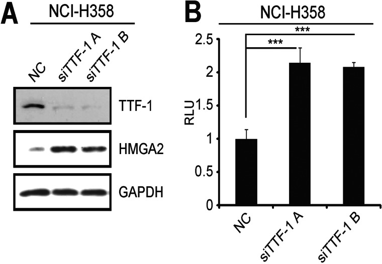
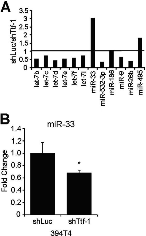
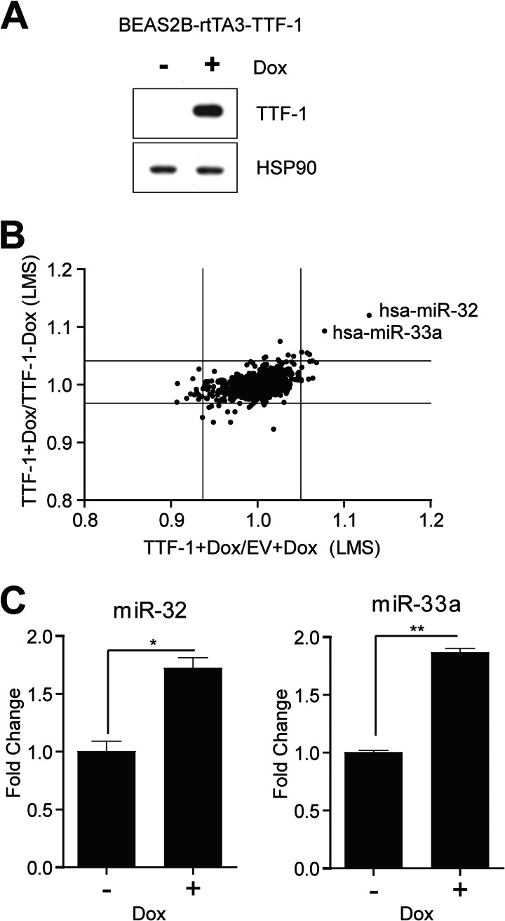
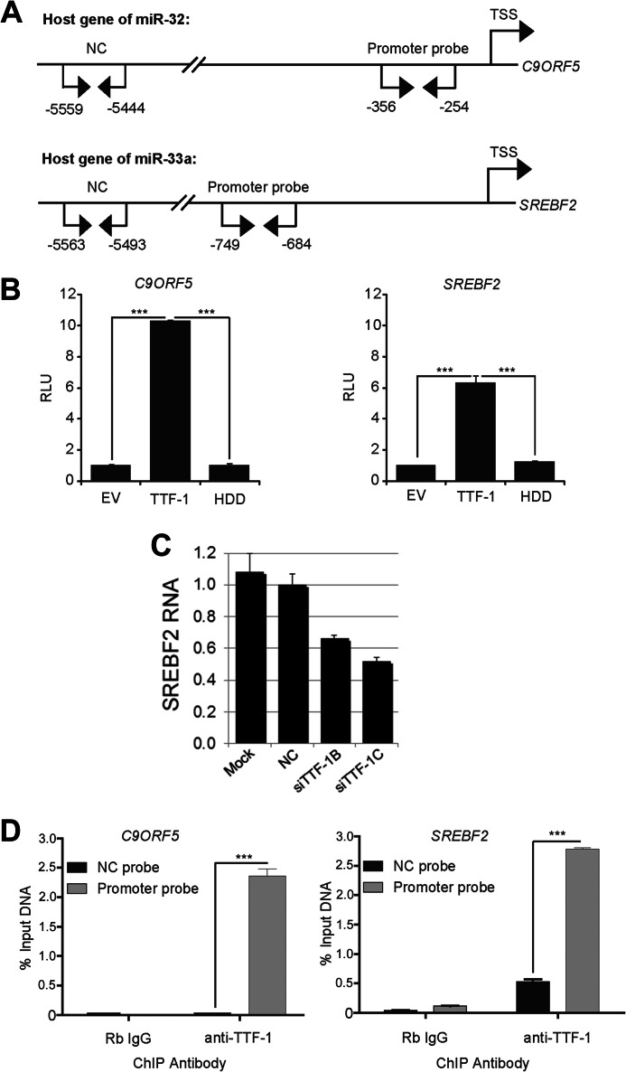
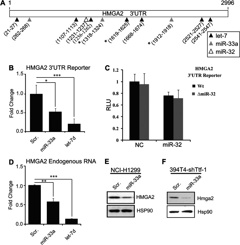
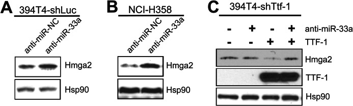
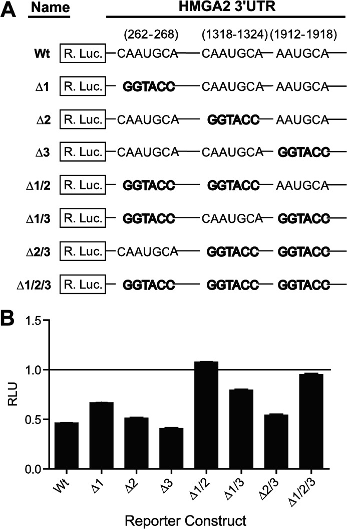
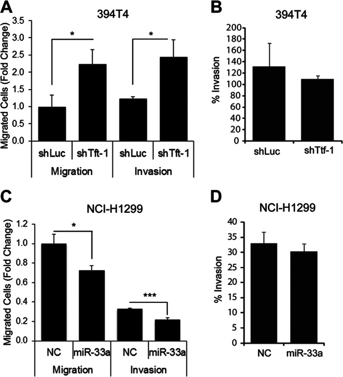
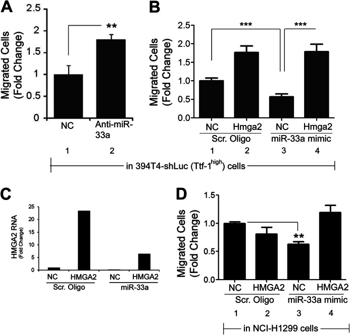
In lung cancers, TTF-1 displays seemingly paradoxical activities. Although TTF-1 is amplified in primary human lung cancers, it inhibits primary lung tumors from metastasizing in a mouse model system. It was reported that the oncogenic proepithelial mesenchymal transition (EMT) high mobility group AT-hook 2 gene (HMGA2) mediates the antimetastatic function of TTF-1. To gain mechanistic insight into the metastasis-critical signaling axis of TTF-1 to HMGA2, we used both reverse and forward strategies and discovered that microRNA-33a (miR-33a) is under direct positive regulation of TTF-1. By chromatin immunoprecipitation, we determined that TTF-1 binds to the promoter of SREBF2, the host gene of miR-33a. The 3'-untranslated region (UTR) of HMGA2 contains three predicted binding sites of miR-33a. We showed that the first two highly conserved sites are conducive to HMGA2 repression by miR-33a, establishing HMGA2 as a genuine target of miR-33a. Functional studies revealed that enforced expression of miR-33a inhibits the motility of lung cancer cells, and this inhibition can be rescued by overexpression of the form of HMGA2 without the 3'-UTR, suggesting that TTF-1 keeps the prometastasis gene HMGA2 in check via up-regulating miR-33a. This study reports the first miRNAs directly regulated by TTF-1 and clarifies how TTF-1 controls HMGA2 expression. Moreover, the documented importance of SREBF2 and miR-33a in regulating cholesterol metabolism suggests that TTF-1 may be a modulator of cholesterol homeostasis in the lung. Future studies will be dedicated to understanding how miRNAs influence the oncogenic activity of TTF-1 and the role of TTF-1 in cholesterol metabolism.

Keywords: Cholesterol Regulation; Gene Regulation; HMGA2; Lung Cancer; Metastasis; MicroRNA; SREBP2; TTF-1; miR-32; miR-33a.


Figures

FIGURE 1.
FIGURE 1.
TTF-1 negatively regulates HMGA2 in NCI-H358 cells. A, NCI-H358 cells were transfected with non-targeting RNA oligonucleotide (negative control; NC) or two separate siRNAs targeting TTF-1 (siTTF-1 A and B), and proteins were harvested after 72 h. Protein levels of TTF-1, HMGA2, and GAPDH (loading control) were determined by immunoblotting. B, NCI-H358 cells were transfected with a negative control or siTTF-1-targeting siRNAs, along with a psiCHECK2 vector containing the HMGA2 3′-UTR fused to the 3′-end of a Renilla luciferase gene. Luciferase assays were performed after 48 h (n = 3). RLU, relative luminescence units. Error bars, S.D. ***, p < 0.001.
FIGURE 2.
FIGURE 2.
Screening murine lung cancer cells for Ttf-1-regulated miRNAs that target HMGA2 using an LNA-QPCR array. A, total RNA isolated from 394T4-shLuc (Ttf-1high) and 394T4-shTtf-1 (Ttf-1low) cells was quantified in duplicate using an LNA-QPCR array designed to include miRNAs predicted to target the 3′-UTR of human HMGA2 gene. Normalized expression values between the two cell lines are expressed as -fold change of shLuc over shTtf-1 cells. B, down-regulation of mmu-miR-33 in 394T4-shTtf-1 cells was confirmed by RT-QPCR using an miRNA detection system and PCR primers (Quanta Biosciences) (n = 3). Error bars, S.D. *, p < 0.05.
FIGURE 3.
FIGURE 3.
A global screen for TTF-1-regulated miRNAs using a microarray platform. A, immortalized human lung epithelial cells with Dox-inducible TTF-1 elements (BEAS-2B-rtTA3-TTF1) were treated with or without dox (0.8 μg/ml) for 24 h prior to protein harvest. Western blot analysis confirmed the induction of TTF-1, with HSP90 protein as the loading control. B, RNA isolated from BEAS-2B-rtTA3-EV or TTF-1 cells treated with or without dox (0.8 μg/ml) for 24 h was evaluated in duplicates for changes in miRNA expression using an LNA-miRNA microarray (Exiqon). Expression of miRNAs between the different groups was compared by log median scores (LMS), and the average miRNA expression changes are graphed as indicated. Lines on the graph indicate the 1st and 99th percentile of the data. C, up-regulation of miR-32 and miR-33a in BEAS-2B-rtTA3-TTF-1 cells was confirmed using RT-QPCR (n = 3). Error bars, S.D. *, p < 0.05; **, p < 0.01.
FIGURE 4.
FIGURE 4.
TTF-1 binds to and activates transcription from the promoters of the miR-32 and miR-33a host genes. A, diagram of the promoter regions of the miR-32 and miR-33a host genes, C9Orf5 and SREBF2, respectively. Locations for negative control (NC) and promoter PCR primer pairs used in the ChIP analyses are shown relative to the respective gene's predicted transcriptional start site (TSS). B, A549 cells were transfected with the indicated expression vector (empty vector (EV), TTF-1, or a TTF-1 homeodomain deletion mutant, HDD) and a luciferase promoter reporter construct for C9Orf5 (miR-32) or SREBF2 (miR-33a). Luciferase assays were performed 24 h after transfection (n = 4). y axis, -fold change in relative luminescence units (RLU). C, TTF-1 knockdown resulted in a decrease of SREBF2 RNA. NCI-H441 cells were transfected with mock or a negative control oligonucleotide or an individual siTTF-1. After 48 h, RT-QPCR was conducted to quantify the expression level of SREBF2 RNA (n = 3). SiTTF-1B and SiTTF-1C were from Dharmacon/Thermo Scientific (catalog nos. D019105-04 and D019105-17, respectively). D, chromatin immunoprecipitation of endogenous TTF-1 in NCI-H441 cells. Sheared chromatins were precipitated with either rabbit immunoglobulin (Rb IgG) or anti-TTF-1 antibody and subsequently analyzed using QPCR location probes shown in A (n = 3). Error bars, S.D. ***, p < 0.001.
FIGURE 5.
FIGURE 5.
HMGA2 is a novel target of miR-33a in mouse and human lung cancer cells. A, diagram depicting the 3′-UTR of the human HMGA2 gene. Locations of predicted miRNA binding sites for let-7 (black), miR-33a (gray), and miR-32 (white) are marked with arrows, and seed site locations are listed below the arrows. B, NCI-H1299 cells were transfected with the HMGA2 3′-UTR reporter and either a scrambled control oligonucleotide (Scr) or an miR-33a mimic (miR-33a). Luciferase activity was read 48 h post-transfection (n = 3). C, HMGA2 3′-UTR is not a target for miR-32. NCI-H1299 cells were cotransfected with an miR-32 mimic or a negative control oligonucleotide (NC). At the same time, the transfections include a wild-type HMGA2 3′-UTR reporter construct (Wt) or an HMGA2 3′-UTR reporter with a deleted miR-32 binding site (ΔmiR-32). After 48 h, Renilla and firefly luciferase activities were assayed. Although miR-32 resulted in a slight inhibition of the HMGA2 reporter, deletion of the sole predicted miR-32 binding site did not cause a derepression of the HMGA2 reporter. RLU, relative luminescence units. D, quantification of endogenous HMGA2 mRNA after transfection of NCI-H1299 cells with a scrambled control oligonucleotide or an miR-33a mimic. HGMA2 expression levels were normalized to GAPDH (n = 3). E, Western blot analysis confirmed a reduced expression of HMGA2 in the human NCI-H1299 cells transfected with miR-33a mimic. F, the Hmga2 expression was knocked down in the murine 394T4-shTtf-1 cells transfected with an miR-33a mimic. *, p < 0.05; **, p < 0.01; ***, p < 0.001.
FIGURE 6.
FIGURE 6.
TTF-1 up-regulation of miR-33a suppresses HMGA2 expression in human and mouse lung epithelial cells. A, Western blot analysis of Hmga2, Ttf-1, and Hsp90 expression in murine 394T4-shLuc cells transfected with either a negative control (anti-miR-NC) or an miR-33a inhibitor (anti-miR-33a). B, Western blot analysis of HMGA2, TTF-1, and Hsp90 expression in the human NCI-H358 cells transfected with either a negative control or an miR-33a inhibitor. C, 394T4-shTtf-1 cells were transfected as indicated, and the corresponding cell lysates were analyzed by immunoblotting for the expression of Ttf-1, Hmga2, and Hsp90.
FIGURE 7.
FIGURE 7.
Characterization of the three predicted miR-33a binding sites located within the HMGA2 3′-UTR. A, diagram depicting the HMGA2 3′-UTR reporter construct mutants used to characterize functional miR-33a sites. The miR-33a seed sequences were mutated by replacement with a KpnI restriction enzyme recognition sequence (GGTACC). B, NCI-H1299 cells, transfected with individual HMGA2 3′-UTR reporter constructs from A and an miR-33a mimetic oligonucleotide or a scrambled control oligonucleotide (each at 20 nm), were assayed for luciferase activities 48 h post-transfection. Relative luminescence units (RLU) were normalized to the corresponding scrambled control for each reporter construct (n = 3). Error bars, S.D.
FIGURE 8.
FIGURE 8.
Knockdown of endogenous Ttf-1 and exogenous miR-33a slow migration but do not alter invasiveness. A, murine 394T4-shLuc and shTtf-1 cells were allowed to migrate through a membrane with 8-μm pores (Migration) or a membrane with 8-μm pores coated with Matrigel (Invasion) for 22 h. B, data from A were used to calculate the percentage invasion (% invasion = (mean number of cells invaded through the Matrigel-coated membrane)/(mean number of cells invaded through the uncoated membrane)). C and D, experiments here were conducted as in A and B except that NCI-H1299 cells transfected with a negative control oligonucleotide (NC) or an miR-33a mimic oligonucleotide were analyzed. Error bars, S.D. *, p < 0.05; ***, p < 0.001.
FIGURE 9.
FIGURE 9.
The TTF-1 → miR-33a ⊣ HMGA2 signaling axis inhibits motility of lung cancer cells. A, the murine 394T4-shLuc (Ttf-1high) cells were transfected with a negative control oligonucleotide (NC) or an miR-33 inhibitor (anti-miR-33a) and allowed to migrate through uncoated transwell inserts for 22 h. Migrated cells were counted and normalized to negative control (n = 3). B, the murine 394T4-Ttf-1 cells stably expressing a transgene Hmga2 lacking 3′-UTR were transfected with a scrambled control oligonucleotide (Scr Oligo) or an miR-33a mimetic oligonucleotide (miR-33a mimic). A transwell migration assay was performed as in A. C, RT-QPCR analysis of HMGA2 RNA expression of NCI-H1299 transfectant cells. Human HMGA2 was stably expressed via retrovirus-mediated gene transfer. Subsequently, a scrambled control oligonucleotide or an miR-33a mimetic oligonucleotide was transfected, and the RNA of total HMGA2 (endogenous plus exogenous) was quantified by RT-QPCR. D, the human NCI-H1299 cells stably expressing a transgene HMGA2 lacking 3′-UTR were transfected with a scrambled control oligonucleotide or an miR-33a mimetic oligonucleotide (miR-33a mimic). Transwell migration assay was performed as in A. Error bars, S.D. **, p < 0.01; ***, p < 0.001.

Similar articles

Cited by

References

    1. Bartel D. P. (2004) MicroRNAs. Genomics, Biogenesis, Mechanism, and Function. Cell 116, 281–297 - PubMed
    1. Croce C. M. (2009) Causes and consequences of microRNA dysregulation in cancer. Nat. Rev. Genet. 10, 704–714 - PMC - PubMed
    1. Esquela-Kerscher A., Slack F. J. (2006) Oncomirs. MicroRNAs with a role in cancer. Nat. Rev. Cancer 6, 259–269 - PubMed
    1. He L., Hannon G. J. (2004) MicroRNAs: small RNAs with a big role in gene regulation. Nat. Rev. Genet. 5, 522–531 - PubMed
    1. Bhaskaran M., Wang Y., Zhang H., Weng T., Baviskar P., Guo Y., Gou D., Liu L. (2009) MicroRNA-127 modulates fetal lung development. Physiol. Genomics 37, 268–278 - PMC - PubMed

Publication types

MeSH terms

LinkOut - more resources