. 2013 Jun 7;288(23):16715-16725.
doi: 10.1074/jbc.M113.462564. Epub 2013 Apr 26.

WDR26 functions as a scaffolding protein to promote Gβγ-mediated phospholipase C β2 (PLCβ2) activation in leukocytes

Affiliations

WDR26 functions as a scaffolding protein to promote Gβγ-mediated phospholipase C β2 (PLCβ2) activation in leukocytes

Zhizeng Sun et al. J Biol Chem. .

Abstract

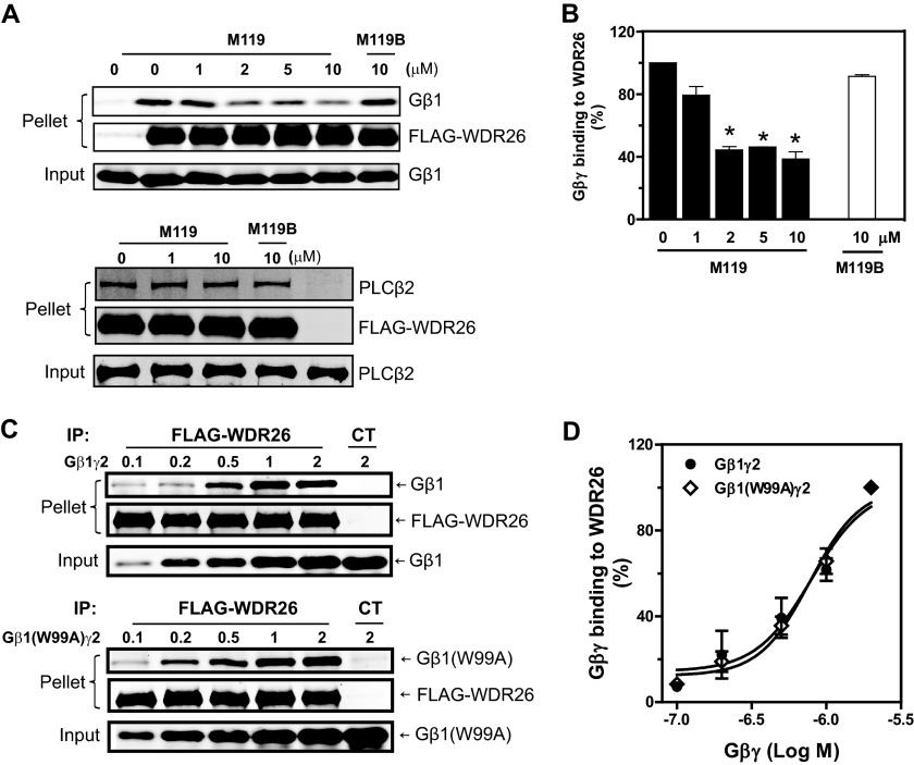
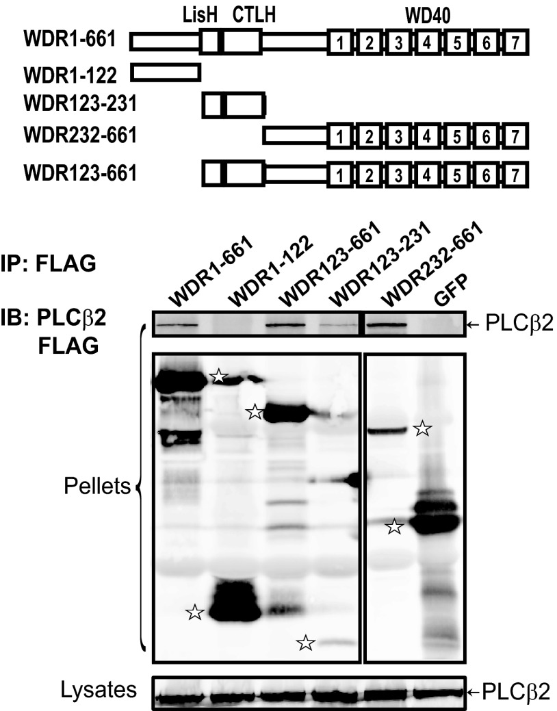
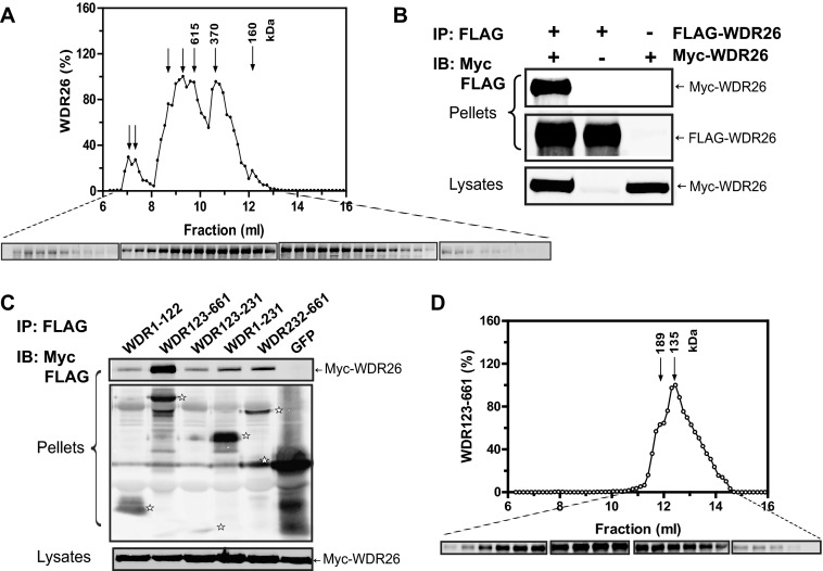
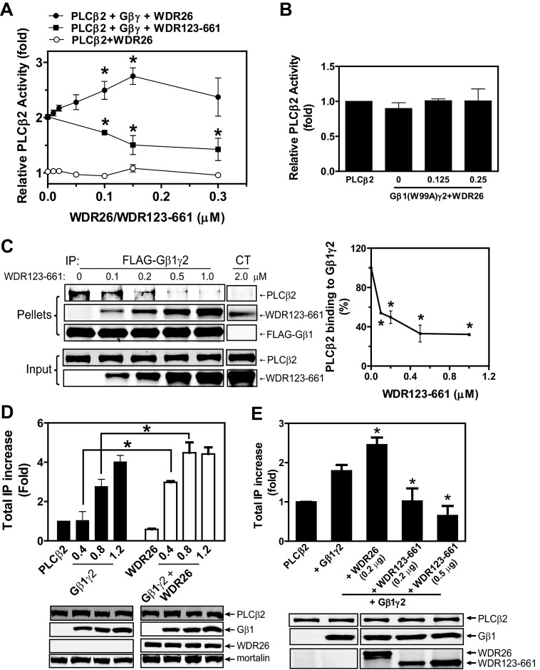
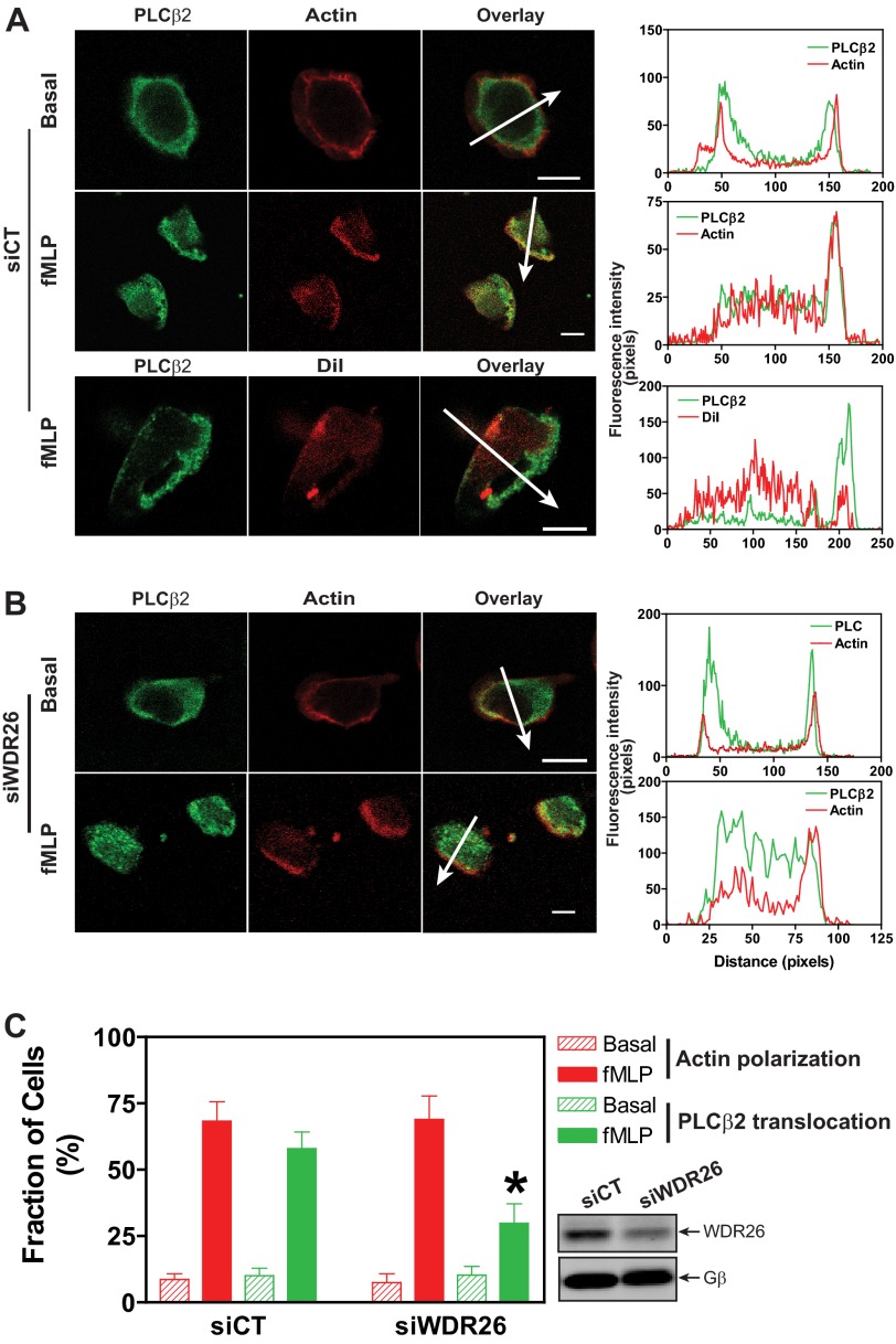
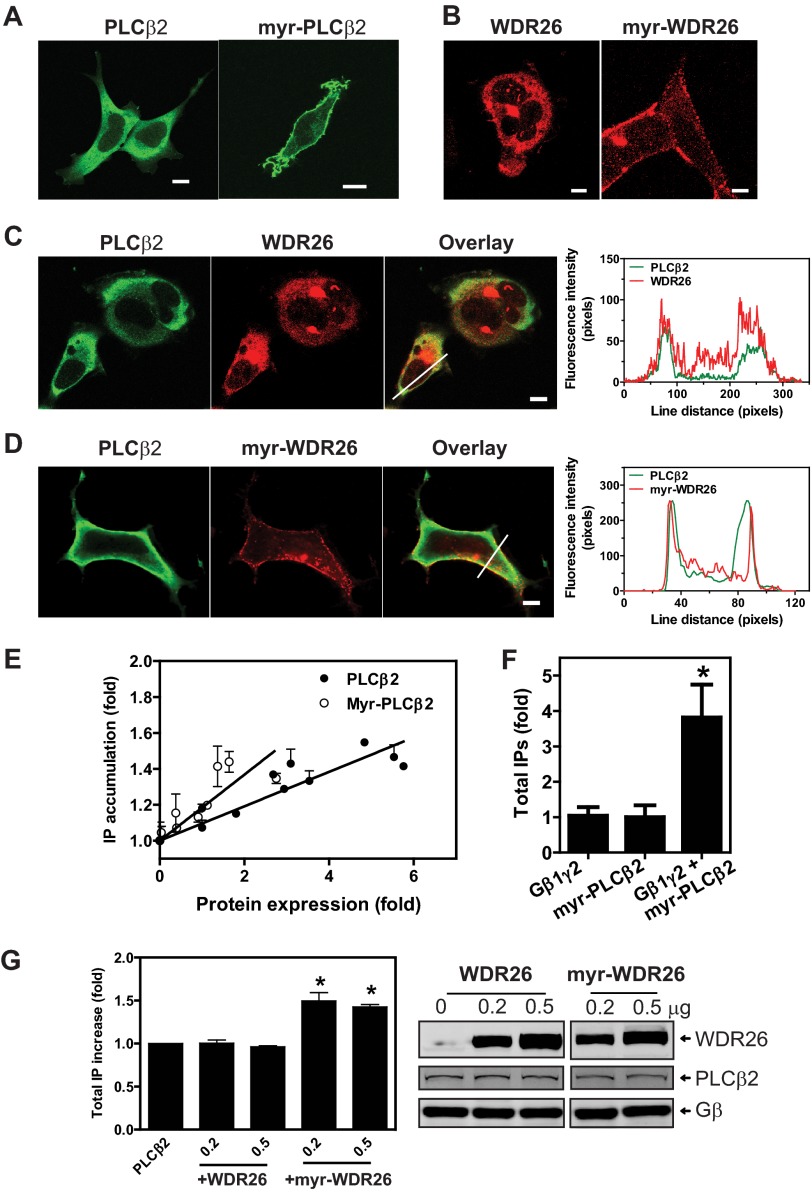
We have recently identified WDR26 as a novel WD40 repeat protein that binds Gβγ and promotes Gβγ signaling during leukocyte migration. Here, we have determined the mechanism by which WDR26 enhances Gβγ-mediated phospholipase C β2 (PLCβ2) activation in leukocytes. We show that WDR26 not only directly bound Gβγ but also PLCβ2. The binding sites of WDR26 and PLCβ2 on Gβ1γ2 were overlapping but not identical. WDR26 used the same domains for binding Gβγ and PLCβ but still formed a signaling complex with Gβγ and PLCβ2 probably due to the fact that WDR26 formed a higher order oligomer through its Lis homology and C-terminal to LisH (LisH-CTLH) and WD40 domains. Additional studies indicated that the formation of higher order oligomers was required for WDR26 to promote PLCβ2 interaction with and activation by Gβγ. Moreover, WDR26 was required for PLCβ2 translocation from the cytosol to the membrane in polarized leukocytes, and the translocation of PLCβ2 was sufficient to cause partial activation of PLCβ2. Collectively, our data indicate that WDR26 functions as a scaffolding protein to promote PLCβ2 membrane translocation and interaction with Gβγ, thereby enhancing PLCβ2 activation in leukocytes. These findings have identified a novel mechanism of regulating Gβγ signaling through a scaffolding protein.

Keywords: Chemotaxis; G Protein βγ; G Protein-coupled Receptors (GPCR); Heterotrimeric G Proteins; Phosphatidylinositol; Phospholipase C; WD40 Repeat Proteins; WDR26.


Figures

FIGURE 1.
FIGURE 1.
WDR26 binds PLCβ2 and enhances its interaction with Gβγ. A, co-immunoprecipitation of WDR26 with endogenous Gβγ and PLCβ2 from Jurkat T cells. FLAG-WDR26 was immunoprecipitated (IP) from Jurkat T cells pretreated with (+PTx) or without (Control) pertussis toxin overnight and stimulated with SDF1α (50 nm) for the indicated time using mouse IgG or an anti-FLAG antibody. The presence of Gβ, Gαi, PLCβ2, and WDR26 in the immunoprecipitates (pellets) and lysates (2.5% of total) was detected with specific antibodies. B, direct interaction of WDR26 with PLCβ2 in vitro. Upper panel, FLAG-WDR26 (0.2 μm) was immunoprecipitated from Sf9 cell lysate by anti-FLAG antibody-conjugated beads and then incubated with increasing concentrations of purified PLCβ2. Lower panel, purified His-PLCβ2 (0.2 μm) was incubated with increasing concentrations of purified WDR26 and then precipitated with Ni-NTA beads as described under “Experimental Procedures.” In the control (CT) samples, purified PLCβ2 or WDR26 alone was incubated with the beads. C and D, WDR26 enhanced Gβγ interaction with PLCβ2. Purified FLAG-Gβ1γ2 (0.2 μm) was incubated with PLCβ2 (0.5 μm) and increasing concentrations of WDR26 and then immunoprecipitated with control beads (CT) or anti-FLAG antibody-conjugated beads. Representative immunoblots are shown in C, and quantitative data from three independent experiments are shown in D. *, p < 0.05 indicates significance versus PLCβ2 binding to Gβγ in the absence of WDR26. E, WDR26 enhanced Gβγ interaction with PLCβ2 in intact cells. HEK293 cells were transfected with the indicated concentrations of FLAG-Gβ1γ2, PLCβ2, and WDR26. Cell lysates were immunoprecipitated with an anti-FLAG antibody. The presence of proteins in the immunoprecipitates (pellets) and lysates was determined by blotting for FLAG, WDR26, and PLCβ2. Unless indicated, representative blots from at least three independent experiments with similar results are shown for all figures. Error bars represent S.E.
FIGURE 2.
FIGURE 2.
The binding sites of WDR26 on Gβ1γ2. A and B, purified FLAG-WDR26 (0.2 μm) was incubated with Gβ1γ2 (0.5 μm) (upper panel) or PLCβ2 (1 μm) (lower panel) in the presence or absence of the indicated concentrations of M119 and M119B and then immunoprecipitated (IP) with an anti-FLAG antibody. Representative immunoblots are shown in A, and quantitative data from at least three independent experiments are shown in B. *, p < 0.05 indicates significance versus binding in the absence of inhibitors. C and D, binding of FLAG-WDR26 to Gβ1γ2 and Gβ1W99Aγ2. Representative immunoblots are shown in C, and quantitative data from three independent experiments are shown in D. Error bars represent S.E. CT, control.
FIGURE 3.
FIGURE 3.
The binding sites of PLCβ2 on WDR26. HEK293 cells were transfected with PLCβ2 and FLAG-tagged GFP, full-length WDR26 (WDR1–661), or the indicated WDR26 deletion mutants. Cell lysates were immunoprecipitated (IP) with an anti-FLAG antibody as described in Fig. 1. The bands corresponding to GFP, WDR26, and its mutants in the immunoprecipitates (pellets) are indicated by stars. A schematic representation of the WDR26 structure and its mutants is shown in the top panel. IB, immunoblot.
FIGURE 4.
FIGURE 4.
Oligomerization of WDR26 and its mutant. A, gel filtration analysis of purified WDR26 was performed as described under “Experimental Procedures.” The elution profile of WDR26 was determined by Western blotting analysis of each fraction (0.15 ml). The molecular mass of each peak fraction calculated from standards (1.35–670 kDa) is indicated in the graph, and representative blots of each fraction are shown under the graph. B, formation of WDR26 oligomers in vivo. HEK293 cells were transfected with FLAG-WDR26 together with or without myc-WDR26. Cell lysates were subjected to immunoprecipitation (IP) with an anti-FLAG antibody. Representative blots are shown. C, interaction of WDR26 with its deletion mutants. Co-immunoprecipitation was performed in HEK293 cells transfected with the full-length myc-WDR26 and FLAG-tagged GFP or WDR26 mutants as described in B. The bands corresponding to GFP and WDR26 mutants in the precipitates are indicated by stars. D, gel filtration analysis of purified WDR123–661 mutant was performed as described in A. IB, immunoblot.
FIGURE 5.
FIGURE 5.
The effect of WDR26 and WDR123–661 on Gβγ-mediated PLCβ2 activation. A, the effect of purified WDR26 or WDR123–661 on the basal activity of PLCβ2 (○) or Gβ1γ2-stimulated PLCβ2 (● and ■, respectively) was determined as described under “Experimental Procedures.” Data are expressed as -fold increases of PLCβ2 activity over its basal activity. *, p < 0.05 indicates significance versus1γ2-stimulated PLCβ2 activity in the absence of WDR26 or WDR123–661. B, WDR26 could not rescue the deficiency of Gβ1W99Aγ2 in stimulating PLCβ2 in vitro. The effect of the indicated concentrations of WDR26 (μm) on Gβ1W99Aγ2 (0.1 μm)-mediated PLCβ2 stimulation was determined as described in A. C, WDR123–661 inhibited PLCβ2 interaction with Gβ1γ2. The effect of purified WDR123–661 on PLCβ2 interaction with Gβ1γ2 was determined in vitro as described in Fig. 1. Representative images are shown in the left panel. Quantitative data shown in the right panel are expressed as percentage of PLCβ2 binding to Gβ1γ2 in the absence of WDR123–661. *, p < 0.05 indicates significance versus PLCβ2 binding to Gβ1γ2 in the absence of WDR123–661. D, WDR26 enhances Gβ1γ2-stimulated PLCβ2 activity in vivo. Total IPs were quantified in HEK293 cells transfected with PLCβ2 alone (PLCβ2; 0.1 μg), PLCβ2 together with WDR26 (0.2 μg) (WDR26), or the indicated concentration of Gβ1γ2 (Gβ1γ2) (0.4, 0.8 and 1.2 μg) or Gβ1γ2 plus WDR26 (Gβ1γ2+WDR26). Data are expressed as -fold increases of total IP over that produced by PLCβ2 alone after subtraction of basal IP accumulation in mock transfected cells. Representative blots of protein expression are shown under the graph. *, p < 0.05 indicates significance (n = 3). E, WDR123–661 inhibited Gβ1γ2-stimulated PLCβ2 activity in vivo. Total IPs were quantified in HEK293 cells transfected with PLCβ2 alone (PLCβ2; 0.1 μg), PLCβ2 together with Gβ1γ2 (0.8 μg) (+Gβ1γ2), or Gβ1γ2 (0.8 μg) plus the indicated concentration of WDR26 or WDR123–661 as described in B. Representative blots of protein expression are shown under the graph. *, p < 0.05 indicates significance versus total IPs generated by Gβ1γ2-stimulated PLCβ2 (n = 3). Error bars represent S.E. IP, immunoprecipitation.
FIGURE 6.
FIGURE 6.
WDR26 regulates PLCβ2 translocation. dHL60 cells were transiently transfected with a control (siCT) (A) or WDR26 siRNA (siWDR26) (B) and stimulated with buffer (Basal) or fMLP (0.2 μm) for 2 min. After fixation and permeabilization, cells were stained with a rabbit anti-PLCβ2 antibody and Alexa Fluor 568-conjugated phalloidin or CM-DiI. Representative images are shown in A and B, and quantitative data from over 100 cells in three separate experiments are shown in C. *, p < 0.05 versus siCT. The graphs in the right panel show the distribution of fluorescence intensity of PLCβ2 and F-actin or CM-DiI along the arrows drawn across the cells. Bar, 10 μm. The level of WDR26 and Gβ expression in control and WDR26 siRNA cells is shown in representative blots in C. Error bars represent S.E.
FIGURE 7.
FIGURE 7.
Membrane translocation of PLCβ2 and WDR26 regulates PLCβ2 activity. A–D, cellular localization of PLCβ2 (A, C, and D), myr-PLCβ2 (A), FLAG-WDR26 (B and C), and myr-FLAG-WDR26 (B and D). HEK293 cells were transiently transfected with the indicated constructs and stained with a rabbit anti-PLCβ2 and/or mouse anti-FLAG. Representative images are shown. The graphs in the right panel of C and D show the distribution of fluorescence intensity of PLCβ2 and WDR26 along the lines drawn across the cells. Bar, 5 μm. E, total IPs in HEK293 cells transfected with increasing concentrations of PLCβ2 or myr-PLCβ2. The levels of protein expression were quantified by Western blotting and expressed as -fold increases over that in cells transfected with the smallest amount of plasmids. IP accumulation is expressed as -fold increases over that in cells expressing the lowest level of proteins (n = 3). F, total IP accumulation in HEK293 cells transfected with Gβ1γ2 (0.4 μg), myr-PLCβ2 (0.2 μg), or Gβ1γ2 (0.4 μg) plus myr-PLCβ2 (0.2 μg). Data are expressed as -fold increases of IPs over that generated by cells expressing myr-PLCβ2 alone. *, p < 0.05 indicates significance versus myr-PLCβ2 alone (n = 4). G, total IP accumulation in HEK293 cells transfected with PLCβ2 (0.1 μg) alone or PLCβ2 together with the indicated concentration (μg) of WDR26 or myr-WDR26. Data are expressed as -fold increases of IPs over that generated by cells expressing PLCβ2 alone. *, p < 0.05 indicates significance versus PLCβ2 alone (n = 3). Representative images in the right panel show the level of protein expression. Error bars represent S.E.

Similar articles

Cited by

References

    1. Oldham W. M., Hamm H. E. (2008) Heterotrimeric G protein activation by G-protein-coupled receptors. Nat. Rev. Mol. Cell Biol. 9, 60–71 - PubMed
    1. Smrcka A. V. (2008) G protein βγ subunits: central mediators of G protein-coupled receptor signaling. Cell. Mol. Life Sci. 65, 2191–2214 - PMC - PubMed
    1. Chen S., Lin F., Shin M. E., Wang F., Shen L., Hamm H. E. (2008) RACK1 regulates directional cell migration by acting on Gβγ at the interface with its effectors PLCβ and PI3Kγ. Mol. Biol. Cell 19, 3909–3922 - PMC - PubMed
    1. Rickert P., Weiner O. D., Wang F., Bourne H. R., Servant G. (2000) Leukocytes navigate by compass: roles of PI3Kγ and its lipid products. Trends Cell Biol. 10, 466–473 - PMC - PubMed
    1. Tang X., Sun Z., Runne C., Madsen J., Domann F., Henry M., Lin F., Chen S. (2011) A critical role of Gβγ in tumorigenesis and metastasis of breast cancer. J. Biol. Chem. 286, 13244–13254 - PMC - PubMed

Publication types

LinkOut - more resources