. 2013 Jun 14;288(24):17111-21.
doi: 10.1074/jbc.M112.447326. Epub 2013 Apr 26.

Engineering A-kinase anchoring protein (AKAP)-selective regulatory subunits of protein kinase A (PKA) through structure-based phage selection

Affiliations

Engineering A-kinase anchoring protein (AKAP)-selective regulatory subunits of protein kinase A (PKA) through structure-based phage selection

Matthew G Gold et al. J Biol Chem. .

Abstract

PKA is retained within distinct subcellular environments by the association of its regulatory type II (RII) subunits with A-kinase anchoring proteins (AKAPs). Conventional reagents that universally disrupt PKA anchoring are patterned after a conserved AKAP motif. We introduce a phage selection procedure that exploits high-resolution structural information to engineer RII mutants that are selective for a particular AKAP. Selective RII (RSelect) sequences were obtained for eight AKAPs following competitive selection screening. Biochemical and cell-based experiments validated the efficacy of RSelect proteins for AKAP2 and AKAP18. These engineered proteins represent a new class of reagents that can be used to dissect the contributions of different AKAP-targeted pools of PKA. Molecular modeling and high-throughput sequencing analyses revealed the molecular basis of AKAP-selective interactions and shed new light on native RII-AKAP interactions. We propose that this structure-directed evolution strategy might be generally applicable for the investigation of other protein interaction surfaces.

Keywords: AKAP; Cell Biology; Compartmentalization; Peptide Arrays; Phage Display; Protein Kinase A (PKA); Structure-based Design.


Figures

FIGURE 1.
FIGURE 1.
Construction of RII variant phage library. A, structural representation of low-variability aliphatic residues on one face of the AKAP anchoring helix (orange) and the reciprocal binding surface in the RII D/D domain (residues 1–45; gray). B, residues in PKA RII that contact AKAP positions of higher variability (orange spheres). C, frequencies of each amino acid at every position in the first 20 amino acids of RII expressed on the surface of the phage in the input library. D, distribution of mutant nucleotides within the RII coding sequences of the input RII phage library. Mutations in wild-type RII coding sequence were restricted to codons 3, 5, 10, and 14. E, distribution of single, double, triple, and quadruple RII mutants in the input phage library. F, high-throughput sequencing statistics of RII variant phage libraries. Rd., round; n/a, not applicable.
FIGURE 2.
FIGURE 2.
Screening of AKAP-selective RII variants by phage selection. A, Coomassie Blue-stained SDS-polyacrylamide gel of GST-AKAP anchoring helix fragments. B, four-step iterative scheme for enrichment of RII variant phage that are selective for a single AKAP. C, predominant RII variant sequences following phage selection with each of the 10 different AKAP baits. High-selection pressure was a 5000-fold molar excess of each competitor AKAP helix, medium-selection pressure was a 250-fold molar excess of the competitors, and None was the absence of competitor fragments.
FIGURE 3.
FIGURE 3.
RSelect subunits exhibit AKAP selectivity in vitro. A, Coomassie Blue-stained SDS-polyacrylamide gel of purified RSelect fragments (residues 1–45). B, RII overlay of a panel of 10 GST-AKAP fusion proteins (Ponceau-stained; first panel) using the D/D domain fragments shown in A: RSelectAKAP2(1–45) (second panel), RSelectAKAP18(1–45) (third panel), RSelectAKAP150(1–45) (fourth panel), and wild-type RII(1–45) (fifth panel). C, AlphaScreen competition binding assay for AKAP18 with D/D domain fragments of either RSelectAKAP18 (red squares) or WT RII (blue triangles) and increasing concentrations of the appropriate untagged R subunit. The Kd (n = 3) for AKAP18 interaction with either RSelectAKAP18 (red squares) or WT RII (blue triangles) is shown. D, competition binding assay for AKAP18 with D/D domain fragments of either RSelectAKAP18 (red squares) or WT RII (blue triangles) in the presence of increasing concentrations of a competitor AKAP peptide mixture. E, the IC50 (n = 3) of the competitor AKAP peptide mixture for AKAP18 associated with either RSelectAKAP18 (red bar) or WT RII (blue bar). F, Coomassie Blue-stained SDS-polyacrylamide gel of the purified full-length RII mutant (RSelectAKAP18FL). G, immunoprecipitations (IP) with HEK293 cell lysates were performed in the presence (lanes 4 and 7) and absence (lanes 1–3, 5, and 6) of RSelectAKAP18FL using IgG (lane 1), anti-V5 antibody (lanes 2–4; for V5-AKAP2), or anti-FLAG antibody (lanes 5–7; for FLAG-AKAP18δ). These were done either with Ht31 (lanes 3, 4, 6, and 7) or without Ht31 (lanes 1, 2, and 5). Anchoring and coprecipitation of the modified PKA holoenzyme were tested by immunoblotting (IB) for the PKA C subunit. Immunoprecipitation of AKAP18δ (anti-FLAG antibody), AKAP2 (anti-V5 antibody), and the PKA C subunit was confirmed by immunoblotting (middle and lower panels). H, AlphaScreen competition binding assay for AKAP2 with either RSelectAKAP2 (red squares) or WT RII (blue triangles) and increasing concentrations of the appropriate untagged R subunit. The Kd (n = 3) for AKAP2 interaction with either RSelectAKAP2 or WT RII is indicted. I, AlphaScreen competition binding assay for AKAP2 with either RSelectAKAP2 (red squares) or WT RII (blue triangles) in the presence of increasing concentrations of a competitor AKAP peptide mixture. J, the IC50 (n = 3) of the competitor AKAP peptide mixture for AKAP2 associated with either RSelectAKAP2 (red bar) or WT RII (blue bar).
FIGURE 4.
FIGURE 4.
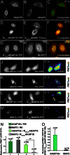
RSelect subunits exhibit AKAP selectivity in cells. A–I, confocal images of full-length AKAPs and RSelect D/D domain fragments show their subcellular location. Immunofluorescent images show HEK293 cells cotransfected with FLAG-AKAP18δ (A; green in C) and AKAP2-V5 (B; red in C). Fluorescent images show RSelectAKAP18-YFP (D; yellow in F) and RSelectAKAP2-Cherry (E; red in F), which were coexpressed with AKAP18δ and AKAP2. Fluorescent images show RSelectAKAP18-YFP (G; yellow in I) and RII-RFP (H; red in I), which were coexpressed with AKAP18δ. J–M, CFP-YFP FRET imaging of the following RII-AKAP or RSelectAKAP18 pairs in HEK293 cells: AKAP18δ-CFP and RII-YFP (J), MAP2-CFP and RII-YFP (K), AKAP18δ-CFP and RSelectAKAP18-YFP (L), and MAP2-CFP and RSelectAKAP18-YFP (M). Images were acquired for donor CFP (left panels) and acceptor YFP (middle columns), and corrected FRET images are presented (right panels). N, ratiometric quantification of intermolecular FRET pairs as described for J–M. **, p < 0.01. O, quantification of FRET signals in AKAP18δ-CFP/RSelectAKAP18-YFP and MAP2-CFP/RSelectAKAP18-YFP pairs normalized to AKAP18δ-CFP/RII-YFP and MAP2-CFP/RII-YFP, respectively.
FIGURE 5.
FIGURE 5.
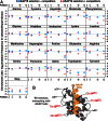
Sequence-function plot of RII variant selection with AKAP18. A, the relative changes in the frequency of each amino acid at the four variable RII positions are shown after three rounds of selection using GST-AKAP18 in either the presence (red triangles) or absence (blue circles) of competitor AKAP peptides. Amino acid mutations that increased in frequency relative to the input library have positive log-transformed enrichment ratios as a consequence of improved binding. B, structural representation of positions 3, 5, 10, and 14 in wild-type RII in relation to an AKAP anchoring helix (orange).
FIGURE 6.
FIGURE 6.
Structural basis of RSelect selectivity A, alignment of AKAP anchoring helices. Variable positions are shown in light gray, and highly conserved small aliphatic residues are shown in dark gray (upper). RII protomer residues 3, 5, 10, and 14 are shown below with dashed lines indicating interaction with variable AKAP positions. B and C, structural representations of a structural model of RSelectAKAP18 in complex with the anchoring helix of AKAP18. D and E, Logo plots of an early round of selection with AKAP18 in the absence (D) or presence (E) of competitor AKAP peptides. Amino acid mutations that increased in frequency after selection were used to generate a Logo plot, where the height of each amino acid indicates its frequency at that position (WebLogo). The selectivity index for each amino acid substitution is shown at positions 3 (F), 5 (G), 10 (H), and 14 (I) in the RII sequence. Black circles indicate selectivity index scores, blue squares denote wild-type RII amino acids, and red squares denote RSelectAKAP18 amino acids.

Similar articles

Cited by

References

    1. Pawson C. T., Scott J. D. (2010) Signal integration through blending, bolstering and bifurcating of intracellular information. Nat. Struct. Mol. Biol. 17, 653–658 - PMC - PubMed
    1. Welch E. J., Jones B. W., Scott J. D. (2010) Networking with AKAPs: context-dependent regulation of anchored enzymes. Mol. Interv. 10, 86–97 - PMC - PubMed
    1. Roy J., Cyert M. S. (2009) Cracking the phosphatase code: docking interactions determine substrate specificity. Sci. Signal. 2, re9. - PubMed
    1. Gold M. G., Barford D., Komander D. (2006) Lining the pockets of kinases and phosphatases. Curr. Opin. Struct. Biol. 16, 693–701 - PubMed
    1. Gold M. G., Lygren B., Dokurno P., Hoshi N., McConnachie G., Taskén K., Carlson C. R., Scott J. D., Barford D. (2006) Molecular basis of AKAP specificity for PKA regulatory subunits. Mol. Cell 24, 383–395 - PubMed

Publication types

MeSH terms

LinkOut - more resources