. 2013 May;12(5):610-20.
doi: 10.1158/1535-7163.MCT-12-0930. Epub 2013 Apr 26.

Small-molecule inhibitors of acetyltransferase p300 identified by high-throughput screening are potent anticancer agents

Affiliations

Small-molecule inhibitors of acetyltransferase p300 identified by high-throughput screening are potent anticancer agents

Heng Yang et al. Mol Cancer Ther. 2013 May.

Erratum in

  • Mol Cancer Ther. 2013 Aug;12(8):1688. Chase, Peter [added]

Abstract

Acetyltransferase p300 (KAT3B) plays key roles in signaling cascades that support cancer cell survival and sustained proliferation. Thus, p300 represents a potential anticancer therapeutic target. To discover novel anticancer agents that target p300, we conducted a high-throughput screening campaign. A library of 622,079 compounds was assayed for cytotoxicity to the triple-negative breast cancer (TNBC) cell line MDA-MB-231 but not to the human mammary epithelial cells. The resulting compounds were tested in a biochemical assay for inhibiting the enzymatic activity of p300. One compound (L002, NSC764414) displayed an IC50 of 1.98 μmol/L against p300 in vitro, inhibited acetylation of histones and p53, and suppressed STAT3 activation in cell-based assays. L002 could be docked to the active site of the p300 catalytic domain. Biochemical tests of a series of related compounds revealed functional groups that may impact inhibitory potency of L002 against p300. Interestingly, these analogs showed inhibitory activities against the cellular paralog of p300 (CBP), p300/CBP-associated factor, and GCN5, but not to other acetyltransferases (KAT5, KAT6B, and KAT7), histone deacetylases, and histone methyltransferases. Among the NCI-60 panel of cancer cell lines, leukemia and lymphoma cell lines were extremely sensitive to L002, whereas it is toxic to only a limited number of cell lines derived from solid tumors. Notably, breast cancer cell lines, especially those derived from TNBC, were highly susceptible to L002. In vivo, it potently suppressed tumor growth and histone acetylation of MDA-MB-468 xenografts. Thus, these new acetyltransferase inhibitors are potential anticancer therapeutics.


Figures

Figure 1
Figure 1
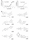
Identification of L002 and its analogs as p300 inhibitors. A, Dose-response curves of p300 inhibition by L002 (black dots; left), and anacardic acid (AA, right). Purified recombinant p300 catalytic domain was incubated in a solution containing 50 μM of acetyl-CoA and a histone H3 N-terminal peptide in the presence of L002 or AA at a specified concentration. CoASH, a product of the acetylation reaction, was detected and quantified through a fluorescence-readout. In separate assays, L002 was added after the completion of the acetylation reaction and the fluorogenic adduct formation to assess whether L002 would quench fluorescence. No quench was observed (gray triangles, left panel). B, The chemical structures of L002 and its analogs are shown. L001, L002, L004, L007 and L008 were distinct hits identified in HTS assays. L003, L004 and L006 were identified based on their structural similarity to L002. Their inhibitory activities to acetyltransferases were tested in vitro (see Table 1).
Figure 2
Figure 2
Docking of L002 to the acetyl-CoA-binding pocket in the p300 catalytic domain. L002 was fitted to the acetyl-CoA-binding pocket of a crystal structure of the p300 catalytic domain. Potential hydrogen bonds between L002 and the indicated residues of p300 are depicted (dotted lines). The residues of interest are labeled in white. A, A cartoon representation of a model of L002-p300 interaction. B, A space-filled representation of the p300 catalytic domain in complex with the inhibitor. C, The L002-p300 complex in a space-filled model. L002 and the p300 catalytic domain are shown in dark and light gray respectively.
Figure 3
Figure 3
Inhibition of p300-dependent functions in cells. A, MDA-MB-468 cells were untreated (NT, lane 1), treated with DMSO (lane 2), TSA (lane 3), L002 (lane 4), and L002 plus TSA (lane 5). The cells cultured in a complete medium with 10% bovine calf serum were exposed to DMSO or L002 for 7 h. TSA was added to 0.2 μM at 1 h before lysing cells for western blotting. A blot was probed with an antibody against acetylated lysine (top panel) or acetylated histone H4 (H4ac, middle panel). The blot was reprobed with an anti-PCNA antibody as a loading control. In a separate gel, an equal amount of the samples was loaded and stained with colloidal blue (bottom panel). B, HCT116 cells were exposed to DMSO or L002 as indicated for 7 h. Etoposide (Etop) was added 1 h after the addition of L002. Cells were lysed for western blotting with the indicated antibodies. C, MIA PaCa-2 cells were exposed to the indicated concentrations of L002 or L004 for 6 h before harvesting cells for western blotting using the indicated antibodies.
Figure 4
Figure 4
In vitro cell growth suppression by L002. A, MDA-MB-231 and MCF7 cells were cultured and exposed to DMSO (control) or the indicated concentrations of L002. The relative abundance of viable cells was determined. Shown are the average values of three assays along with SD. B, L002 suppressed clonogenic growth of breast cancer cell lines. The cells were exposed to DMSO or the indicated concentrations of L002. Surviving colonies were stained with methylene blue. The images shown are representative of three experiments. C, Reversibility of L002-mediated cytotoxicity. MDA-MB-231 or HCT116 cells were exposed to the indicated concentrations of L002. At 24 h of its addition to a cell culture, L002 was washed out in one set of the experiments as indicated. Cell viability was determined at 96 h after L002 addition.
Figure 5
Figure 5
L002 induces cell cycle arrest and apoptosis. A, MDA-MB-231 cells were exposed to DMSO, 10 μM of L002 or AA for 24 h. Cells were then processed for flow cytometry analysis. Percentage of cells in different phases of the cell cycle as well as cells with sub-G1 DNA content (apoptotic cells) is plotted. The P value was calculated based on a Student’s t-test. B, HCT116 cells were exposed to DMSO or the indicated concentrations of L002 for 24 h. Percent thymidine incorporation is plotted against L002 concentration.
Figure 6
Figure 6
In vivo anticancer efficacy of L002. Mice bearing MDA-MB-468 xenografts were untreated, or treated with the vehicle (DMSO) or L002. Each treatment group consisted of five mice (n=5). The vehicle or L002 (0.5 mg per injection) was injected intraperitoneally twice weekly for three weeks. Tumor volumes (A) and the percent change of body weights (B) of tumor-bearing mice along with the standard error of the mean (SEM) are plotted. The arrow in the left graph denotes the treatment endpoint. C, Tumor sections of mice treated with DMSO or L002 were subjected to hematoxylin and eosin (H&E) staining or immunohistochemical analysis with an anti-H4ac antibody. Three different magnifications of the tumor sections stained with the anti-H4ac antibody are shown.

Similar articles

Cited by

References

    1. Eckner R, Ewen ME, Newsome D, Gerdes M, DeCaprio JA, Lawrence JB, et al. Molecular cloning and functional analysis of the adenovirus E1A-associated 300-kD protein (p300) reveals a protein with properties of a transcriptional adaptor. Genes Dev. 1994;8:869–84. - PubMed
    1. Bedford DC, Kasper LH, Fukuyama T, Brindle PK. Target gene context influences the transcriptional requirement for the KAT3 family of CBP and p300 histone acetyltransferases. Epigenetics. 2010;5:9–15. - PMC - PubMed
    1. Gu W, Roeder RG. Activation of p53 sequence-specific DNA binding by acetylation of the p53 C-terminal domain. Cell. 1997;90:595–606. - PubMed
    1. Heemers HV, Debes JD, Tindall DJ. The role of the transcriptional coactivator p300 in prostate cancer progression. Adv Exp Med Biol. 2008;617:535–40. - PMC - PubMed
    1. Isharwal S, Miller MC, Marlow C, Makarov DV, Partin AW, Veltri RW. p300 (histone acetyltransferase) biomarker predicts prostate cancer biochemical recurrence and correlates with changes in epithelia nuclear size and shape. Prostate. 2008;68:1097–104. - PMC - PubMed

Publication types

MeSH terms

Substances