. 2013 Jul;41(12):e124.
doi: 10.1093/nar/gkt340. Epub 2013 Apr 26.

Multi-chromatic control of mammalian gene expression and signaling

Affiliations

Multi-chromatic control of mammalian gene expression and signaling

Konrad Müller et al. Nucleic Acids Res. 2013 Jul.

Abstract

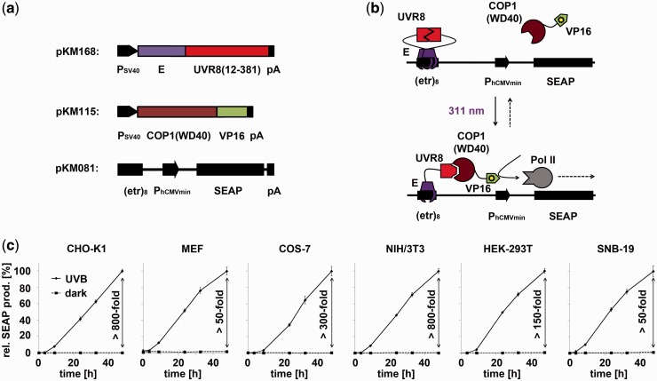
The emergence and future of mammalian synthetic biology depends on technologies for orchestrating and custom tailoring complementary gene expression and signaling processes in a predictable manner. Here, we demonstrate for the first time multi-chromatic expression control in mammalian cells by differentially inducing up to three genes in a single cell culture in response to light of different wavelengths. To this end, we developed an ultraviolet B (UVB)-inducible expression system by designing a UVB-responsive split transcription factor based on the Arabidopsis thaliana UVB receptor UVR8 and the WD40 domain of COP1. The system allowed high (up to 800-fold) UVB-induced gene expression in human, monkey, hamster and mouse cells. Based on a quantitative model, we determined critical system parameters. By combining this UVB-responsive system with blue and red light-inducible gene control technology, we demonstrate multi-chromatic multi-gene control by differentially expressing three genes in a single cell culture in mammalian cells, and we apply this system for the multi-chromatic control of angiogenic signaling processes. This portfolio of optogenetic tools enables the design and implementation of synthetic biological networks showing unmatched spatiotemporal precision for future research and biomedical applications.


Figures

Figure 1.
Figure 1.
Design and implementation of the UVB-inducible gene expression system. (a) Molecular building blocks for UVB-inducible gene expression. The expression vectors (pKM168 and pKM115) for the UVB-inducible split transcription factor were constructed by (i) fusing the gene of the macrolide-responsive repressor E to the coding sequence of the A. thaliana UVR8 core domain and (ii) by linking the gene for the COP1-derived WD40 motif to the DNA sequence encoding the VP16 transactivation domain. The reporter construct (pKM081) was assembled by cloning an octameric E-specific operator site (etr)8 upstream of the minimal human cytomegalovirus promoter PhCMVmin. As reporter gene, human placental secreted alkaline phosphatase (SEAP) was applied. pA, polyadenylation signal; PSV40, simian virus 40 promoter. (b) Mode of function. In the dark, UVR8 is tethered to the operator sequence in the closed configuration but cannot interact with COP1(WD40). On illumination with 311-nm light, the UVR8 transits from the closed to the open state and recruits COP1(WD40)-VP16, to result in the activation of PhCMVmin. On shifting to the dark, UVR8 spontaneously assumes the closed configuration, thereby resulting in a gradual shut down of gene expression. (c) Characterization of UVB-inducible gene expression in mammalian cell lines. The indicated cell lines were transfected with the UVB-inducible expression system (plasmids pKM168, pKM115 and pKM081) and kept in the dark or were illuminated with 311-nm light for 48 h. SEAP production was determined at the indicated points in time and is represented normalized to the values obtained after 48 h under UVB illumination. The corresponding induction factors are indicated. The absolute values for this condition are CHO-K1, 50.2 [U/L]; MEF, 1.6 [U/L]; COS-7, 12.4 [U/L]; NIH/3T3, 17.1 [U/L]; HEK-293T, 687.5 [U/L]; SNB-19, 1.5 [U/L]. Data are means ± standard deviation (SD) (n = 4).
Figure 2.
Figure 2.
Model-based quantitative characterization of UVB-inducible gene expression. (a and b) Light-inducible expression kinetics. CHO-K1 cells were engineered for UVB-inducible SEAP expression (plasmids pKM168, pKM115 and pKM081). After 24 h, the medium was exchanged, and the cells were illuminated for 6 h at 311 nm and were then either kept under 311 nm, moved to darkness or were supplemented with erythromycin (arrow). The SEAP mRNA (a) and protein (b) levels were determined at the indicated points in time. The curves represent the model fit to the data, and the shaded error bands are estimated by a simple error model with a constant Gaussian error. (c) Model-based analysis of expression kinetics. The SEAP mRNA production rate per promoter is shown for cells subjected to the stimuli described in Figure 2a. The shaded bands indicate the 95% prediction confidence interval. (d) Model prediction for SEAP production and cell viability under pulsed light. For this prediction, it was assumed that CHO-K1 cells were transfected with plasmids pKM168, pKM115 and pKM081, cultivated for 24 h in the dark, followed by an 24 h illumination period under pulsed UVB light (indicated in min light per 30 min cycle). The predictions for SEAP production and cell viability are represented by the lines, whereas experimental validation data are shown as circles.
Figure 3.
Figure 3.
Multi-chromatic multi-gene expression control. (a) Spectral responses of the UVB, blue and red light-inducible gene expression systems. CHO-K1 cells were transfected for UVB-inducible SEAP production (left), blue light-inducible firefly luciferase (FLuc) expression (middle) or red/far-red light-switchable gene expression (right). Twenty-four hours post-transfection, the cell culture medium was replaced with fresh medium that was supplemented with 15 µM phycocyanobilin (PCB) for the red/far-red light-switchable gene expression system. After incubation in the dark for 1 h, the cells were illuminated with light of the indicated wavelengths for 24 h before the quantification of the reporter proteins. (b) Multi-chromatic multi-gene control. CHO-K1 cells were transfected for red light-inducible SEAP production (plasmids pKM022 and pKM006), blue light-inducible FLuc expression (plasmids pKM085 and pFR-LUC) and UVB-responsive angiopoietin 1 (Ang1) synthesis (plasmids pKM168, pKM115 and pKM172). Twenty-four hours post-transfection, the medium was exchanged to DMEMcomplete that was supplemented with 15 µM PCB, and after incubation for 1 h in the dark, the cells were illuminated with 660-nm light. After 24 h, illumination was changed to 465 nm, and another 24 h later, the cells were exposed to 311-nm light for another day. Control cells (open symbols) were kept in the dark for the entire experiment. At the indicated points in time SEAP, FLuc and Ang1 production was determined. (c) Avoiding cross-talk between red light and blue/UVB light-inducible gene expression. CHO-K1 cells were transfected for red light-inducible SEAP expression, blue light-responsive FLuc production or UVB-inducible SEAP expression. After 24 h, the medium was replaced, and the cells were either illuminated with pulses of UVB light (2.7 µmol m−2 s−1, 5 min every 30 min; left) or with pulses of blue light (3.5 µmol m−2 s−1, 5 min every 30 min, right) in the absence or presence of constant 740-nm light. Twenty-four hours after illumination start, the reporter production was quantified. The reporter activities are normalized to the samples that received only UVB or blue light, respectively. Data are means ± SD (n = 4).
Figure 4.
Figure 4.
Multi-chromatically controlled angiogenic signaling. (a) Schematic experimental set-up. HEK-293T cells, transgenic for blue light-inducible VEGF165 production (plasmids pKM085 and pKM181) and UVB-responsive Ang1 expression (plasmids pKM085, pKM115 and pKM172) were co-cultured with a monolayer of endothelial Ea.hy926 cells in a transwell system. Blue light-induced production of VEGF165 triggers enhanced permeability of the endothelial cell monolayer, whereas UVB-induced Ang1 signaling induces the formation of tight cell junctions. (b and c) Multi-chromatically controlled VEGF165 and Ang1 production profiles. The angiogenic signaling set-up (a) was illuminated for 24 h with blue (465 nm) light, and subsequently for 24 h with UVB (311 nm) light. Control cells were continuously kept under 465-nm light or in the dark. The concentrations of the signaling molecules VEGF165 (b) and Ang1 (c) were quantified at the indicated points in time. (d) Effect of multi-chromatically controlled angiogenic signaling on endothelial cells. The effect of VEGF165 and Ang1 signaling on the permeability of the endothelial cell layer was determined by quantifying the trans-layer permeation of fluorescently labeled dextran. The permeability is normalized to the control value obtained from monolayers of endothelial cells that were co-cultivated with mock-transfected HEK-293T cells. Data are means ± SD (n = 4). Statistics were performed by the two-tailed t-test. *P < 0.01; ns, not significant.

Similar articles

Cited by

References

    1. Deans TL, Cantor CR, Collins JJ. A tunable genetic switch based on RNAi and repressor proteins for regulating gene expression in mammalian cells. Cell. 2007;130:363–372. - PubMed
    1. Kramer BP, Viretta AU, Daoud-El-Baba M, Aubel D, Weber W, Fussenegger M. An engineered epigenetic transgene switch in mammalian cells. Nat. Biotechnol. 2004;22:867–870. - PubMed
    1. Kramer BP, Fussenegger M. Hysteresis in a synthetic mammalian gene network. Proc. Natl Acad. Sci. USA. 2005;102:9517–9522. - PMC - PubMed
    1. Weber W, Stelling J, Rimann M, Keller B, Daoud-El Baba M, Weber CC, Aubel D, Fussenegger M. A synthetic time-delay circuit in mammalian cells and mice. Proc. Natl Acad. Sci. USA. 2007;104:2643–2648. - PMC - PubMed
    1. Tigges M, Marquez-Lago TT, Stelling J, Fussenegger M. A tunable synthetic mammalian oscillator. Nature. 2009;457:309–312. - PubMed

Publication types

Substances