

The inhibitory effect of small interference RNA protein kinase C-alpha on the experimental proliferative vitreoretinopathy induced by dispase in mice
- PMID: 23626468
- PMCID: PMC3632628
- DOI: 10.2147/IJN.S37635
The inhibitory effect of small interference RNA protein kinase C-alpha on the experimental proliferative vitreoretinopathy induced by dispase in mice
Abstract
Aim: To evaluate the effects of small interference RNA protein kinase C-alpha (siRNA-PKCα) on experimental proliferative vitreoretinopathy (PVR) induced by dispase in mice.
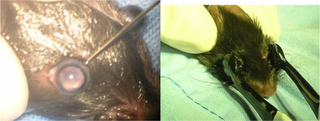
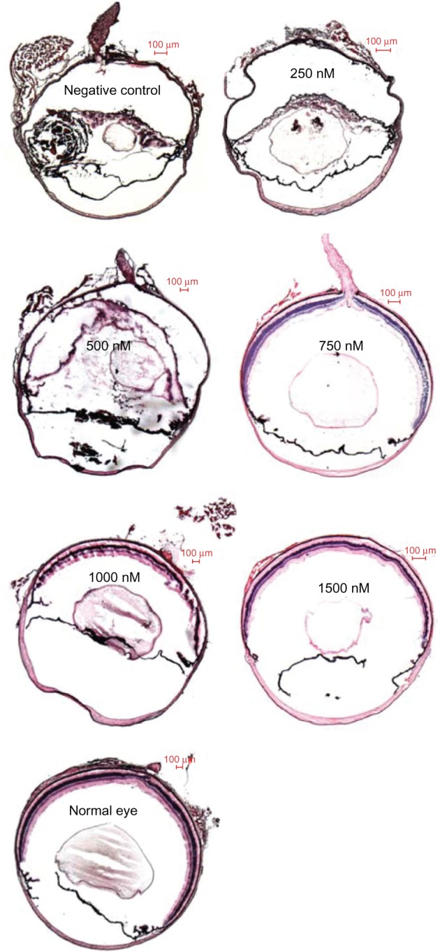
Methods: C57BL/6 mice PVR models (4-6 weeks old) were induced by intravitreal injection of dispase and then equally divided into six groups. After 1 week, the five treatment groups received 2 μL, intravitreal injections of siRNA-PKCα at a concentration of 250 nM, 500 nM, 750 nM, 1000 nM, and 1500 nM, respectively, while the negative control group received 2 μL of 500 nM no-silencing siRNA. SiRNA-PKCα was transfected by a square wave electroporator. Postoperative ophthalmic observations of lens clarity and the fundus of the eyes were performed periodically. The eyeballs of the mice were enucleated and imbedded in optimal cutting temperature to perform histological and immunofluorescence analysis at the end of a 4-week observation period.
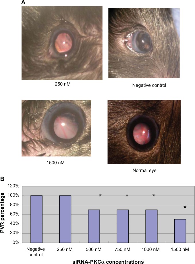
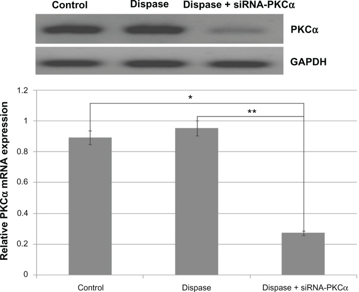
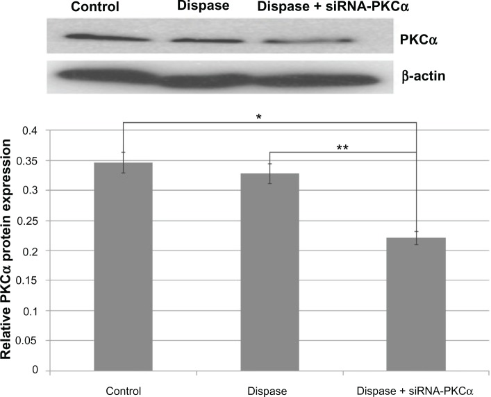
Results: Four weeks after the siRNA-PKCα injections, there are 100% lens dissolution and 100% PVR in the 250 nM group and 70%, 70%, 70%, and 50% PVR in the 500 nM, 750 nM, 1000 nM, and 1500 nM groups, respectively, which is significantly different from the negative group. Abnormalities in fundus appearance were related to the concentrations of siRNA-PKCα; a higher concentration of siRNA-PKCα resulted in a more normal fundus. Histological sections by hematoxylin-eosin staining of the eyes support the clinical observation. Immunofluorescence analysis showed that RPE65, glutamine synthase, glial acidic fibrillary protein, and α-smooth muscle actin were increasing in the retina with the decreasing concentration of siRNA-PKCα, indicating that intraocular siRNA-PKCα can partly inhibit changes of markers for glia cells, fibroblast cells, retinal pigment epithelium cells, and Müller cells in the process of PVR.
Conclusion: Gene therapy with siRNA-PKCα could effectively inhibit PVR in mice and provide us with a novel therapeutic target on PVR.
Keywords: dispase; proliferative vitreoretinopathy; protein kinase Cα; small interference RNA.
Figures


References
-
- Grigoropoulos VG, Benson S, Bunce C, Charteris DG. Functional outcome and prognostic factors in 304 eyes managed by retinectomy. Graefes Arch Clin Exp Ophthalmol. 2007;245(5):641–649. - PubMed
-
- Quiram PA, Gonzales CR, Hu W, et al. Outcomes of vitrectomy with inferior retinectomy in patients with recurrent rhegmatogenous retinal detachments and proliferative vitreoretinopathy. Ophthalmology. 2006;113(11):2041–2047. - PubMed
-
- Pastor JC, de la Rúa ER, Martin F. Proliferative vitreoretinopathy: risk factors and pathobiology. Prog Retin Eye Res. 2002;21(1):127–144. - PubMed
-
- Ryan SJ. The pathophysiology of proliferative vitreoretinopathy in its management. Am J Ophthalmol. 1985;100(1):188–193. - PubMed
-
- Agrawal RN, He S, Spee C, Cui JZ, Ryan SJ, Hinton DR. In vivo models of proliferative vitreoretinopathy. Nat Protoc. 2007;2(1):67–77. - PubMed
Publication types
MeSH terms
Substances
LinkOut - more resources
Full Text Sources
Other Literature Sources
Research Materials
