

Neurotransmitter signaling in the pathophysiology of microglia
- PMID: 23626522
- PMCID: PMC3630369
- DOI: 10.3389/fncel.2013.00049
Neurotransmitter signaling in the pathophysiology of microglia
Erratum in
- Front Cell Neurosci. 2013;7:107
Abstract
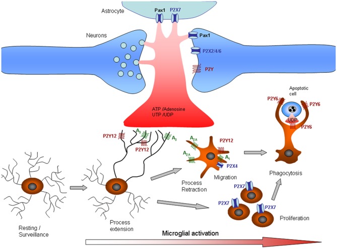
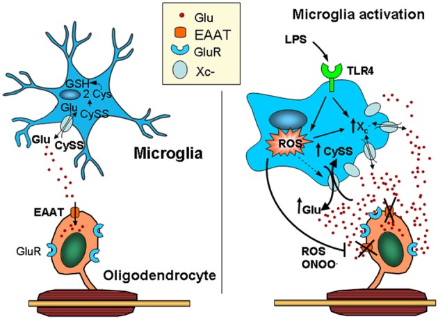
Microglial cells are the resident immune cells of the central nervous system. In the resting state, microglia are highly dynamic and control the environment by rapidly extending and retracting motile processes. Microglia are closely associated with astrocytes and neurons, particularly at the synapses, and more recent data indicate that neurotransmission plays a role in regulating the morphology and function of surveying/resting microglia, as they are endowed with receptors for most known neurotransmitters. In particular, microglia express receptors for ATP and glutamate, which regulate microglial motility. After local damage, the release of ATP induces microgliosis and activated microglial cells migrate to the site of injury, proliferate, and phagocytose cells, and cellular compartments. However, excessive activation of microglia could contribute to the progression of chronic neurodegenerative diseases, though the underlying mechanisms are still unclear. Microglia have the capacity to release a large number of substances that can be detrimental to the surrounding neurons, including glutamate, ATP, and reactive oxygen species. However, how altered neurotransmission following acute insults or chronic neurodegenerative conditions modulates microglial functions is still poorly understood. This review summarizes the relevant data regarding the role of neurotransmitter receptors in microglial physiology and pathology.
Keywords: ATP; glutamate; microglia; purinergic and glutamatergic receptors.
Figures
Similar articles
-
Microglial morphology and dynamic behavior is regulated by ionotropic glutamatergic and GABAergic neurotransmission.PLoS One. 2011 Jan 25;6(1):e15973. doi: 10.1371/journal.pone.0015973. PLoS One. 2011. PMID: 21283568 Free PMC article.
-
Physiology of microglia.Physiol Rev. 2011 Apr;91(2):461-553. doi: 10.1152/physrev.00011.2010. Physiol Rev. 2011. PMID: 21527731 Review.
-
Regulation of Microglial Functions by Purinergic Mechanisms in the Healthy and Diseased CNS.Cells. 2020 Apr 29;9(5):1108. doi: 10.3390/cells9051108. Cells. 2020. PMID: 32365642 Free PMC article. Review.
-
Deciphering Resting Microglial Morphology and Process Motility from a Synaptic Prospect.Front Integr Neurosci. 2016 Jan 19;9:73. doi: 10.3389/fnint.2015.00073. eCollection 2015. Front Integr Neurosci. 2016. PMID: 26834588 Free PMC article. Review.
-
Norepinephrine modulates the motility of resting and activated microglia via different adrenergic receptors.J Biol Chem. 2013 May 24;288(21):15291-302. doi: 10.1074/jbc.M113.458901. Epub 2013 Apr 2. J Biol Chem. 2013. PMID: 23548902 Free PMC article.
Cited by
-
The impaired distribution of adenosine deaminase isoenzymes in multiple sclerosis plasma and cerebrospinal fluid.Front Mol Neurosci. 2022 Sep 20;15:998023. doi: 10.3389/fnmol.2022.998023. eCollection 2022. Front Mol Neurosci. 2022. PMID: 36204140 Free PMC article.
-
Modulation of Microglial Function by ATP-Gated P2X7 Receptors: Studies in Rat, Mice and Human.Cells. 2024 Jan 16;13(2):161. doi: 10.3390/cells13020161. Cells. 2024. PMID: 38247852 Free PMC article. Review.
-
Pathophysiology of blood brain barrier dysfunction during chronic cerebral hypoperfusion in vascular cognitive impairment.Theranostics. 2022 Jan 16;12(4):1639-1658. doi: 10.7150/thno.68304. eCollection 2022. Theranostics. 2022. PMID: 35198062 Free PMC article. Review.
-
Temporal alteration of microglia to microinfarcts in rat brain induced by the vascular occlusion with fluorescent microspheres.Front Cell Neurosci. 2022 Aug 3;16:956342. doi: 10.3389/fncel.2022.956342. eCollection 2022. Front Cell Neurosci. 2022. PMID: 35990892 Free PMC article.
-
Trovafloxacin attenuates neuroinflammation and improves outcome after traumatic brain injury in mice.J Neuroinflammation. 2018 Feb 13;15(1):42. doi: 10.1186/s12974-018-1069-9. J Neuroinflammation. 2018. PMID: 29439712 Free PMC article.
References
LinkOut - more resources
Full Text Sources
Other Literature Sources