

The role of VEGF in melanoma progression
- PMID: 23626629
- PMCID: PMC3634290
The role of VEGF in melanoma progression
Abstract
Background: Melanoma is the most serious skin cancer. There is an established correlation between thickness and aggressiveness of the tumor. Nevertheless, the potential value of vascular endothelial growth factor (VEGF) in correlation with tumor progression remains unresolved.
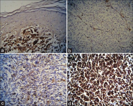
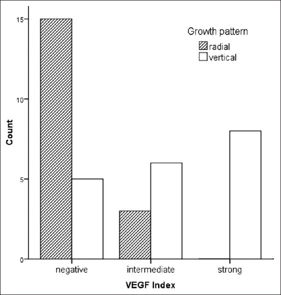
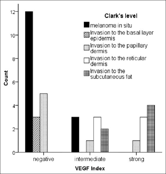
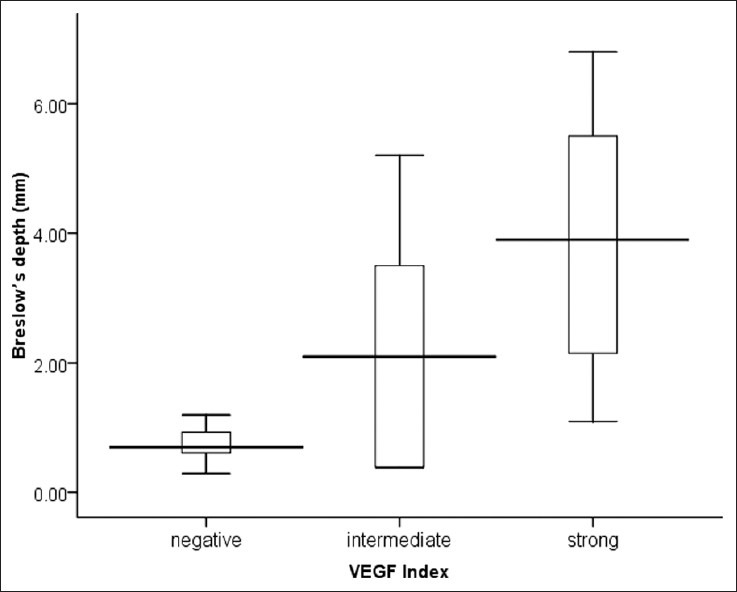
Materials and methods: Thirty seven paraffin blocks of cutaneous melanoma were obtained from Pathology department of Al-zahra hospital between 2005 and 2010. The sections were stained with monoclonal mouse antibodies (mAbs) against vascular endothelial growth factor A and evaluated by distribution of expression of VEGF in tumor cells as 0, 0%; 1, 1%--25%; 2, 25%--50%; 3, >50% and the staining intensity from 0 (negative) to 3 (strong). The sum of intensity score and distribution score was then calculated as the VEGF index. The relationship between VEGF expression (distribution, intensity, and index) and tumor progression (vertical and radial growth, Clark's level, and Breslow's depth) was studied. SPSS software was used to analyze the data by ANOVA, and chi-square tests.
Results: 51.4% of the patients showed vertical growth pattern. Mean Breslow's depth was 1.84 ± 1.79 mm. There was a significant association between growth pattern and VEGF distribution, intensity and index (P = 0.006, P = 0.005, and P = 0.001 respectively). VEGF distribution, intensity, and index all had correlation with Breslow's depth as well (ANOVA test: P = 0.003, P < 0.001, and P < 0.001 respectively) VEGF index had also correlation with Clark's level, but this was not seen for VEGF distribution and intensity.
Conclusion: VEGF expression (both VEGF distribution and intensity) is associated with progression of malignant melanoma. VEGF index can explain this association better.
Keywords: Breslow's depth; Melanoma; vascular endothelial growth factor.
Conflict of interest statement
Figures
Similar articles
-
Association between tumor size and Breslow's thickness in malignant melanoma: a cross-sectional, multicenter study.Melanoma Res. 2015 Oct;25(5):450-2. doi: 10.1097/CMR.0000000000000184. Melanoma Res. 2015. PMID: 26237766
-
Association of vascular endothelial growth factor expression with patohistological parameters of cutaneous melanoma.Vojnosanit Pregl. 2016 May;73(5):449-57. doi: 10.2298/vsp140804027g. Vojnosanit Pregl. 2016. PMID: 27430109
-
Hypoxia-inducible factors 1alpha and 2alpha are related to vascular endothelial growth factor expression and a poorer prognosis in nodular malignant melanomas of the skin.Melanoma Res. 2003 Oct;13(5):493-501. doi: 10.1097/00008390-200310000-00008. Melanoma Res. 2003. PMID: 14512791
-
Angiogenesis in cutaneous melanoma: pathogenesis and clinical implications.Microsc Res Tech. 2003 Feb 1;60(2):208-24. doi: 10.1002/jemt.10259. Microsc Res Tech. 2003. PMID: 12539175 Review.
-
Malignant melanoma: basic approach to clinicopathologic correlation.Mayo Clin Proc. 1997 Mar;72(3):267-72. doi: 10.4065/72.3.267. Mayo Clin Proc. 1997. PMID: 9070204 Review.
Cited by
-
A Review of Key Biological and Molecular Events Underpinning Transformation of Melanocytes to Primary and Metastatic Melanoma.Cancers (Basel). 2019 Dec 17;11(12):2041. doi: 10.3390/cancers11122041. Cancers (Basel). 2019. PMID: 31861163 Free PMC article. Review.
-
NIR-Responsive injectable hydrogel cross-linked by homobifunctional PEG for photo-hyperthermia of melanoma, antibacterial wound healing, and preventing post-operative adhesion.Mater Today Bio. 2024 Apr 24;26:101062. doi: 10.1016/j.mtbio.2024.101062. eCollection 2024 Jun. Mater Today Bio. 2024. PMID: 38706729 Free PMC article.
-
Epigenetic strategies synergize with PD-L1/PD-1 targeted cancer immunotherapies to enhance antitumor responses.Acta Pharm Sin B. 2020 May;10(5):723-733. doi: 10.1016/j.apsb.2019.09.006. Epub 2019 Sep 25. Acta Pharm Sin B. 2020. PMID: 32528824 Free PMC article. Review.
-
[Efficacy of combined treatment with pirfenidone and PD-L1 inhibitor in mice bearing ectopic bladder cancer xenograft].Nan Fang Yi Ke Da Xue Xue Bao. 2024 Feb 20;44(2):210-216. doi: 10.12122/j.issn.1673-4254.2024.02.02. Nan Fang Yi Ke Da Xue Xue Bao. 2024. PMID: 38501405 Free PMC article. Chinese.
-
Resistance of melanoma cells to anticancer treatment: a role of vascular endothelial growth factor.Postepy Dermatol Alergol. 2020 Feb;37(1):11-18. doi: 10.5114/ada.2020.93378. Epub 2020 Mar 9. Postepy Dermatol Alergol. 2020. PMID: 32467677 Free PMC article. Review.
References
-
- Rigel DS, Friedman RJ, Kopf AW. The incidence of malignant melanoma in the United States: Issues as we approach the 21st century. J Am Acad Dermatol. 1996;34:839–47. - PubMed
-
- Berwick M, Wiggins C. The current epidemiology of cutaneous malignant melanoma. Front Biosci. 2006;11:1244–54. - PubMed
-
- Liu V, Mihm MC. Pathology of malignant melanoma. Surg Clin North Am. 2003;83:31–60. v. - PubMed
-
- Homsi J, Kashani-Sabet M, Messina JL, Daud A. Cutaneous melanoma: Prognostic factors. Cancer Control. 2005;12:223–9. - PubMed
-
- Buzaid AC, Ross MI, Balch CM, Soong S, McCarthy WH, Tinoco L, et al. Critical analysis of the current American Joint Committee on Cancer staging system for cutaneous melanoma and proposal of a new staging system. J Clin Oncol. 1997;15:1039–51. - PubMed
LinkOut - more resources
Full Text Sources
Other Literature Sources