. 2013 Apr 23;8(4):e58239.
doi: 10.1371/journal.pone.0058239. Print 2013.

ATM deficiency results in accumulation of DNA-topoisomerase I covalent intermediates in neural cells

Affiliations

ATM deficiency results in accumulation of DNA-topoisomerase I covalent intermediates in neural cells

Meryem Alagoz et al. PLoS One. .

Abstract

Accumulation of peptide-linked DNA breaks contributes to neurodegeration in humans. This is typified by defects in tyrosyl DNA phosphodiesterase 1 (TDP1) and human hereditary ataxia. TDP1 primarily operates at single-strand breaks (SSBs) created by oxidative stress or by collision of transcription machinery with topoisomerase I intermediates (Top1-CCs). Cellular and cell-free studies have shown that Top1 at stalled Top1-CCs is first degraded to a small peptide resulting in Top1-SSBs, which are the primary substrates for TDP1. Here we established an assay to directly compare Top1-SSBs and Top1-CCs. We subsequently employed this assay to reveal an increased steady state level of Top1-CCs in neural cells lacking Atm; the protein mutated in ataxia telangiectasia. Our data suggest that the accumulation of endogenous Top1-CCs in Atm-/- neural cells is primarily due to elevated levels of reactive oxygen species. Biochemical purification of Top1-CCs from neural cell extract and the use of Top1 poisons further confirmed a role for Atm during the formation/resolution of Top1-CCs. Finally, we report that global transcription is reduced in Atm-/- neural cells and fails to recover to normal levels following Top1-mediated DNA damage. Together, these data identify a distinct role for ATM during the formation/resolution of neural Top1-CCs and suggest that their accumulation contributes to the neuropathology of ataxia telangiectasia.


Conflict of interest statement

Competing Interests: The authors have declared that no competing interests exist.

Figures

Figure 1
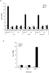
Figure 1. ATM deficiency does not impact on the accumulation of Top1-single-strand breaks.
(a) Human lymphoblastoid cells (LCLs) derived from a normal individual ‘WT’, spinocerebellar ataxia with axonal neuropathy ‘SCAN1’, or ataxia telangiectasia ‘A–T’ patients, and mouse embryonic fibroblasts (MEFs) or quiescent cortical astrocytes from control ‘WT’ or Tdp1-/- mice were incubated with DMSO (Mock) or 30 µM camptothecin (CPT) for 40 min with or without pre-incubation with 10 µM ATM inhibitor KU-55933 (ATMi) for 2 hours at 37°C. Top1-single-strand breaks ‘Top1-SSBs’ were quantified by alkaline comet assays (ACAs). Mean tail moments were calculated for 50 cells/sample/experiment and data are the average of n = 3 biological replicates ± s.e.m. (b) Top1-SSBs were analysed in quiescent cortical astrocytes derived from wild-type ‘WT’, Atm-/- or Tdp1-/- mice following incubation with DMSO (Mock) or 30 µM camptothecin (CPT) and quantified as described above.
Figure 2
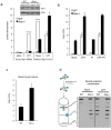
Figure 2. Modification of the alkaline comet assay uncovers un-degraded Top1-DNA cleavage complexes (Top1-CCs).
(a) Scheme depicting the major differences between Top1-CCs and Top1-SSBs: Top1 relaxes DNA supercoiling by introducing a reversible nick to which Top1 becomes covalently attached (Top1-CCs). Stalling of Top1-CCs through collision with the transcription machinery or oxidative DNA damage triggers proteasomal degradation of Top1, resulting in Top1 single-strand breaks (Top1-SSBs). Repair of Top1-SSBs is initiated by removal of Top1 peptide by TDP1 followed by subsequent ligation. Note that un-degraded ‘Top1-CCs’ are not detected by the ‘classical’ alkaline comet assays (ACA) due to the reversible nature of these intermediates and the reduced ability of covalently bound Top1 on DNA to produce measurable tail upon electrophoresis. (b) Control ‘WT’ or SCAN1 LCLs ‘SCAN1’ harbouring the TDP1 catalytic mutation H493R were incubated with 20 µM camptothecin “CPT” with or without a prior 2-hr incubation with 30 µM proteasome inhibitor MG132 ‘PI’. Cells were divided into two fractions for the comparative detection of Top1-SSBs and Top1-CCs using the ACAs and modified ACAs ‘MACA’, respectively. Mean tail moments were calculated for 50 cells/sample/experiment and data are the average of n = 3 biological replicates ± s.e.m. Note that inhibiting the proteasome resulted in a reduction of Top1-SSBs (as measured by ACA) to near background levels with minimal impact on Top1-CCs (as measured by MACA).
Figure 3
Figure 3. Loss of Atm results in accumulation of Top1-CCs in cortical neural cells.
(a) Endogenous steady-state level of Top1-SSBs and Top1-CCs were quantified in quiescent wild type ‘WT’ and Atm-/- cortical astrocytes by ACAs and MACAs, respectively. Neural cells were subsequently subjected to 30 µM CPT for 40 min at 37 °C and the level of Top1-SSBs and Top1-CCs were quantified as above from 50 cells/sample/experiment. Data are the average of n = 3 biological replicates ± s.e.m. Inset: astrocytes were incubated with DMSO ‘Mock’ or CPT ‘CPT’ and the expression of Top1 was measured by anti-Top1 immunoblotting. Anti-actin was employed as a loading control. (b) WT or Atm-/- quiescent astrocytes were mock incubated with DMSO ‘Mock’ or with the 30 µM CPT for 40 min at 37°C with or without a prior 2-hour incubation with the proteasome inhibitor MG132 ‘PI’. Top1-CCs were quantified by MACA from 50 cells/sample/experiment and the average of n = 3 biological replicates ± s.e.m is presented (c) Cortices from WT or Atm-/- mice were harvested at P6 and cells were dissociated and immediately subjected to MACA analyses. Data are the average of n = 3 biological replicates ± s.e.m. (d) Left: scheme depicting the biochemical fractionation of Top1-CCs. Blue circles are Top1 covalently bound to DNA (Top1-CCs) and yellow circles are Top1 non-covalently bound to DNA. Right: Wild-type ‘WT’ or Atm-/- quiescent cortical astrocytes were mock incubated or incubated with 30 µM CPT for 60 min at 37 °C. Neural cells were lysed in denaturing buffer and lysates fractionated on CsCl gradients. Fractions were slot blotted onto nitrocellulose and immunblotted with anti-Top1 monoclonal antibodies. A representative experiment from 3 biological replicates is shown. P values indicate the statistical difference between WT and Atm-/- cells (student t-test).
Figure 4
Figure 4. ATM deficiency results in accumulation of Top1-CCs in human cells.
(a) Top1-SSBs and Top1-CCs were quantified in wild-type 1BR3 ‘WT’ or ATM deficient AT-1BR ‘A–T’ human primary fibroblasts by ACAs and MACAs, respectively. Cells were mock incubated with DMSO ‘Mock’ or with the 30 µM CPT for 40 min at 37°C and DNA strand breaks quantified from 50 cells/sample/experiment. Data are the average of n = 3 biological replicates ± s.e.m. (b) Primary human fibroblasts were grown to confluency and serum starved for 3 days, and Top1-SSBs/Top1-CCs were quantified as described in (a).
Figure 5
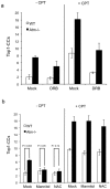
Figure 5. Reactive oxygen species scavengers reduce the accumulation of Top1-CCs in Atm-/- cells.
(a) Top1-CCs were analysed in WT or Atm-/- quiescent astrocytes with and without incubation with 30 µM CPT ± a prior 2-hour incubation with 50 µM of the transcription inhibitor 5,6-dichloro-1-beta-D-ribofuranosylbenzimidazole ‘DRB’. Top1-CCs were quantified by MACA from 50 cells/sample/experiment and data represent the average of n = 3 biological replicates ± s.e.m. (b) Top1-CCs were analysed as in (a) with or without prior incubation with the reactive oxygen species (ROS) scavengers mannitol (50 mM) or N-Acetyl cysteine ‘NAC’ (10 mM) for 17-hours. 50 cells/sample/experiment were analysed and data represent the average of n = 3 biological replicates ± s.e.m. P values indicate the statistical difference between WT and Atm-/- cells (student t-test). Note that prior incubation with ROS scavengers reduces the endogenous level of Top1-CCs in Atm-/- cells to that observed in control cells .
Figure 6
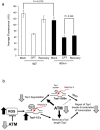
Figure 6. Atm-/- neural cells fail to recover transcription following Top1-mediated DNA damage.
(a) Quiescent wild-type ‘WT’ or Atm-/- cortical astrocytes grown on coverslips were treated with 30 µM CPT for 1 hour and either harvested immediately after treatment or incubated in CPT-free media for a subsequent 3 hour to allow for transcription recovery. Cells were incubated with 0.1 mM 5-ethynl uridine (EU) for 30 min to label newly synthesized RNA, which was visualised by utilising the Click iT reaction with Alexa Flour azide 488. EU-labelled RNA was subjected to immuofluorescence analyses and data represent the average fluorescence signal (arbitrary units ‘AU’) from n = 3 biological replicates ± s.e.m, quantified from 200–300 cells using Corel Photo Draw software. P values indicate the statistical difference between the indicated bars (student t-test). (b) A model for the role of ATM during Top1-CC formation/resolution: ATM deficiency results in elevated levels of ROS, which leads to oxidative DNA breaks that are ‘masked’ from detection by the ‘classical’ comet due to trapping of Top1 on DNA, resulting in elevated steady state level of Top1-CCs. Stalling of Top1 on DNA can also occur through collision of Top1-CCs with elongating RNA polymerases ‘RNA Pol’. Top1 is first degraded by the proteasome to a small peptide, which is a substrate for the tyrosyl DNA phosphodiesterase activity of TDP1. We suggest that ATM facilitates the resolution of Top1-CCs by promoting Top1 degradation. ATM may also enhance the activity of one or more TDP1-independent processes to remove ‘un-degraded’ Top1 from DNA. Failure to remove Top1 from stalled Top1-CCs in ATM deficient cells results in a failure to maintain normal transcriptional activity following Top1-mediated DNA damage, and may contribute to the neuropathology of A–T.

References

    1. Pommier Y, Redon C, Rao VA, Seiler JA, Sordet O, et al. (2003) Repair of and checkpoint response to topoisomerase I-mediated DNA damage. Mutat Res 532: 173–203. - PubMed
    1. Pourquier P, Pilon AA, Kohlhagen G, Mazumder A, Sharma A, et al. (1997) Trapping of mammalian topoisomerase I and recombinations induced by damaged DNA containing nicks or gaps. Importance of DNA end phosphorylation and camptothecin effects. J Biol Chem 272: 26441–26447. - PubMed
    1. Takashima H, Boerkoel CF, John J, Saifi GM, Salih MAM, et al. (2002) Mutation of TDP1, encoding a topoisomerase I–dependent DNA damage repair enzyme, in spinocerebellar ataxia with axonal neuropathy. Nat Genet 32: 267–272. - PubMed
    1. Biton S, Dar I, Mittelman L, Pereg Y, Barzilai A, et al. (2006) Nuclear ataxia-telangiectasia mutated (ATM) mediates the cellular response to DNA double strand breaks in human neuron-like cells. J Biol Chem 281: 17482–17491. - PubMed
    1. El-Khamisy SF (2011) To live or to die: a matter of processing damaged DNA termini in neurons. EMBO Mol Med 3: 78–88. - PMC - PubMed

Publication types

MeSH terms