. 2013 Apr 23;8(4):e61394.
doi: 10.1371/journal.pone.0061394. Print 2013.

Ecrg4 attenuates the inflammatory proliferative response of mucosal epithelial cells to infection

Affiliations

Ecrg4 attenuates the inflammatory proliferative response of mucosal epithelial cells to infection

Arwa Kurabi et al. PLoS One. .

Abstract

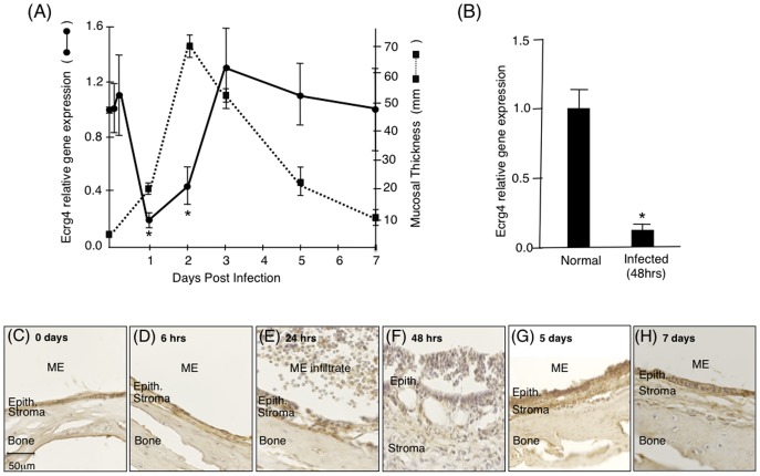
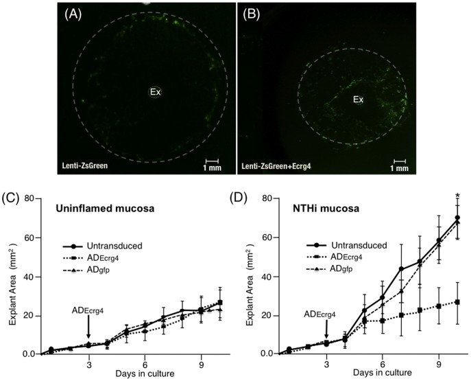
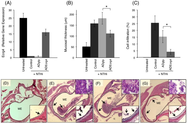
We report an inverse relationship between expression of the orphan candidate tumor suppressor gene esophageal cancer related gene 4 (Ecrg4), and the mucosal epithelial cell response to infection in the middle ear (ME). First, we found constitutive Ecrg4 mRNA expression in normal, quiescent ME mucosa that was confirmed by immunostainning of mucosal epithelial cells and immunoblotting of tissue lysates for the 14 kDa Ecrg4 protein. Upon experimental ME infection, Ecrg4 gene expression rapidly decreased by over 80%, between 3 to 48 hrs, post infection. When explants of this infected mucosa were placed in culture and transduced with an adenovirus (AD) encoding Ecrg4 gene (ADEcrg4), the proliferative and migratory responses of mucosal cells were significantly inhibited. ADEcrg4 transduction of control explants from uninfected MEs had no effect on basal growth and migration. Over-expression of Ecrg4 in vivo, by pre-injecting MEs with ADEcrg4 48 hrs prior to infection, prevented the natural down-regulation of Ecrg4, reduced mucosal proliferation and prevented inflammatory cell infiltration normally observed after infection. Taken together, these data support a hypothesis that Ecrg4 plays a role in coordinating the inflammatory and proliferative response to infection of mucosal epithelium suggesting a possible mechanism for its putative anti-tumor activity.


Conflict of interest statement

Competing Interests: The authors have declared that no competing interests exist.

Figures

Figure 1
Figure 1. Ecrg4 is present in normal rat ME mucosa.
Panel A: Ecrg4 PCR product amplified from uninfected rat ME mucosa cDNAs. A 100 bp molecular size marker is shown on the left lane. Panel B: Organization of the rat Ecrg4 gene shows the 4 exons and the start (set to +1) and stop codons of the open reading frame (ORF). The mRNA isolated from rat ME mucosa was subjected to 5′RACE analysis using a primer annealing to exon 4 (solid arrow). Following primer elongation, cloning and sequencing of the 5′ RACE products (clear bars), translation start sites (TSS) at −42 bp and −21 bp upstream of the ATG translation start site produce the identical ORF. An asterisk denotes that the −21 bp TSS was the most frequently amplified. Panel C: Immunolocalization of Ecrg4 (red) in the ME epithelium compared to a background immunofluorescence signal using pre-immune IgY (insert). Nuclei are counterstained with DAPI (blue). Epith.  =  epithelial cells (D) Immunoblotting of rat ME mucosa revealed the presence of a prominent 14 kDa Ecrg4 protein band.
Figure 2
Figure 2. Characterization of changes in Ecrg4 gene expression in ME mucosal after NTHi infection.
Panel A: Mining a genechip microarray showed time-dependent decreased Ecrg4 expression levels after NTHi infection (solid line) of mouse ME. The decrease was compared to the thickness of the ME mucosa (dashed line). Mouse Ecrg4 expression is down-regulated within 24 hrs, while mucosal hyperplasia increases beginning 24 hrs after infection and peaking at 48 hrs. Ecrg4 expression also recovers just prior to return of the mucosa to normal thickness. Each gene expression data point represents gene arrays obtained from 2 independent sets of 20 C57BL/6J mice and expressed as fold change from the expression levels measured at time 0 hr (see , for details). *P<0.05. Panel B: RT-PCR confirmed that Ecrg4 mRNA is expressed in normal rat ME mucosa and that it is down-regulated 48 hrs after NTHi infection. Bars represent the mean ± SEM (n = 4 MEs per time point). *Significantly different from normal (P<0.05). Panels C–H Immunohistochemistry of rat ear tissue harvested at 0 hrs, 6 hrs, 24 hrs, 48 hrs, 5 days, and 7 days after NTHi infection showed changes in Ecrg4 immunostaining in the ME mucosa. Epith.  =  epithelial cells.
Figure 3
Figure 3. Changes in gene expression of epithelial cell genes in the responding mucosal epithelium after NTHi infection.
Panel A, Unchanged. The expression of some epithelial genes like E-cadherin (Cdh1), occludin (Ocln) and collagen-1A1 (Col1A1) were minimally affected by the inflammatory response after NTHi infection. Panel B, Epithelial cytokeratins: The expression of several cytokeratins in the ME increased in correlation to the inflammatory response and mucosal thickening at 24 to 48 hrs. cytokeratin 19 (Krt-19), cytokeratin 14 (Krt-14), cytokeratin 18 (Krt-18), cytokeratin-5 (Krt-5) and cytokeratin 8 (Krt-8). Panel C, Tight junction genes: Expression of the claudins family that have been shown to specifically target tight junctions. Claudin-4 (Cldn-4) and claudin-1 (Cladn-1) gene expression was up-regulated early in response to NTHi infection. In contrast, changes in claudin-3 (Cladn-3) and β-catenin gene expression were moderate and up-regulated later on in the course of OM.
Figure 4
Figure 4. The effect of Ecrg4 expression on mucosal epithelial growth in vitro.
Panel A: Fluorescent images of representative NTHi-infected ME explant outgrowth after transduction with Lenti-ZsGreen or (Panel B) lenti-ZsGreen+Ecrg4 and culture for 10 days. The NTHi-induced hyperplastic growth response is decreased by Ecrg4 gene expression in vitro suggesting that Ecrg4 is a regulatory component of the cellular response to inflammation. Explants of mucosa from MEs were harvested and cultured in vitro for 2 days then transduced. Panel C: Surface area quantification of control (uninfected) explant expansion showing no effect of transduction with ADEcrg4. Panel D: ADEcrg4 transduction dramatically decreases the growth of mucosal explants harvested from NTHi-infected MEs, when compared to those transduced with ADgfp or non-transduced infected explants. In panels C and D, n>6 explants per group per time point, bars represent mean ± SEM with *P<0.05.
Figure 5
Figure 5. The impact of Ecrg4 over-expression on ME inflammation after NTHi infection in vivo.
Panel A: RT-PCR shows the relative Ecrg4 gene expression in ME mucosal tissue 4 days after AD injection into the ME in vivo and 48 hrs post-infection. Untreated animals received no AD or NTHi injection (n = 4 ears) while control animals received NTHi with no follow-up AD injection. Panel B: Mucosal thickness measured in untreated animals (untreated) or 48 hrs after they received NTHi only (control), or NTHi after ADgfp (ADgfp + NTHi) or ADEcrg4 (ADEcrg4 + NTHi). Results are expressed as means ± SEM of six MEs, sampled at standardized locations. Panel C: Area of the ME cavity occupied by inflammatory cells was used to quantify leukocyte infiltration (n = 6 ears). Leukocytes were substantially reduced with Ecrg4 transduction. Panels D–G: Representative histological sections at 48 hrs post-NTHi challenge showing the MEs of untreated (Panel D), NTHi (Panel E), ADgfp + NTHi (Panel F), and ADEcrg4 + NTHi (Panel G) in rats. There is considerable inflammation in the ME space filling the cavity with fluid and inflammatory cells in the NTHi and ADgfp + NTHi groups. ME mucosa thickening is seen at a higher magnification in the inserts. Arrows denote the epithelial mucosa (Panels D–G) and mucosa and the inflammatory infiltrate (Panels E–G) within the ME space. Scale bars indicate magnification.

References

    1. Kagnoff MF, Eckmann L (1997) Epithelial cells as sensors for microbial infection. J Clin Invest 100: 6–10. - PMC - PubMed
    1. Holmgren J, Czerkinsky C (2005) Mucosal immunity and vaccines. Nat Med 11: S45–53. - PubMed
    1. Gill N, Wlodarska M, Finlay BB (2010) The future of mucosal immunology: studying an integrated system-wide organ. Nat Immunol 11: 558–560. - PubMed
    1. Beutler BA (2009) TLRs and innate immunity. Blood 113: 1399–1407. - PMC - PubMed
    1. Istomin AY, Godzik A (2009) Understanding diversity of human innate immunity receptors: analysis of surface features of leucine-rich repeat domains in NLRs and TLRs. BMC Immunol 10: 48. - PMC - PubMed

Publication types

MeSH terms