. 2013 Apr 23;8(4):e61616.
doi: 10.1371/journal.pone.0061616. Print 2013.

The cytokine ciliary neurotrophic factor (CNTF) activates hypothalamic urocortin-expressing neurons both in vitro and in vivo

Affiliations

The cytokine ciliary neurotrophic factor (CNTF) activates hypothalamic urocortin-expressing neurons both in vitro and in vivo

Matthew J Purser et al. PLoS One. .

Abstract

Ciliary neurotrophic factor (CNTF) induces neurogenesis, reduces feeding, and induces weight loss. However, the central mechanisms by which CNTF acts are vague. We employed the mHypoE-20/2 line that endogenously expresses the CNTF receptor to examine the direct effects of CNTF on mRNA levels of urocortin-1, urocortin-2, agouti-related peptide, brain-derived neurotrophic factor, and neurotensin. We found that treatment of 10 ng/ml CNTF significantly increased only urocortin-1 mRNA by 1.84-fold at 48 h. We then performed intracerebroventricular injections of 0.5 mg/mL CNTF into mice, and examined its effects on urocortin-1 neurons post-exposure. Through double-label immunohistochemistry using specific antibodies against c-Fos and urocortin-1, we showed that central CNTF administration significantly activated urocortin-1 neurons in specific areas of the hypothalamus. Taken together, our studies point to a potential role for CNTF in regulating hypothalamic urocortin-1-expressing neurons to mediate its recognized effects on energy homeostasis, neuronal proliferaton/survival, and/or neurogenesis.


Conflict of interest statement

Competing Interests: The authors have declared that no competing interests exist.

Figures

Figure 1
Figure 1. Expression profile of CNTF receptor and appetite-regulating neuropeptides in the mHypoE-20/2 neuronal cell line.
(A) mHypoE-20/2 neurons were imaged using a phase contrast microscope at 100× magnification. (B) RNA harvested from mHypoE-20/2 neurons and hypothalamus control was used as a template for semi-quantitative RT-PCR. Listed in the table is the presence (+) or absence (−) of specific genes, including NPY, neuropeptide Y; AgRP, agouti-related peptide; POMC, proopiomelanocortin; urocortin-1; urocortin-2; neurotensin; BDNF, brain-derived neurotrophic factor; Gad 67, glutamate decarboxylase 67; CNTFRa, ciliary neurotrophic factor receptor alpha.
Figure 2
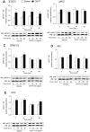
Figure 2. Effect of CNTF on phosphorylation of signaling proteins in the mHypoE-20/2 neuronal cells.
(A–E) mHypoE-20/2 neurons were serum starved for 12–16 h before treatment with 10 ng/ml (0.45 nM) CNTF (+) or vehicle alone (−) over a 60 min time course. Western blot analysis of cell lysates was performed using phospho-specific antibodies directed against (A) STAT3, (B) JAK2, (C) ERK1/2, (D) Akt, and (E) ACC. All results shown are normalized to G-protein β subunit relative to corresponding control protein levels at each time point and are expressed as mean ± SEM (n = 4 independent experiments, *P<0.05). Representative Western blots are shown.
Figure 3
Figure 3. Effect of CNTF on regulation of neuropeptides linked to feeding in mHypoE-20/2 neuronal cells.
Regulation of (A) urocortin-1, (B) urocortin-2, (C) neurotensin, (D) AgRP, and (E) BDNF mRNA expression by CNTF in the mHypoE-20/2 neuronal cell line. mHypoE-20/2 neuronal cells were exposed to 10 ng/ml (0.45 nM) CNTF, and real-time RT-PCR was performed over a 48 h timecourse. At the indicated time points, total RNA was extracted and used as a template for real-time RT-PCR with primers specifically designed to amplify each mRNA. mRNA levels were quantified using the standard curve method and normalized to the internal control (γ-actin). All results shown are relative to corresponding control mRNA levels at each time point. Data are represented as mean ± SEM (n = 5; *P<0.05; **P<0.01; ***P<0.001).
Figure 4
Figure 4. Acute CNTF treatment activates hypothalamic neurons.
Immunohistochemistry was performed to assess neuronal activation by c-Fos-immunoreactivity (ir) in wild-type mice treated with intracerebroventricular (i.c.v.) saline or CNTF (0.5 µg/ml). A–F: Representative photomicrographs showing expression of c-Fos-ir in the hypothalamic ARC, VMH, LH, dDMH, vDMH, PeV, and PVN regions in coronal sections of mouse hypothalami (as indicated on the images). Scale bar: 1 mm. Inset in each image represents a higher magnification of the boxed area. Scale bar: 100 µm. A and D represent negative control images for the anti-c-Fos antibody. DAB staining: nuclear brown (c-Fos). 3V, third ventricle. G: Bar graph showing the number of c-Fos-ir neurons in the hypothalamic regions at 2 h post-treatment. Data in the bar graph are expressed as mean ± SEM (n = 4 animals/group; *P<0.05; **P<0.01).
Figure 5
Figure 5. Acute CNTF treatment increases the number of hypothalamic neurons co-expressing c-Fos-immunoreactivity (ir) with urocortin-1-ir.
Representative photomicrographs are shown of neurons co-expressing c-Fos-ir with (A–D) urocortin-1-ir in coronal sections of the hypothalami from wild-type mice at 2 h following i.c.v. administration of saline or CNTF (0.5 µg/ml). A–D: low magnification (×50) images of the hypothalamic regions (scale bar: 1 mm); A′–D′: High magnification (×400) images representative of the selected regions in the images A–D, respectively (scale bar: 100 µm). A–D: Co-expression of c-Fos-ir with urocortin-1-ir in the PVN (A, B) and ARC (C, D). Black arrowheads represent neurons expressing only nuclear c-Fos-ir, yellow arrowheads represent neurons expressing only cytoplasmic perinuclear neuropeptide-ir, and red arrowheads represent double-labeled neurons with co-expression of c-Fos-ir and neuropeptide-ir. (E–F) Graphical representation showing the number of neurons expressing c-Fos and neuropeptide-immunoreactivity (ir) in the ARC, VMH, LH, dDMH, vDMH, PeV and PVN of the hypothalami from wild-type mice at 2 h following i.c.v. administration of saline or CNTF (0.5 µg/ml). Double-labeled immunohistochemistry for (F) c-Fos-ir and urocortin-1-ir indicates that intracerebroventricular CNTF activates hypothalamic neuropeptidergic neurons. No significant change in the number of neurons expressing only (E) urocortin-1-ir was observed in the hypothalamic regions of saline- or CNTF-treated animals. Data are represented as mean ± SEM (n = 4 mice/group; *P<0.05; **P<0.01; ***P<0.001 vs. saline treatment).

Similar articles

Cited by

References

    1. Sleeman MW, Anderson KD, Lambert PD, Yancopoulos GD, Wiegand SJ (2000) The ciliary neurotrophic factor and its receptor, CNTFR alpha. Pharm Acta Helv 74: 265–272. - PubMed
    1. Kokoeva MV, Yin H, Flier JS (2005) Neurogenesis in the hypothalamus of adult mice: potential role in energy balance. Science 310: 679–683. - PubMed
    1. Ettinger MP, Littlejohn TW, Schwartz SL, Weiss SR, McIlwain HH, et al. (2003) Recombinant variant of ciliary neurotrophic factor for weight loss in obese adults: a randomized, dose-ranging study. Jama 289: 1826–1832. - PubMed
    1. Lambert PD, Anderson KD, Sleeman MW, Wong V, Tan J, et al. (2001) Ciliary neurotrophic factor activates leptin-like pathways and reduces body fat, without cachexia or rebound weight gain, even in leptin-resistant obesity. Proc Natl Acad Sci U S A 98: 4652–4657. - PMC - PubMed
    1. Kozicz T, Bittencourt JC, May PJ, Reiner A, Gamlin PD, et al. (2011) The Edinger-Westphal nucleus: a historical, structural, and functional perspective on a dichotomous terminology. J Comp Neurol 519: 1413–1434. - PMC - PubMed

Publication types

MeSH terms