Global and ocular hypothermic preconditioning protect the rat retina from ischemic damage
- PMID: 23626711
- PMCID: PMC3633982
- DOI: 10.1371/journal.pone.0061656
Global and ocular hypothermic preconditioning protect the rat retina from ischemic damage
Abstract
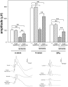
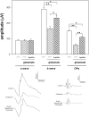
Retinal ischemia could provoke blindness. At present, there is no effective treatment against retinal ischemic damage. Strong evidence supports that glutamate is implicated in retinal ischemic damage. We investigated whether a brief period of global or ocular hypothermia applied 24 h before ischemia (i.e. hypothermic preconditioning, HPC) protects the retina from ischemia/reperfusion damage, and the involvement of glutamate in the retinal protection induced by HPC. For this purpose, ischemia was induced by increasing intraocular pressure to 120 mm Hg for 40 min. One day before ischemia, animals were submitted to global or ocular hypothermia (33°C and 32°C for 20 min, respectively) and fourteen days after ischemia, animals were subjected to electroretinography and histological analysis. Global or ocular HPC afforded significant functional (electroretinographic) protection in eyes exposed to ischemia/reperfusion injury. A marked alteration of the retinal structure and a decrease in retinal ganglion cell number were observed in ischemic retinas, whereas global or ocular HPC significantly preserved retinal structure and ganglion cell count. Three days after ischemia, a significant decrease in retinal glutamate uptake and glutamine synthetase activity was observed, whereas ocular HPC prevented the effect of ischemia on these parameters. The intravitreal injection of supraphysiological levels of glutamate induced alterations in retinal function and histology which were significantly prevented by ocular HPC. These results support that global or ocular HPC significantly protected retinal function and histology from ischemia/reperfusion injury, probably through a glutamate-dependent mechanism.
Conflict of interest statement
Figures


Similar articles
-
Involvement of glutamate in retinal protection against ischemia/reperfusion damage induced by post-conditioning.J Neurochem. 2009 Oct;111(2):488-98. doi: 10.1111/j.1471-4159.2009.06334.x. Epub 2009 Aug 13. J Neurochem. 2009. PMID: 19682205
-
Post-ischemic environmental enrichment protects the retina from ischemic damage in adult rats.Exp Neurol. 2013 Feb;240:146-56. doi: 10.1016/j.expneurol.2012.11.017. Epub 2012 Nov 27. Exp Neurol. 2013. PMID: 23195592
-
Retinal neuroprotection against ischemia-reperfusion damage induced by postconditioning.Invest Ophthalmol Vis Sci. 2009 Aug;50(8):3922-30. doi: 10.1167/iovs.08-3344. Epub 2009 Apr 1. Invest Ophthalmol Vis Sci. 2009. PMID: 19339733
-
Neuroprotective effect of transcorneal electrical stimulation on ischemic damage in the rat retina.Exp Eye Res. 2011 Nov;93(5):753-60. doi: 10.1016/j.exer.2011.09.022. Epub 2011 Oct 12. Exp Eye Res. 2011. PMID: 22008240
-
Molecular imaging of retinal disease.J Ocul Pharmacol Ther. 2013 Mar;29(2):275-86. doi: 10.1089/jop.2012.0279. Epub 2013 Feb 19. J Ocul Pharmacol Ther. 2013. PMID: 23421501 Free PMC article. Review.
Cited by
-
Adaptive Plasticity in the Retina: Protection Against Acute Injury and Neurodegenerative Disease by Conditioning Stimuli.Cond Med. 2018 Feb;1(2):85-97. Epub 2018 Feb 15. Cond Med. 2018. PMID: 31423482 Free PMC article.
-
Effect of hypothermic perfusion on phacoemulsification in cataract patients complicated with uveitis: a randomised trial.BMC Ophthalmol. 2020 Jun 16;20(1):232. doi: 10.1186/s12886-020-01507-9. BMC Ophthalmol. 2020. PMID: 32546211 Free PMC article. Clinical Trial.
-
Diminished apoptosis in hypoxic porcine retina explant cultures through hypothermia.Sci Rep. 2019 Mar 20;9(1):4898. doi: 10.1038/s41598-019-41113-4. Sci Rep. 2019. PMID: 30894574 Free PMC article.
-
Microglial Activation Promotes Cell Survival in Organotypic Cultures of Postnatal Mouse Retinal Explants.PLoS One. 2015 Aug 7;10(8):e0135238. doi: 10.1371/journal.pone.0135238. eCollection 2015. PLoS One. 2015. PMID: 26252475 Free PMC article.
-
PhTx3-4, a Spider Toxin Calcium Channel Blocker, Reduces NMDA-Induced Injury of the Retina.Toxins (Basel). 2016 Mar 11;8(3):70. doi: 10.3390/toxins8030070. Toxins (Basel). 2016. PMID: 26978403 Free PMC article.
References
-
- Osborne NN, Casson RJ, Wood JP, Chidlow G, Graham M, et al. (2004) Retinal ischemia: mechanisms of damage and potential therapeutic strategies. Prog Retin Eye Res 23: 91–147. - PubMed
-
- Fernandez DC, Chianelli MS, Rosenstein RE (2009) Involvement of glutamate in retinal protection against ischemia/reperfusion damage induced by post-conditioning. J Neurochem 111: 488–498. - PubMed
-
- Ikeda H, Hankins MW, Asai T, Dawes EA (1992) Electrophysiological properties of neurones following mild and acute retinal ischaemia. Exp Eye Res 55: 435–442. - PubMed
-
- Iversen LL (1991) Pharmacological approaches to the treatment of ischaemic neuronal damage. Eye (Lond) 5: 193–197. - PubMed
-
- Cazevieille C, Osborne NN (1997) Retinal neurones containing kainate receptors are influenced by exogenous kainate and ischaemia while neurones lacking these receptors are not – melatonin counteracts the effects of ischaemia and kainate. Brain Res 755: 91–100. - PubMed
Publication types
MeSH terms
Substances
LinkOut - more resources
Full Text Sources
Other Literature Sources
Medical

