. 2013 Apr 23;8(4):e61690.
doi: 10.1371/journal.pone.0061690. Print 2013.

In vitro effects of a small-molecule antagonist of the Tcf/ß-catenin complex on endometrial and endometriotic cells of patients with endometriosis

Affiliations

In vitro effects of a small-molecule antagonist of the Tcf/ß-catenin complex on endometrial and endometriotic cells of patients with endometriosis

Sachiko Matsuzaki et al. PLoS One. .

Abstract

Background: Our previous studies suggested that aberrant activation of Wnt/ß-catenin signaling might be involved in the pathophysiology of endometriosis. We hypothesized that inhibition of Wnt/ß-catenin signaling might result in inhibition of cell proliferation, migration, and/or invasion of endometrial and endometriotic epithelial and stromal cells of patients with endometriosis.

Objectives: The aim of the present study was to evaluate the effects of a small-molecule antagonist of the Tcf/ß-catenin complex (PKF 115-584) on cell proliferation, migration, and invasion of endometrial and endometriotic epithelial and stromal cells.

Methods: One hundred twenty-six patients (78 with and 48 without endometriosis) with normal menstrual cycles were recruited. In vitro effects of PKF 115-584 on cell proliferation, migration, and invasion and on the Tcf/ß-catenin target genes were evaluated in endometrial epithelial and stromal cells of patients with and without endometriosis, and in endometrial and endometriotic epithelial and stromal cells of the same patients.

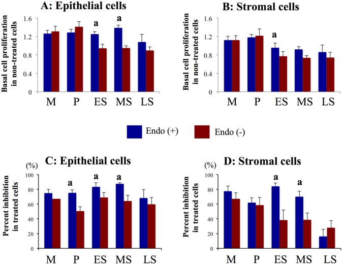
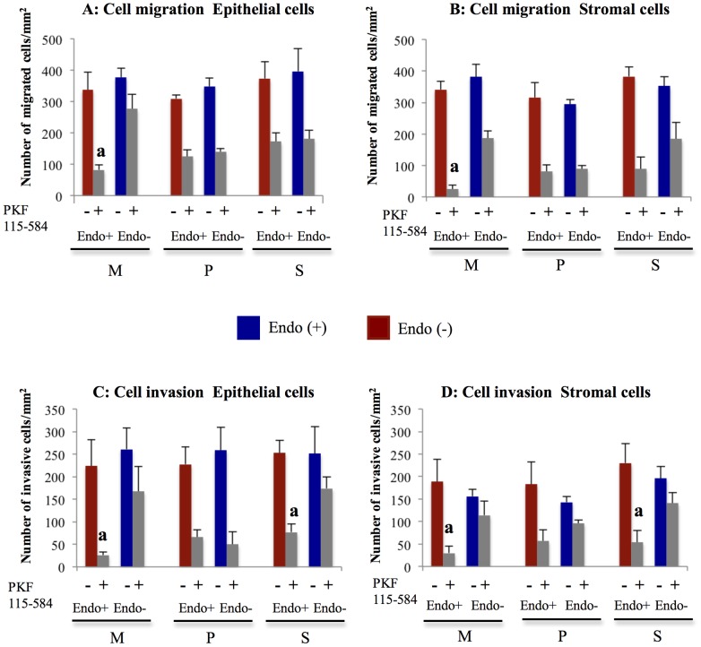
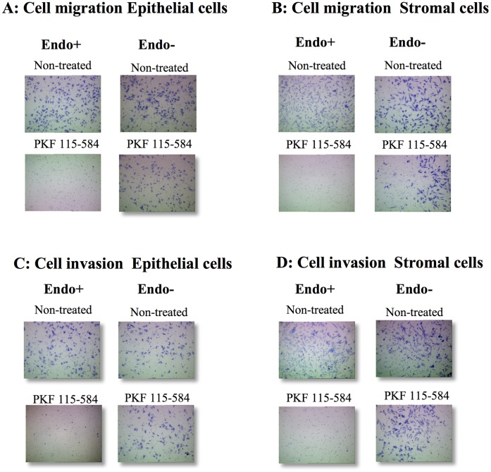
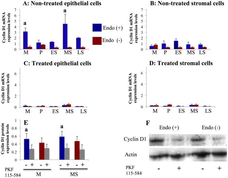
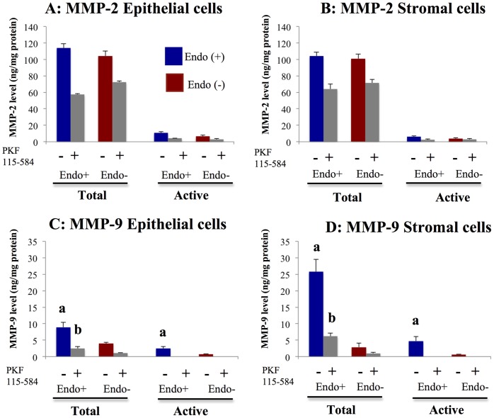
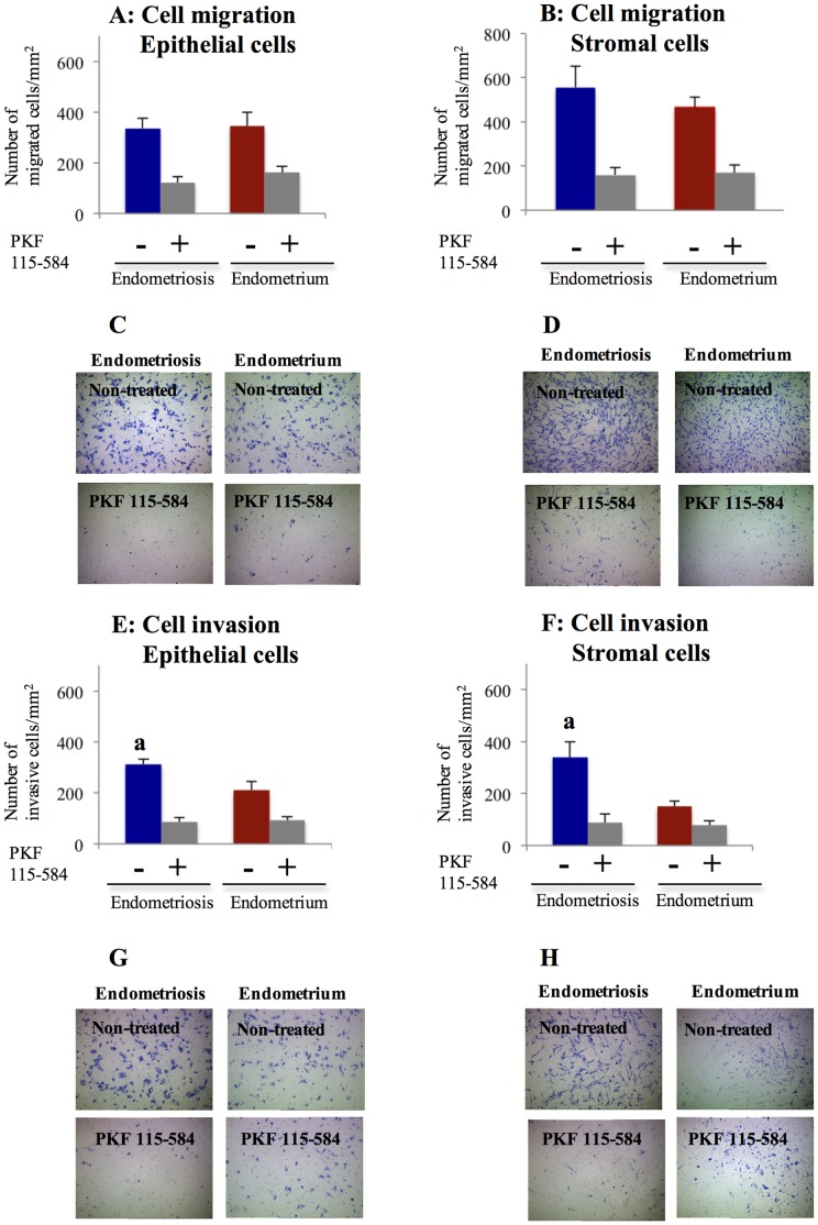
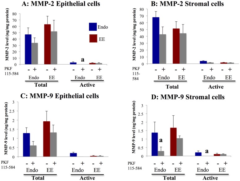
Results: The inhibitory effects of PKF 115-584 on cell migration and invasion in endometrial epithelial and stromal cells of patients with endometriosis prepared from the menstrual phase were significantly higher than those of patients without endometriosis. Levels of total and active forms of MMP-9 were significantly higher in epithelial and stromal cells prepared from menstrual endometrium in patients with endometriosis compared to patients without endometriosis. Treatment with PKF 115-584 inhibited MMP-9 activity to undetectable levels in both menstrual endometrial epithelial and stromal cells of patients with endometriosis. The number of invasive cells was significantly higher in epithelial and stromal cells of endometriotic tissue compared with matched eutopic endometrium of the same patients. Treatment with PKF 115-584 decreased the number of invasive endometriotic epithelial cells by 73% and stromal cells by 75%.

Conclusions: The present findings demonstrated that cellular mechanisms known to be involved in endometriotic lesion development are inhibited by targeting the Wnt/β-catenin pathway.


Conflict of interest statement

Competing Interests: The authors declare that this study was supported in part by Karl Storz Endoscopy & GmbH (Tuttlingen, Germany). There are no patents, products in development or marketed products to declare. This does not alter the authors’ adherence to all the PLOS ONE policies on sharing data and materials.

Figures

Figure 1
Figure 1. Effects of PKF 115–584 on cell proliferation.
A, B: Basal cell proliferation in non-treated endometrial epithelial (A) and stromal (B) cells of patients with and without endometriosis. C, D: Percent inhibition of cell proliferation in endometrial epithelial (C) and stromal (D) cells of patients with and without endometriosis treated with PKF 115–584. Results are presented as the mean+SEM. Basal cell proliferation is presented as OD. Percent inhibition of cell proliferation is calculated as percent of vehicle control. M: menstrual phase, P: proliferative phase, ES: early secretory phase, MS: mid- secretory phase, LS: late secretory phase. Endo (+): Endometrium of patients with endometriosis (M: n = 6, P: n = 20, ES: n = 7, MS: n = 15, LS: n = 6). Endo (–): endometrium of patients without endometriosis (M: n = 4, P: n = 11, ES: n = 8, MS: n = 8, LS: n = 4). a: p<.05 versus patients without endometriosis.
Figure 2
Figure 2. Effects of PKF 115–584 on cell migration and invasion.
A, B: Number of migrated cells/mm2 in non-treated and PKF 115–584–treated endometrial epithelial (A) and stromal (B) cells of patients with and without endometriosis. C, D: Number of invasive cells/mm2 in non-treated and PKF 115–584–treated endometrial epithelial (C) and stromal (D) cells of patients with and without endometriosis. Results are presented as the mean+SEM. M: menstrual phase, P: proliferative phase, S: secretory phase. Endo (+): Endometrium of patients with endometriosis (M: n = 4, P: n = 8, S: n = 8). Endo (–): endometrium of patients without endometriosis (M: n = 4, P: n = 5, S: n = 5). a: p<.05 versus PKF 115–584–treated endometrial epithelial or stromal cells of patients without endometriosis.
Figure 3
Figure 3. Representative photomicrographs of cell migration and invasion.
A, B: Representative photomicrographs of migration of non-treated and PKF 115–584–treated menstrual endometrial epithelial (A) and stromal (B) cells of patients with and without endometriosis (magnification x100). C, D: Representative photomicrographs of invasion of non-treated and PKF 115–584–treated menstrual endometrial epithelial (C) and stromal (D) cells of patients with and without endometriosis (magnification x100). Endo (+): Endometrium of patients with endometriosis prepared from the menstrual phase. Endo (–): endometrium of patients without endometriosis prepared from the menstrual phase.
Figure 4
Figure 4. Effects of PKF 115–584 on Cyclin D1 expression.
A, B: Cyclin D1 mRNA expression in non-treated endometrial epithelial (A) and stromal (B) cells of patients with and without endometriosis. Endo (+) (M: n = 6, P: n = 20, ES: n = 7, MS: n = 15, LS: n = 6). Endo (–) (M: n = 4, P: n = 11; ES: n = 8, MS: n = 8; LS: n = 4). C, D: Cyclin D1 mRNA expression in PKF 115–584–treated endometrial epithelial (C) and stromal (D) cells of patients with and without endometriosis. Endo (+):(M: n = 6, P: n = 20, ES: n = 7, MS: n = 15, LS: n = 6). Endo (–):(M: n = 4, P: n = 11, ES: n = 8, MS: n = 8, LS: n = 4). E: Cyclin D1 protein expression in non-treated and PKF 115–584–treated endometrial epithelial cells from the mid-secretory and menstrual phases. Endo (+):(M: n = 4, MS: n = 5). Endo (–):(M: n = 4, MS: n = 5). F: Representative photomicrographs of western blot analysis in non-treated and PKF 115–584–treated endometrial epithelial from the mid-secretory phase. Numerical values are presented as the mean+SEM. Expression levels of Cyclin D1 mRNA are given relative to the expression levels of the reference gene, GAPDH. Relative density is density of Cyclin D1 relative to that of Actin. M: menstrual phase, P: proliferative phase, ES: early secretory phase, MS: mid- secretory phase, LS: late secretory phase. a: p<.05 versus patients without endometriosis.
Figure 5
Figure 5. Effects of PKF 115–584 on total and active forms of MMP-2 and MMP-9.
A, B: Total and active forms of MMP-2 in non-treated and PKF 115–584–treated endometrial epithelial (A) and stromal (B) cells of patients with and without endometriosis. C, D: Total and active forms of MMP-9 in non-treated and PKF 115–584–treated endometrial epithelial (C) and stromal (D) cells of patients with and without endometriosis. Endo (+): Endometrium of patients with endometriosis (epithelial cells: n = 4, stromal cells: n = 4). Endo (–): endometrium of patients without endometriosis (epithelial cells: n = 3, stromal cells: n = 3). Values are normalized to the total protein content of the culture supernatants. Results are presented as the mean+SEM. a: p<.05 versus non-treated endometrial epithelial or stromal cells of patients without endometriosis. b: p<.05 versus PKF 115–584–treated endometrial epithelial or stromal cells of patients without endometriosis.MMP-9
Figure 6
Figure 6. Effects of PKF 115–584 on cell proliferation.
A, B: Basal cell proliferation in non-treated epithelial (A) and stromal (B) cells of endometriotic tissue and matched eutopic endometrium of the same patients. C, D: Percent inhibition of cell proliferation in epithelial (C) and stromal (D) cells of endometriotic tissue and matched eutopic endometrium of the same patients treated with PKF 115–584. Results are presented as the mean+SEM. Basal cell proliferation is presented as OD. Percent inhibition of cell proliferation is calculated as percent of vehicle control. P: proliferative phase, S: secretory phase. DE: deep infiltrating endometriosis (epithelial cells: P: n = 7, S: n = 8; stromal cells: P: n = 7, S:n = 9 ). OE: ovarian endometriosis (epithelial cells: P: n = 7 S: n = 6; stromal cells: P: n = 7, S: n = 8 ). SE: superficial peritoneal endometriosis (epithelial cells: P: n = 6, S: n = 6; stromal cells: P: n = 6, S: n = 7 ). A: p<.05 versus matched eutopic endometrium of the same patients.
Figure 7
Figure 7. Effects of PKF 115–584 on cell migration and invasion.
A, B: Number of migrated cells/mm2 in non-treated and PKF 115–584–treated epithelial (A) and stromal (B) cells of endometriotic tissue and matched eutopic endometrium of the same patient. C, D: Representative photomicrographs of migration of non-treated and PKF 115–584–treated epithelial (C) and stromal (D) cells of endometriotic tissue and matched eutopic endometrium of a same patient (magnification x100). E, F: Number of invasive cells/mm2 in non-treated and PKF 115–584–treated epithelial (E) and stromal (F) cells of endometriotic tissue and matched eutopic endometrium of the same patients. G, H: Representative photomicrographs of invasion of non-treated and PKF 115–584–treated epithelial (G) and stromal (H) cells of endometriotic tissue and matched eutopic endometrium of a same patient (magnification x100). Results are presented as the mean+SEM. Epithelial cells of endometriotic tissue and matched eutopic endometrium of the same patients (n = 16). Stromal cells of endometriotic tissue and matched eutopic endometrium (n = 16). a: p<.05 versus matched eutopic endometrium of the same patients.
Figure 8
Figure 8. Effects of PKF 115–584 on Cyclin D1 expression.
A, B: Cyclin D1 mRNA expression in non-treated epithelial (A) and stromal (B) cells of endometriotic tissue and matched eutopic endometrium of the same patients. C, D: Cyclin D1 mRNA expression in PKF 115–584–treated epithelial (C) and stromal (D) cells of endometriotic tissue and matched eutopic endometrium of the same patients. Numerical values are presented as the mean+SEM. Expression levels of Cyclin D1 mRNA are given relative to the expression levels of the reference gene, GAPDH. P: proliferative phase, S: secretory phase. DE: deep infiltrating endometriosis (epithelial cells: P: n = 6, S: n = 6, stromal cells: P: n = 6, S: n = 6). OE: ovarian endometriosis (epithelial cells: P: n = 6, S: n = 6; stromal cells: P: n = 6, S: n = 6). SE: superficial peritoneal endometriosis (epithelial cells: P: n = 4, S: n = 4; stromal cells: P: n = 4, S: n = 4). a: p<.05 versus matched eutopic endometrium of the same patients.
Figure 9
Figure 9. Effects of PKF 115–584 on total and active forms of MMP-2 and MMP-9.
A, B: Total and active forms of MMP-2 in non-treated and PKF 115–584–treated epithelial (A) and stromal (B) cells of endometriotic tissue and matched eutopic endometrium of patients with endometriosis. C, D: Total and active forms of MMP-9 in non-treated and PKF 115–584–treated epithelial (C) and stromal (D) cells of endometriosis and matched eutopic endometrium of patients with endometriosis. Values are normalized to the total protein content of the culture supernatants. Results are presented as the mean +SEM. Endo: endometriosis, EE: matched eutopic endometrium. Endo: (epithelial cells: n = 6, stromal cells: n = 6). EE: (epithelial cells: n = 6, stromal cells: n = 6). a: p<.05 versus PKF 115–584–treated endometrial epithelial or stromal cells.

References

    1. Giudice LC, Kao LC (2004) Endometriosis. Lancet 364: 1789–1799. - PubMed
    1. Matsuzaki S, Darcha C, Maleysson E, Canis M, Mage G (2010) Impaired down-regulation of E-cadherin and beta-catenin protein expression in endometrial epithelial cells in the mid-secretory endometrium of infertile patients with endometriosis. J Clin Endocrinol Metab 95: 3437–3445. - PubMed
    1. Matsuzaki S, Darcha C (2012) Epithelial to mesenchymal transition-like and mesenchymal to epithelial transition-like processes might be involved in the pathogenesis of pelvic endometriosis. Hum Reprod 27: 712–721. - PubMed
    1. Klaus A, Birchmeier W (2008) Wnt signalling and its impact on development and cancer. Nat Rev Cancer 8: 387–398. - PubMed
    1. Clevers H (2006) Wnt/beta-catenin signaling in development and disease. Cell 127: 469–480. - PubMed

Publication types

MeSH terms