. 2013 Apr 23;8(4):e62187.
doi: 10.1371/journal.pone.0062187. Print 2013.

Lipopolysaccharide disrupts the milk-blood barrier by modulating claudins in mammary alveolar tight junctions

Affiliations

Lipopolysaccharide disrupts the milk-blood barrier by modulating claudins in mammary alveolar tight junctions

Ken Kobayashi et al. PLoS One. .

Abstract

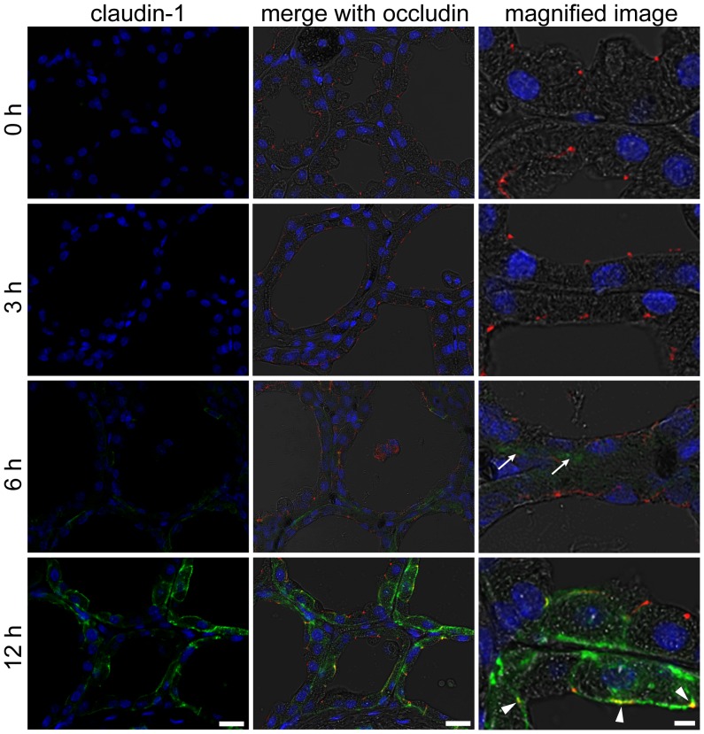
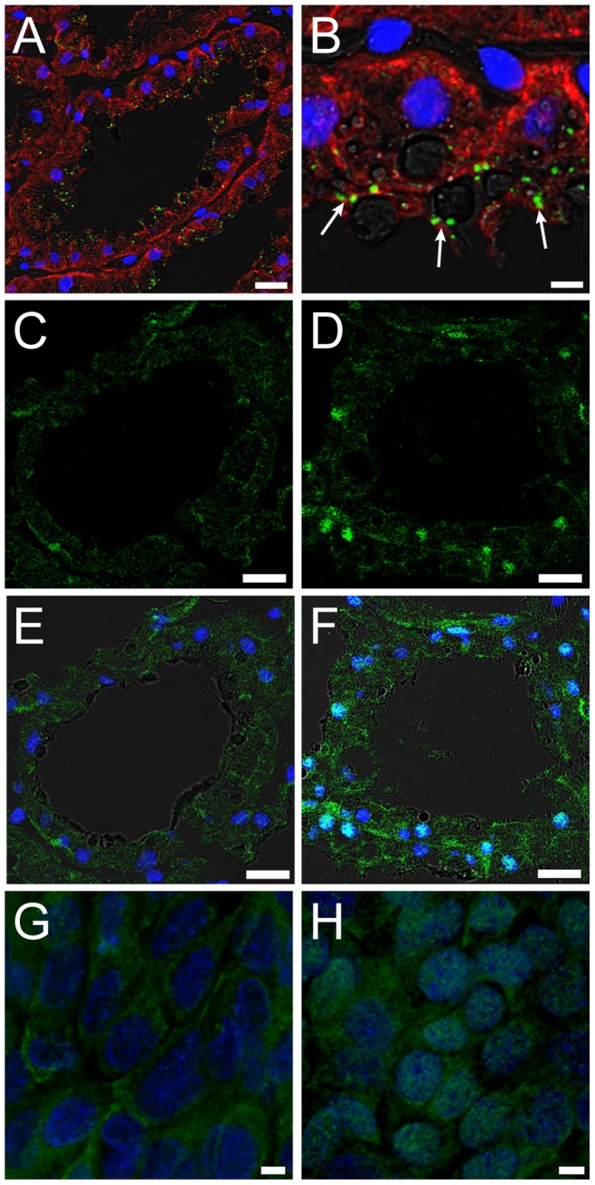
Mastitis, inflammation of the mammary gland, is the most costly common disease in the dairy industry, and is caused by mammary pathogenic bacteria, including Escherichia coli. The bacteria invade the mammary alveolar lumen and disrupt the blood-milk barrier. In normal mammary gland, alveolar epithelial tight junctions (TJs) contribute the blood-milk barrier of alveolar epithelium by blocking the leakage of milk components from the luminal side into the blood serum. In this study, we focused on claudin subtypes that participate in the alveolar epithelial TJs, because the composition of claudins is an important factor that affects TJ permeability. In normal mouse lactating mammary glands, alveolar TJs consist of claudin-3 without claudin-1, -4, and -7. In lipopolysaccharide (LPS)-induced mastitis, alveolar TJs showed 2-staged compositional changes in claudins. First, a qualitative change in claudin-3, presumably caused by phosphorylation and participation of claudin-7 in alveolar TJs, was recognized in parallel with the leakage of fluorescein isothiocyanate-conjugated albumin (FITC-albumin) via the alveolar epithelium. Second, claudin-4 participated in alveolar TJs with claudin-3 and claudin-7 12 h after LPS injection. The partial localization of claudin-1 was also observed by immunostaining. Coinciding with the second change of alveolar TJs, the severe disruption of the blood-milk barrier was recognized by ectopic localization of β-casein and much leakage of FITC-albumin. Furthermore, the localization of toll-like receptor 4 (TLR4) on the luminal side and NFκB activation by LPS was observed in the alveolar epithelial cells. We suggest that the weakening and disruption of the blood-milk barrier are caused by compositional changes of claudins in alveolar epithelial TJs through LPS/TLR4 signaling.


Conflict of interest statement

Competing Interests: The authors have declared that no competing interests exist.

Figures

Figure 1
Figure 1. LPS increases the permeability of the alveolar epithelium.
Mammary glands non-treated (0 h) and 3, 6, and 12 h after lipopolysaccharide (LPS) injection were immersed in FITC-albumin containing 0.5 mM CaCl2 and 0.5 mM MgCl2–containing phosphate-buffered saline (mPBS), and the localization of FITC-albumin was observed after cutting the frozen sections (A). Green and blue show FITC-albumin and nuclei (4′,6-diamidino-2-phenylindole, DAPI), respectively. FITC-albumin was observed in the alveolar lumen of LPS-injected mammary glands. Arrows show the localization of FITC-albumin in the interfacing regions of alveolar epithelial cells. (B) The paraffin sections were stained with antibodies to β-casein (red). Arrowheads show localization of β-casein on the interstitial side. Scale bars: 20 µm.
Figure 2
Figure 2. Expression changes of claudin-1, -3, -4, and -7 after LPS injection.
(A) Results of a western blot analysis of claudin-1, -3, -4, and -7 and β-actin in the mammary glands non-treated (0 h) and 3, 6, and 12 h after LPS injection. (B) The relative expression levels of claudin-1, -3 (total, upper band, lower band), -4, and -7 were analyzed by densitometry. The upper band of claudin-3 gradually disappeared after LPS injection. Beta-actin was used as a normalization control. Data represent mean (SD) (n  = 6). *, p<0.05; **, p<0.005 vs. 0 h.
Figure 3
Figure 3. LPS induces claudin-1 in the mammary alveolar epithelium.
The left column shows the immunostaining images of claudin-1 (green) and nuclear staining with DAPI (blue) in mammary glands non-treated (0 h) and 3, 6, and 12 h after LPS injection. The middle and right columns show the merged images with occludin (red) and bright field. Arrows indicate claudin-1–positive regions in basolateral membranes 6 h after LPS injection. Arrowheads indicate the localization of claudin-1 in the apical membrane 12 h after LPS injection. Scale bars: 20 µm (left and middle columns) and 5 µm (right column).
Figure 4
Figure 4. Claudin-3 localization around the apical-most regions before and after LPS injection.
The left column shows the immunostaining images of claudin-3 (green) and nuclear staining with DAPI (blue) in mammary glands non-treated (0 h) and 3, 6, and 12 h after LPS injection. The middle and right columns show the merged images with occludin (red) and bright field. Arrows indicate the localization of claudin-3 in the absence of occludin around the apical membrane after LPS injection. Scale bars: 20 µm (left and middle columns) and 5 µm (right column).
Figure 5
Figure 5. LPS induces claudin-4 expression in the mammary alveolar epithelium.
The left column shows the immunostaining images of claudin-4 (green) and nuclear staining with DAPI (blue) in mammary glands non-treated (0 h) and 3, 6, and 12 h after LPS injection. The middle and right columns show the merged images with occludin (red) and bright field, respectively. Scale bars: 20 µm (left and middle columns) and 5 µm (right column). Arrows indicate claudin-4–positive cells, which are rarely observed in the mammary alveolar epithelium. Arrowheads indicate the colocalization of claudin-4 and occludin at the apical-most regions.
Figure 6
Figure 6. LPS induces the translocation of claudin-7 from the basolateral membrane to the apical-most regions.
The left column shows the immunostaining images of claudin-7 (green) and nuclear staining with DAPI (blue) in mammary glands non-treated (0 h) and 3, 6, and 12 h after LPS injection. The middle and right columns show the merged images with occludin (red) and bright field, respectively. Scale bars: 20 µm (left and middle columns) and 5 µm (right column). Arrows indicate claudin-7–positive regions at the apical-most regions in the mammary alveolar epithelial cells with LPS injection. Arrowheads indicate the apical-most regions without claudin-7.
Figure 7
Figure 7. Influences of LPS on the detergent solubility of claudin-1, -3, -4, and -7.
(A) LPS was injected into the mammary glands on day 10 of lactation; the detergent-soluble (S) and detergent-insoluble (P) fractions of the mammary glands non-treated (0 h) and 3, 6 and 12 h after injection of LPS were isolated; and western blotting of claudin-1, -3, -4, and -7 was performed. Beta-actin (mixture of equal parts of the detergent-soluble and -insoluble fractions) was used as the normalization control. (B, D, E) The bands of the insoluble fractions were analyzed by densitometry. (C) The ratio of the upper band to the lower band of insoluble fractions of claudin-3 was calculated using the formula S/P. Data are represented as mean (SD) (n  = 4). *, p<0.05; **, p<0.005 vs. 0 h.
Figure 8
Figure 8. Influences of LPS on shedding of alveolar epithelial cells.
Tight junctions (TJs) around shedding cells from the alveolar epithelium by LPS injection were observed by immunostaining for claudin-3 (green) and occludin (red). Continuous TJs were observed between detaching cells and undetached cells during the shedding process in mammary glands 3 (top), 6 (middle), and 12 h (bottom) after LPS injection. Scale bars: 20 µm (left column) and 5 µm (right column).
Figure 9
Figure 9. Localization of TLR4 and NFκB in mammary alveolar epithelial cells.
(A, B) The mammary glands on day 10 of lactation were immunostained for TLR4 (green) and pan-keratin (red). The localization of TLR4 in the apical membrane was observed (arrow). (C, D) Immunostaining images of NFκB (green) in the mammary gland non-treated and 3 h after LPS injection, respectively. (E, F) Merged images of C and D, with DAPI and bright field, respectively. Cultured mammary epithelial cells without (G) or with LPS treatment for 1 h (H) were immunostained for NFκB (green). Scale bars: 20 µm (A, C–F) and 5 µm (B, G, H).

Similar articles

Cited by

References

    1. Akers RM, Nickerson SC (2011) Mastitis and its impact on structure and function in the ruminant mammary gland. J Mammary Gland Biol Neoplasia 16: 275–289. - PubMed
    1. Spencer JP (2008) Management of mastitis in breastfeeding women. Am Fam Physician 78: 727–731. - PubMed
    1. Barlow J (2011) Mastitis therapy and antimicrobial susceptibility: a multispecies review with a focus on antibiotic treatment of mastitis in dairy cattle. J Mammary Gland Biol Neoplasia 16: 383–407. - PubMed
    1. McFadden TB, Akers RM, Capuco AV (1988) Relationship of milk proteins in blood with somatic cell counts in milk of dairy cows. J Dairy Sci 71: 826–834. - PubMed
    1. McFadden TB, Akers RM, Kazmer GW (1987) Alpha-lactalbumin in bovine serum: relationships with udder development and function. J Dairy Sci 70: 259–264. - PubMed

Publication types

MeSH terms

LinkOut - more resources