. 2013 Apr 23;8(4):e62188.
doi: 10.1371/journal.pone.0062188. Print 2013.

ATM influences the efficiency of TCRβ rearrangement, subsequent TCRβ-dependent T cell development, and generation of the pre-selection TCRβ CDR3 repertoire

Affiliations

ATM influences the efficiency of TCRβ rearrangement, subsequent TCRβ-dependent T cell development, and generation of the pre-selection TCRβ CDR3 repertoire

Karen S Hathcock et al. PLoS One. .

Abstract

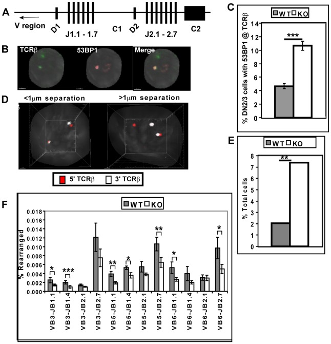
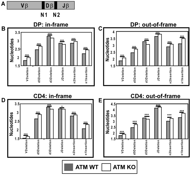
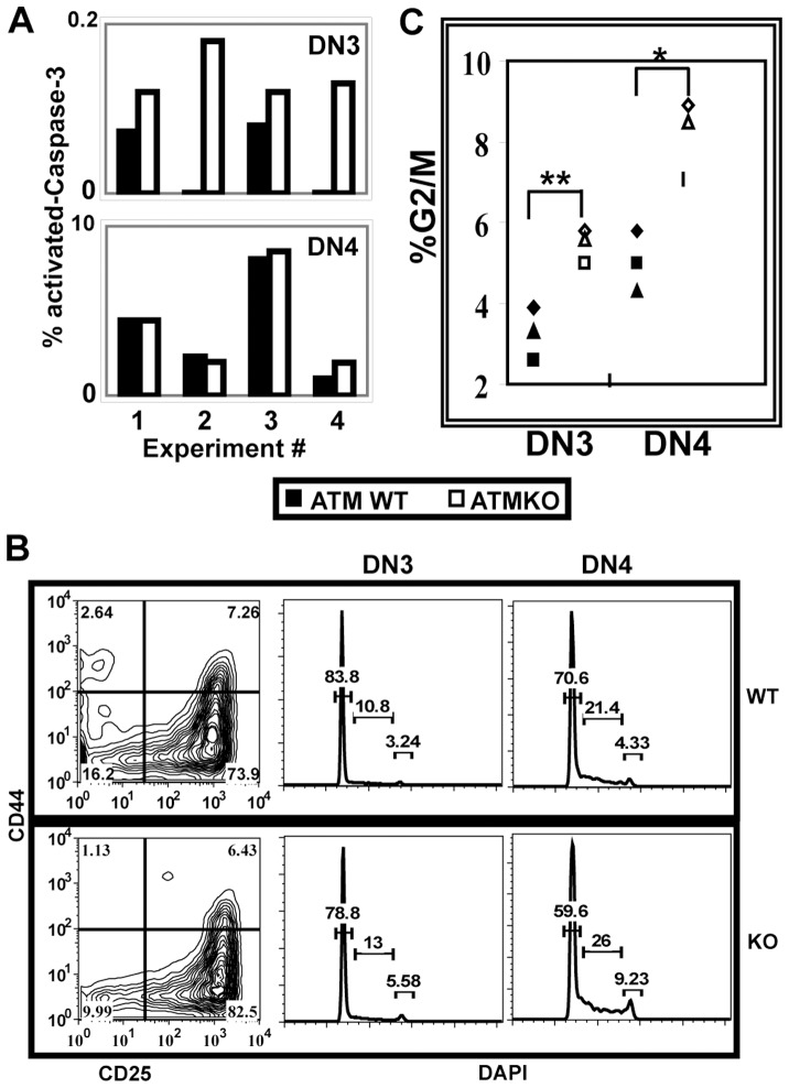
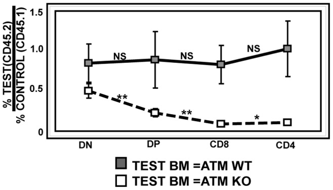
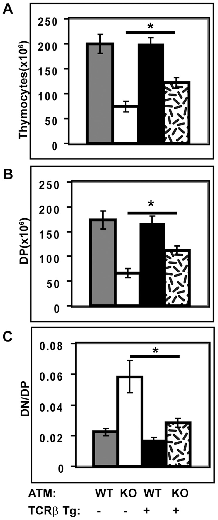
Generation and resolution of DNA double-strand breaks is required to assemble antigen-specific receptors from the genes encoding V, D, and J gene segments during recombination. The present report investigates the requirement for ataxia telangiectasia-mutated (ATM) kinase, a component of DNA double-strand break repair, during TCRβ recombination and in subsequent TCRβ-dependent repertoire generation and thymocyte development. CD4(-)CD8(-) double negative stage 2/3 thymocytes from ATM-deficient mice have both an increased frequency of cells with DNA break foci at TCRβ loci and reduced Vβ-DJβ rearrangement. Sequencing of TCRβ complementarity-determining region 3 demonstrates that ATM-deficient CD4(+)CD8(+) double positive thymocytes and peripheral T cells have altered processing of coding ends for both in-frame and out-of-frame TCRβ rearrangements, providing the unique demonstration that ATM deficiency alters the expressed TCRβ repertoire by a selection-independent mechanism. ATMKO thymi exhibit a partial developmental block in DN cells as they negotiate the β-selection checkpoint to become double negative stage 4 and CD4(+)CD8(+) thymocytes, resulting in reduced numbers of CD4(+)CD8(+) cells. Importantly, expression of a rearranged TCRβ transgene substantially reverses this defect in CD4(+)CD8(+) cells, directly linking a requirement for ATM during endogenous TCRβ rearrangement to subsequent TCRβ-dependent stages of development. These results demonstrate that ATM plays an important role in TCRβ rearrangement, generation of the TCRβ CDR3 repertoire, and efficient TCRβ-dependent T cell development.


Conflict of interest statement

Competing Interests: The authors have declared that no competing interests exist.

Figures

Figure 1
Figure 1. Rearrangement of TCRβ is impaired in ATMKO DN2/3 cells.
(A) Genomic organization of the murine TCRβ locus. (B) A representative immuno-FISH image of an ATMKO DN2/3 cell showing a 53 BP1 focus (red) at one of two TCRβ loci (green). White bar denotes 1.5 micron. (C) Graph shows mean frequency (±SEM) of ATMWT (WT) and ATMKO (KO) DN2/3 cells expressing 53 BP1 foci at the TCRβ locus. 200 cells of each genotype were analyzed in two experiments. (D) Representative 3D-FISH images of two ATMKO DN2/3 cells hybridized with 5′TCRβ (red) and 3′TCRβ probes (white). In left image probes are separated by less than 1 µm at both TCRβ loci; whereas, in right image probes are separated by more than 1 µm at one of two TCRβ loci. (E) Summary of the frequency of ATMWT and KO DN2/3 cells in which the 5′ and 3′TCRβ probes are separated by more than 1 µm one of two TCRβ loci. More than 300 cells of each genotype were collected in two experiments. (F) Vβ-DJβ rearrangement in genomic DNA from ATMWT and KO DN2/3 cells was quantified using real-time PCR and normalized to invariant Cα. This plot is mean Vβ-DJβ rearrangements±SEM of seven ATMWT and KO data sets analyzed in five real-time PCR experiments. P values: Student’s unpaired (C) and paired (F) 1-tailed t-test and Fisher’ exact test (E). *p<0.05; **<0.01, ***p<0.001.
Figure 2
Figure 2. ATM deficiency alters in-frame and out-of-frame junctional sequences of TCRβ CDR3 in pre- and post-selection T cells.
(A) Diagram of TCRβ CDR3 junctions. Nucleotide deletions and additions were sequenced from in-frame and out-of-frame TCRβ CDR3 regions isolated from ATMWT (grey bars) or KO (white bars) cells that were either TCRα KO DP thymocytes (B and C) or naïve CD4+ T cells (D and E). TCRαKO DP and naïve CD4+ T cells were sorted from two mice of each ATM genotype. Each data point represents >55,000 sequences. P values were calculated using Student’s unpaired 2-tailed t-test. *p<0.05; **p<0.01; ***p<0.001.
Figure 3
Figure 3. ATM deficiency alters thymic development.
(A) Thymocytes from four pairs of ATMWT and KO mice were analyzed by flow cytometry to enumerate cells in each CD4/CD8 subpopulation. (B) The DN3/DN4 ratio is elevated in ATMKO thymi. Representative CD44 and CD25 staining profiles of lineage negative ATMWT and KO DN cells and the calculated DN3/DN4 ratios are shown. Quantification of DN3/DN4 ratios measured in ATMWT and KO thymi (ATMWT vs KO p<0.025). (C) The DN/DP ratio is elevated in ATMKO thymi. Images show CD4/CD8 staining profiles and corresponding DN/DP ratios from representative ATMWT and KO thymi. Quantification of DN/DP ratios measured in ATMWT and KO thymi (ATMWT vs KO p<0.003). Data are mean (± SEM) of six separate experiments analyzing nine pairs of ATMWT and KO thymi. P values were calculated using Student’s paired 1-tailed t test.
Figure 4
Figure 4. ATM deficiency alters cell survival and proliferation in DN cells.
Freshly explanted thymocytes were stained for surface molecules, fixed, and permeabilized prior to intracellular staining. (A) As compared to ATMWT, ATMKO DN3 but not DN4 cells have an increased frequency of cleaved-Caspase-3+ cells (ATMWT vs KO DN3 p<0.03). Graphs show frequencies of ATMWT (grey bar) and KO (white bar) lineage negative DN3 and DN4 cells that express cleaved-Caspase-3. Data from four independent experiments analyzing ATMWT and KO pairs is shown. (B) Panels show representative lineage negative CD25 CD44 DN staining profiles for ATMWT (top panel) and KO (lower panel) thymi and DAPI staining profiles gated on DN3 and DN4 cells. (C) Frequency of DN3 and DN4 cells from individual ATMWT (black symbols) and KO (open symbols) mice that are in G2/M of the cell cycle. ATM deficiency results in increased cycling cells in both DN3 and DN4 stages (p<0.002 and 0.02, respectively). P values were calculated using Student’s paired 1-tailed t test from data collected from three-four ATMWT and KO pairs analyzed in three independent experiments.
Figure 5
Figure 5. Competitive chimeras demonstrate that the ATMKO defect in DN to DP development is T cell-intrinsic.
This graph shows the ratio of thymocytes derived from test (ATMWT or KO CD45.2+) and control (ATMWT CD45.1+) bone marrow for each thymic subset. When both test (CD45.2+) and control (CD45.1+) bone marrows are ATMWT (solid black line), the ratio of test/control cells is not significantly changed during thymic development. In contrast, when test bone marrow is ATMKO (CD45.2+) (dashed black line) the ratio of test/control (ATMKO/ATMWT) cells decreases as thymocytes develop from DN to DP cells (p<0.007) and from DP to CD8 (p<0.005) or CD4 (p<0.02) SP cells. Data from two independent sets of chimeras consisting of 16–20 mice in each group were combined for this analysis. P values were calculated using Student’s unpaired 1-tailed t-test.
Figure 6
Figure 6. Introduction of a rearranged TCRβ TG significantly improves the ATMKO defect in DN to DP development.
Effects of TCRβ TG expression on ATMWT (WT) and ATMKO (KO) thymic cellularity (A), numbers of DP cells (B), and DN/DP ratios (C). Results in panels A–C are mean (± SEM) for three ATMWT and KO pairs analyzed in three independent experiments. For the parameters that were tested, ATMWT thymi with and without the TCRβ TG did not differ significantly. In contrast, ATMKO thymi with and without the TCRβ TG did differ significantly (p<0.05). P values were calculated using Student’s paired 1-tailed t test.

Similar articles

Cited by

References

    1. Hesslein DG, Schatz DG (2001) Factors and forces controlling V(D)J recombination. Adv Immunol 78: 169–232. - PubMed
    1. Fugmann SD, Lee AI, Shockett PE, Villey IJ, Schatz DG (2000) The RAG proteins and V(D)J recombination: complexes, ends, and transposition. Annu Rev Immunol 18: 495–527. - PubMed
    1. Katayama CD, Eidelman FJ, Duncan A, Hooshmand F, Hedrick SM (1995) Predicted complementarity determining regions of the T cell antigen receptor determine antigen specificity. Embo J 14: 927–938. - PMC - PubMed
    1. Goyarts EC, Vegh Z, Kalergis AM, Horig H, Papadopoulos NJ, et al. (1998) Point mutations in the beta chain CDR3 can alter the T cell receptor recognition pattern on an MHC class I/peptide complex over a broad interface area. Mol Immunol 35: 593–607. - PubMed
    1. Godfrey DI, Kennedy J, Mombaerts P, Tonegawa S, Zlotnik A (1994) Onset of TCR-beta gene rearrangement and role of TCR-beta expression during CD3–CD4–CD8- thymocyte differentiation. J Immunol 152: 4783–4792. - PubMed

Publication types

MeSH terms

Substances