. 2013 Apr 23;8(4):e62252.
doi: 10.1371/journal.pone.0062252. Print 2013.

Crystal structures of a plant trypsin inhibitor from Enterolobium contortisiliquum (EcTI) and of its complex with bovine trypsin

Affiliations

Crystal structures of a plant trypsin inhibitor from Enterolobium contortisiliquum (EcTI) and of its complex with bovine trypsin

Dongwen Zhou et al. PLoS One. .

Abstract

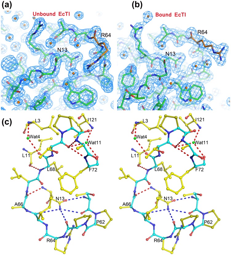
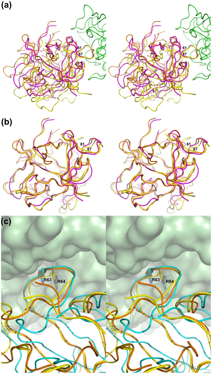
A serine protease inhibitor from Enterolobium contortisiliquum (EcTI) belongs to the Kunitz family of plant inhibitors, common in plant seeds. It was shown that EcTI inhibits the invasion of gastric cancer cells through alterations in integrin-dependent cell signaling pathway. We determined high-resolution crystal structures of free EcTI (at 1.75 Å) and complexed with bovine trypsin (at 2 Å). High quality of the resulting electron density maps and the redundancy of structural information indicated that the sequence of the crystallized isoform contained 176 residues and differed from the one published previously. The structure of the complex confirmed the standard inhibitory mechanism in which the reactive loop of the inhibitor is docked into trypsin active site with the side chains of Arg64 and Ile65 occupying the S1 and S1' pockets, respectively. The overall conformation of the reactive loop undergoes only minor adjustments upon binding to trypsin. Larger deviations are seen in the vicinity of Arg64, driven by the needs to satisfy specificity requirements. A comparison of the EcTI-trypsin complex with the complexes of related Kunitz inhibitors has shown that rigid body rotation of the inhibitors by as much as 15° is required for accurate juxtaposition of the reactive loop with the active site while preserving its conformation. Modeling of the putative complexes of EcTI with several serine proteases and a comparison with equivalent models for other Kunitz inhibitors elucidated the structural basis for the fine differences in their specificity, providing tools that might allow modification of their potency towards the individual enzymes.


Conflict of interest statement

Competing Interests: The authors have declared that no competing interests exist.

Figures

Figure 1
Figure 1. Preparation of the EcTI-trypsin complex.
Free EcTI and its complex with trypsin were run on the same Sephacryl S-100 HR column, with the same buffer. The two curves were overlaid, with the blue one representing EcTI-trypsin and the red one representing free EcTI.
Figure 2
Figure 2. Sequence alignment of EcTI and related serine protease inhibitors, partially structure-based (the structures of Acacia, DE5, and Bauhinia (rows 2–4) have not been determined).
The top two sets of rows correspond to the long chain in EcTI, and the third set of rows represents the short chain. The secondary structure elements of EcTI are shown on top of the alignment in orange. Residues identical in all inhibitors are colored green and those identical in some, red. The reactive loop regions are highlighted in yellow, and a blue triangle indicates the position of P1 residue.
Figure 3
Figure 3. EcTI and its complex with trypsin.
(a) Ribbon representation of the overall three-dimensional structure of EcTI. β-strands labeled β1–12 are shown in red, and loops labeled L1–13 are in green. Loop L10 is only marked for consistency with the other related structures, since the main chain is broken in this region. (b) Pseudo-dimer of the two crystallographically independent molecules of EcTI in the asymmetric unit. The main contact loops are labeled. (c) Overall structure of the EcTI-trypsin complex shown as a cartoon diagram. EcTI is colored green, while trypsin is red, and the reactive loop is labeled.
Figure 4
Figure 4. Superposition of EcTI in free and bound state.
(a) Interface between EcTI (yellow ribbon) and trypsin (green space filling representation) in the inhibitor-trypsin complex. The side chains of Arg64 and Leu115 of EcTI are shown as sticks. A superimposed tracing of molecule B of free EcTI is shown in red, clearly indicating that the conformational changes in the reactive loop around Arg64 are needed in order to avoid clashes and allow the proper fit of its side chain into the S1 pocket of the enzyme. (b) Steroview of the superposition of trypsin-bound EcTI and STI (PDB ID: 1AVW). Color highlights are for the loops which are most different between these two proteins, blue for EcTI and red for STI, whereas other parts of the chain are gray.
Figure 5
Figure 5. The reactive loop of EcTI.
(a,b) 2Fo-Fc electron density maps contoured at 1.2σ of the reactive loop area of (a) free EcTI and (b) its complex with trypsin. (c) A steroview of the hydrogen networks of the reactive loop region of a molecule of free EcTI. Blue dashed lines indicate the hydrogen bond network formed by the side chain of Asn13 and the interacting residues, and red dashed lines indicate the hydrogen bond network formed by the reactive loop region residues and their partners. Green spheres represent water molecules.
Figure 6
Figure 6. Rigid body movement of the Kunitz inhibitors bound to trypsin.
(a) Superposition of the trypsin complexes with EcTI, STI, and TKI based on the Cα coordinates of trypsin reveals overall rigid body shifts of the inhibitors. The tracing of the trypsin chain in the complex with EcTI is shown as green ribbon, EcTI is yellow, STI red, and TKI magenta. (b) Superposition of EcTI, STI, and TKI based on their Cα coordinates (colored as in panel (a)) reveals a small change in the orientation of the reactive loop (residues 61–67). (c) Superposition of EcTI bound to trypsin with the STI complex. STI is shown in orange and trypsin in space-filling green. In order to show the reason for the rigid body movement of EcTI it is shown twice, in yellow when superimposed directly on STI, and in blue when the trypsin molecules of the two complexes were superimposed.
Figure 7
Figure 7. Close-up of the interactions of EcTI and trypsin.
(a) Steroview of the region of interactions, with residues belonging to trypsin shown as cyan sticks and the disulfide bond colored yellow. The residues of EcTI are shown as green sticks and the gray spheres represent water molecules. Hydrogen bonds are indicated by red dashed lines. (b) Interface of the EcTI-trypsin complex, with EcTI shown as a green ribbon with selected side chains in stick representation, whereas the surface of trypsin is colored according to charge (blue positive, red negative, gray uncharged).
Figure 8
Figure 8. Structural basis for the differences in the inhibitory properties of Kunitz inhibitors towards different serine proteases.
(a) Two possible areas of conflict between EcTI and Factor Xa. Factor Xa (gray) is superimposed on trypsin (green) complexed with EcTI (yellow). Complexes of STI and TKI are superimposed on the basis of their trypsin components (orange and magenta, respectively). (b) The third possible area of conflict between EcTI and Factor Xa. A modeled surface of Factor Xa (PDB ID: 2JKH) is gray. The colors for other molecules are as in (a). (c) Different orientation of the N terminus in EcTI (yellow) compared to STI (magenta) and TKI (orange).

References

    1. Murzin AG, Chothia C (1992) Protein architecture: new super families. Curr Opin Struct Biol 2: 895–903.
    1. Renko M, Sabotic J, Turk D (2012) Beta trefoil inhibitors from the work of Kunitz onwards. Biol Chem 393: 1043–1054. - PubMed
    1. Horigome D, Satoh H, Itoh N, Mitsunaga K, Oonishi I, et al. (2007) Structural mechanism and photoprotective function of water-soluble chlorophyll-binding protein. J Biol Chem 289: 6525–6531. - PubMed
    1. Cagan RH (1973) Chemostimulatory protein: A new type of taste stimulus. Science 181: 32–35. - PubMed
    1. van Oostrum J, Priestle JP, Grütter MG, Schmitz A (1991) The structure of murine interleukin-1 beta at 2.8 Å resolution. J Struct Biol 107: 189–195. - PubMed

Publication types

LinkOut - more resources