. 2013 Apr 23;8(4):e62621.
doi: 10.1371/journal.pone.0062621. Print 2013.

Generation of inducible immortalized dendritic cells with proper immune function in vitro and in vivo

Affiliations

Generation of inducible immortalized dendritic cells with proper immune function in vitro and in vivo

Cornelia Richter et al. PLoS One. .

Abstract

Dendritic cells are the professional antigen presenting cells of innate immunity and key players in maintaining the balance of immune responses. Studies with dendritic cells are mainly limited by their low numbers in vivo and their difficult maintenance in vitro. We differentiated bone marrow cells from transgenic mice expressing an inducible SV40 large T-antigen into dendritic cells. When immortalized by dexamethasone and doxycycline, these cells were stable in long-term culture. In the absence of dexamethasone and doxycycline (de-induction), dendritic cells displayed properties of primary cells, characterized by expression of classical dendritic cell surface markers CD11c, CD11b, MHCII, CD40 and CD86. Furthermore, de-induced lipopolysaccharide activated dendritic cells secreted IL-1β, IL-6, TNFα and IL-12. De-induced, Ovalbumin-loaded dendritic cells polarize CD4(+) T cells into Th1, Th17 and Th2 cells, indicating their correct antigen presenting property. Consistent with intratracheal application of Ovalbumin-loaded primary dendritic cells into mice, the application of de-induced dendritic cells resulted in recruitment of lymphocytes to the lungs. In summary, we successfully expanded dendritic cells using conditional immortalization. The generated dendritic cells demonstrate the characteristic immunophenotype of primary dendritic cells and will facilitate further studies on immunomodulatory properties of dendritic cells.


Conflict of interest statement

Competing Interests: The authors have declared that no competing interests exist.

Figures

Figure 1
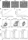
Figure 1. Morphology, cell cycle and proliferation.
(A) Microscopic images of BM-DCs, iniDCs and de-iniDCs 3-days after de-induction at 10× magnification. Both, BM-DCs and de-iniDCs show an adherent phenotype with the typical formation of dendrites. (B) Proliferation of iniDCs and de-iniDCs was analyzed by counting the cells in a haemocytometer over a time period of 6 days. (C) Percentage of dead cells counted over a time period of 6 days. (D) Apoptosis and necrosis of iniDCs, 3- and 5-days cultured de-iniDCs were analyzed using anti-AnnexinV-PE antibody and DAPI. Dot blots display AnnexinV and DAPI stained cells. (E) For cell cycle analysis, iniDCs, 3- and 5-days cultured de-iniDCs were stained with PI and analyzed by flow cytometry. Cell cycle stages G1 (left peak), S (middle) and G2 (right peak) were calculated with the Dean-Jett-Fox model using FlowJo software. Proliferation, apoptosis and cell cycle analyses were performed in three independent experiments. For apoptosis and cell cycle analysis the result of a representative experiment is given.
Figure 2
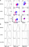
Figure 2. Dendritic cell surface marker expression.
(A) BM-DCs, iniDCs and 3-days cultured de-iniDCs were stained with antibodies against the dendritic cell subset markers CD11c, CD8α, CD11b, B220 and Ly6C. CD11c+ cells (black curve) were further gated for CD8α and CD11b, Ly6C and B220 (contour blots). Gates for CD8α and CD11b, Ly6C and B220 were set on the respective unstained control (red). (B) Immature and mature BM-DCs, iniDCs and de-iniDCs were stained for MHCII, CD40, and CD86. Dead cells (DAPI staining) and cell doublets were excluded. Histograms show the isotype control (grey, dotted), immature cells (grey) and LPS-matured cells (black). The result of one representative experiment is given.
Figure 3
Figure 3. Cytokine profile of dendritic cells.
BM-DCs, iniDCs and de-iniDCs were stimulated with LPS (1 µg/mL) for 24 hours. Secretion of IL-1β, IL-6, IL-12p70 and TNFα was measured in the supernatant of non-induced BM-DCs (white bars), iniDCs (black bars) and de-iniDCs (grey bars) via CBA. For each cytokine the mean ± SEM of three independent experiments is given.
Figure 4
Figure 4. CD11c expression and IL-12 production in single cell clones.
De-iniDC single cell clones were stimulated with LPS or left untreated for 24 hours in the presence of the protein transport inhibitor Monensin. Afterwards, cells were stained for the surface marker CD11c, permeabilized and stained for intracellular IL-12. (A) CD11c expression (black) of LPS stimulated cells is displayed. (B) Intracellular IL-12 expression level of CD11c+ LPS stimulated (black) and non-stimulated cells (grey) are shown. Isotype control is displayed as grey, dotted curve (A, B).
Figure 5
Figure 5. Antigen presentation of de-iniDC clone #1 and BM-DCs to T cells.
De-iniDC clone #1 or BM-DCs were incubated with OVA (13.5 µg/mL) for 24 hours prior to co-culture with OTII/CD45.1 CD4+ T cells or OTI CD8+ T cells. (A) Proliferation of CD4+ T cells was measured using CFSE staining and analyzed by flow cytometry. (B) Secretion of IL-2 was measured with CBA. (C) CD4+ T cell secreted cytokines IFNγ, IL-13 and IL-17 were measured in the cell culture supernatant using CBA after 48 hours. (D) Proliferation of CD8+ T cells was measured using CFSE staining and flow cytometry. (E) CD8+ T cell secreted cytokines IL-2 and IFNγ were measured in the supernatant using CBA after 48 hours. Results of three to four independent experiments are given as mean ± SEM, (n.d.) not detectable. Statistical significance is indicated, *(P<0.05), **(P<0.01) and ***(P<0.001).
Figure 6
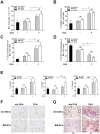
Figure 6. In vivo immune response induced by de-iniDCs and BM-DCs.
(A) 48 hours after intratracheal application of cells, BAL fluid was collected and cells were counted in a haemocytometer. (B) The percentage of the CD66a+ neutrophils in the BAL fluid was analyzed by flow cytometry. (C) Percentage of CD3+ T cells in the BAL fluid of provoked mice were analyzed by flow cytometry. (D) Numbers of F4/80+ macrophages in the BAL fluid were analyzed by flow cytometry. (E) T cell cytokine secretion was measured in the BAL fluid by CBA. (F) May-Grünwald-Giemsa stained cytospin preparations demonstrate recruited eosinophils. (G) Paraffin-embedded lung sections were stained with Hematoxylin and Eosin. Results are expressed as mean ± SEM from 5 mice per group. Statistical significance is indicated, *(P<0.05) and **(P<0.01).
Figure 7
Figure 7. Lentiviral vector mediated transgene expression in iniDCs.
(A) RFP expression level was measured in untransduced (grey dotted) and lentiviral vector particle-transduced iniDCs before (grey) and after (black) puromycin selection. (B) Expression level of maturation markers MHCII, CD40 and CD86 were determined in transduced iniDCs and after their deinduction (de-iniDCs) using flow cytometry. Transduced iniDCs and de-iniDCs (grey) and LPS-stimulated transduced iniDCs and de-iniDCs (black) are shown. Isotype controls are displayed as grey dotted lines. One representative experiment out of 3 is shown.

References

    1. Steinman RM, Cohn ZA (1973) Identification of a novel cell type in peripheral lymphoid organs of mice. I. Morphology, quantitation, tissue distribution. J Exp Med 137: 1142–1162. - PMC - PubMed
    1. Guilliams M, Henri S, Tamoutounour S, Ardouin L, Schwartz-Cornil I, et al. (2010) From skin dendritic cells to a simplified classification of human and mouse dendritic cell subsets. Eur J Immunol 40: 2089–2094. - PubMed
    1. Ginhoux F, Liu K, Helft J, Bogunovic M, Greter M, et al. (2009) The origin and development of nonlymphoid tissue CD103+ DCs. J Exp Med 206: 3115–3130. - PMC - PubMed
    1. Sancho D, Mourao-Sa D, Joffre OP, Schulz O, Rogers NC, et al. (2008) Tumor therapy in mice via antigen targeting to a novel, DC-restricted C-type lectin. J Clin Invest 118: 2098–2110. - PMC - PubMed
    1. Shortman K, Heath WR (2010) The CD8+ dendritic cell subset. Immunol Rev 234: 18–31. - PubMed

Publication types

MeSH terms