

A novel two-component response regulator links rpf with biofilm formation and virulence of Xanthomonas axonopodis pv. citri
- PMID: 23626857
- PMCID: PMC3633832
- DOI: 10.1371/journal.pone.0062824
A novel two-component response regulator links rpf with biofilm formation and virulence of Xanthomonas axonopodis pv. citri
Abstract
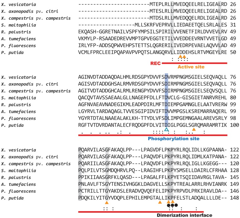
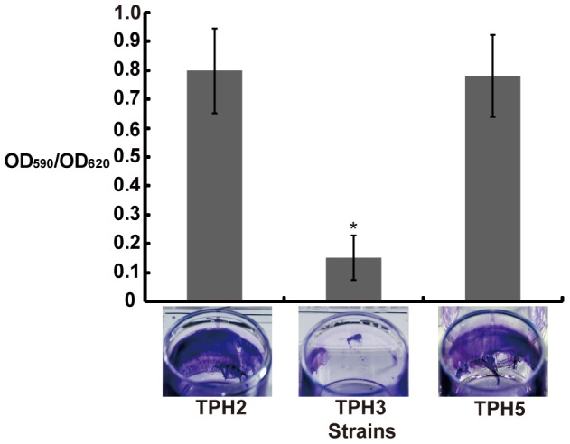
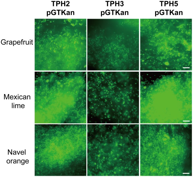
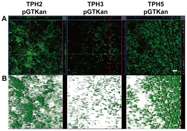
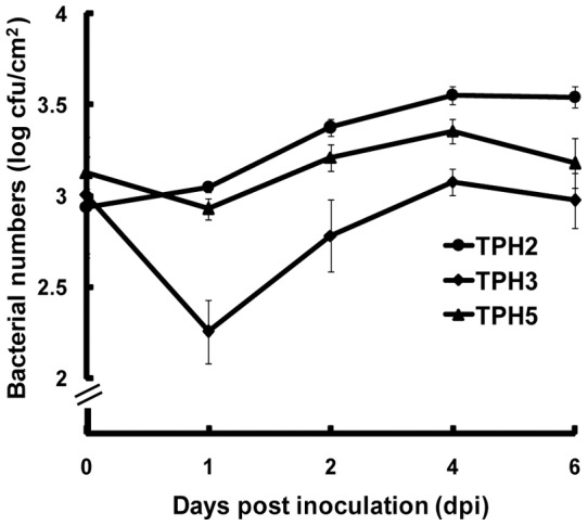
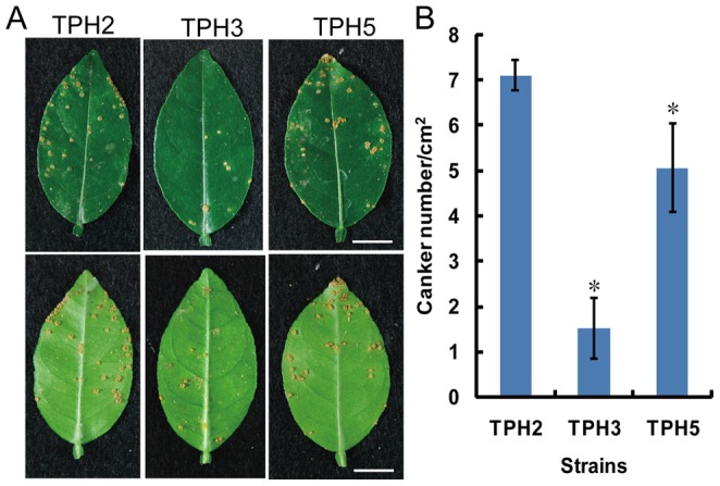
Citrus bacterial canker caused by Xanthomonas axonopodis pv. citri is a serious disease that impacts citrus production worldwide, and X. axonopodis pv. citri is listed as a quarantine pest in certain countries. Biofilm formation is important for the successful development of a pathogenic relationship between various bacteria and their host(s). To understand the mechanisms of biofilm formation by X. axonopodis pv. citri strain XW19, the strain was subjected to transposon mutagenesis. One mutant with a mutation in a two-component response regulator gene that was deficient in biofilm formation on a polystyrene microplate was selected for further study. The protein was designated as BfdR for biofilm formation defective regulator. BfdR from strain XW19 shares 100% amino acid sequence identity with XAC1284 of X. axonopodis pv. citri strain 306 and 30-100% identity with two-component response regulators in various pathogens and environmental microorganisms. The bfdR mutant strain exhibited significantly decreased biofilm formation on the leaf surfaces of Mexican lime compared with the wild type strain. The bfdR mutant was also compromised in its ability to cause canker lesions. The wild-type phenotype was restored by providing pbfdR in trans in the bfdR mutant. Our data indicated that BfdR did not regulate the production of virulence-related extracellular enzymes including amylase, lipase, protease, and lecithinase or the expression of hrpG, rfbC, and katE; however, BfdR controlled the expression of rpfF in XVM2 medium, which mimics cytoplasmic fluids in planta. In conclusion, biofilm formation on leaf surfaces of citrus is important for canker development in X. axonopodis pv. citri XW19. The process is controlled by the two-component response regulator BfdR via regulation of rpfF, which is required for the biosynthesis of a diffusible signal factor.
Conflict of interest statement
Figures
Similar articles
-
Biofilm formation, epiphytic fitness, and canker development in Xanthomonas axonopodis pv. citri.Mol Plant Microbe Interact. 2007 Oct;20(10):1222-30. doi: 10.1094/MPMI-20-10-1222. Mol Plant Microbe Interact. 2007. PMID: 17918624
-
A LOV protein modulates the physiological attributes of Xanthomonas axonopodis pv. citri relevant for host plant colonization.PLoS One. 2012;7(6):e38226. doi: 10.1371/journal.pone.0038226. Epub 2012 Jun 4. PLoS One. 2012. PMID: 22675525 Free PMC article.
-
Insights into xanthomonas axonopodis pv. citri biofilm through proteomics.BMC Microbiol. 2013 Aug 7;13:186. doi: 10.1186/1471-2180-13-186. BMC Microbiol. 2013. PMID: 23924281 Free PMC article.
-
Genome-wide mutagenesis of Xanthomonas axonopodis pv. citri reveals novel genetic determinants and regulation mechanisms of biofilm formation.PLoS One. 2011;6(7):e21804. doi: 10.1371/journal.pone.0021804. Epub 2011 Jul 5. PLoS One. 2011. PMID: 21750733 Free PMC article.
-
Bacteria causing important diseases of citrus utilise distinct modes of pathogenesis to attack a common host.Appl Microbiol Biotechnol. 2010 Jun;87(2):467-77. doi: 10.1007/s00253-010-2631-2. Epub 2010 May 7. Appl Microbiol Biotechnol. 2010. PMID: 20449739 Review.
Cited by
-
CitB is required for full virulence of Xanthomonas oryzae pv. oryzae.World J Microbiol Biotechnol. 2015 Oct;31(10):1619-27. doi: 10.1007/s11274-015-1914-2. Epub 2015 Aug 7. World J Microbiol Biotechnol. 2015. PMID: 26250545
-
Genome-Wide Screening for Novel Candidate Virulence Related Response Regulator Genes in Xanthomonas oryzae pv. oryzicola.Front Microbiol. 2018 Aug 7;9:1789. doi: 10.3389/fmicb.2018.01789. eCollection 2018. Front Microbiol. 2018. PMID: 30131784 Free PMC article.
-
Antibacterial and biofilm inhibition activity of biofabricated silver nanoparticles against Xanthomonas oryzae pv. oryzae causing blight disease of rice instigates disease suppression.World J Microbiol Biotechnol. 2020 Mar 16;36(4):55. doi: 10.1007/s11274-020-02826-1. World J Microbiol Biotechnol. 2020. PMID: 32180020
-
Citrus Canker Pathogen, Its Mechanism of Infection, Eradication, and Impacts.Plants (Basel). 2022 Dec 26;12(1):123. doi: 10.3390/plants12010123. Plants (Basel). 2022. PMID: 36616252 Free PMC article. Review.
-
A LysR-Type Transcriptional Regulator LcrX Is Involved in Virulence, Biofilm Formation, Swimming Motility, Siderophore Secretion, and Growth in Sugar Sources in Xanthomonas axonopodis Pv. glycines.Front Plant Sci. 2020 Jan 10;10:1657. doi: 10.3389/fpls.2019.01657. eCollection 2019. Front Plant Sci. 2020. PMID: 31998344 Free PMC article.
References
-
- Schaad NW, Postnikova E, Lacy GH, Sechler A, Agarkova I, et al. (2005) Reclassification of Xanthomonas campestris pv. citri (ex Hasse 1915) Dye 1978 forms A, B/C/D, and E as X. smithii subsp. citri (ex Hasse) sp. nov. nom. rev. comb. nov., X. fuscans subsp. aurantifolii (ex Gabriel 1989) sp. nov. nom. rev. comb. nov., and X. alfalfae subsp. citrumelo (ex Riker and Jones) Gabriel, et al., 1989 sp. nov. nom. rev. comb. nov.; X. campestris pv. malvacearum (ex smith 1901) Dye 1978 as X. smithii subsp. smithii nov. comb. nov. nom. nov.; X. campestris pv. alfalfae (ex Riker and Jones, 1935) dye 1978 as X. alfalfae subsp. alfalfae (ex Riker, et al., 1935) sp. nov. nom. rev.; and “var. fuscans” of X. campestris pv. phaseoli (ex Smith, 1987) Dye 1978 as X. fuscans subsp. fuscans sp. nov. Syst Appl Microbiol 28: 494–518. - PubMed
-
- Ryan RP, Vorhölter F-J, Potnis N, Jones JB, Van Sluys M-A, et al. (2011) Pathogenomics of Xanthomonas: understanding bacterium–plant interactions. Nat Rev Microbiol 9: 344–355. - PubMed
-
- Brunings AM, Gabriel DW (2003) Xanthomonas citri: breaking the surface. Mol Plant Pathol 4: 141–157. - PubMed
-
- Characklis WG, Marshall KC (1990) Biofilms: a basis for an interdisciplinary approach. In: Characklis WG, Marshall KC, editors. Biofilms. New York: A Wiley-Interscience Publication, John Wiley & Sons, Inc. 3–16.
-
- Rigano LA, Siciliano F, Enrique R, Sendin L, Filippone P, et al. (2007) Biofilm formation, epiphytic fitness, and canker development in Xanthomonas axonopodis pv. citri . Mol Plant-Microbe Interact 20: 1222–1230. - PubMed
Publication types
MeSH terms
Substances
Associated data
- Actions
- Actions
LinkOut - more resources
Full Text Sources
Other Literature Sources
Molecular Biology Databases
Research Materials
